Growth of ZnO nanorods by hydrothermal method under different temperatures
Bạn đang xem bản rút gọn của tài liệu. Xem và tải ngay bản đầy đủ của tài liệu tại đây (1.9 MB, 4 trang )
Growth Of ZnO Nanorods by Hydrothermal
Method Under Different Temperatures
T. H. Meen, W. Water, Y. S. Chen*, W. R. Chen, L. W. Ji and C. J. Huang
Abstract –In this study, the aqueous solution
method was employed to synthesize one-dimensional
well-aligned ZnO nano-array on ITO glass substrate.
We can find that the dimension of ZnO nanorod will
changes with different growth temperature. X-ray
diffraction patterns show that the nanorods are
high-quality crystals growing along [001] direction
with a high consistent orientation perpendicular to the
substrate while the growth temperature is equal to 80.
SEM images show that the average diameters of ZnO
nanorods are about 60-90 nm by changing growth
temperature. The smallest diameter of ZnO nanorods
is observed while the growth temperature is equal to
75 ℃. The UV/Vis spectra analyses show the
absorption peaks appear at 330nm, 370nm and 390nm
while growth temperature increases from 65 ℃ to 85
℃.
I.
INTRODUCTION
ZnO nanostructure has been envisioned to enhance
performance of various technologically important devices
such as short-wavelength lasers[1], Gratzel-type solar cell
[2],[3], and chemical sensors [4],[5]. The interest in
synthesis of well-aligned ZnO nanowires or nanorods on
substrates keeps growing. ZnO has shown a great deal of
research in DSSCs [6]–[9] due to some of its fascinating
properties. Comparing with other semiconductors, ZnO
has unique excellent properties, such as higher binding
energy (60meV), wide band gap (3.37 eV), high
breakdown strength, cohesion, and exciton stability.
Moreover, ZnO is one of the hardest materials in the
family of II–VI semiconductors. Electron mobility in
ZnO is more than that in TiO2 making the former suitable
for DSSCs. Recently, it has become possible to form
vertical nanowires of ZnO [10]. Such nanowires
expectedly provide morphology for better electron
transport. The vertical geometry also provides a more
open structure for filling with hole-transporting
materials[11]-[14]. The preparation of 1D ZnO
nanostructures has been demonstrated by various
methods, including vapor–liquid–solid (VLS) growth
T. H. Meen, W. Water, Y. S. Chen W. R. Chen and L. W. Ji
are with the Institute of Electro-Optical and Materials Science,
and Department of Electronic Engineering, National Formosa
University, Yunlin 632, Taiwan, R.O.C. C. J. Huang is with
the Department of Applied Physics, National University
of Kaohsiung, Nan-Tzu 811, Kaohsiung, R. O. C.
E-mail:
1-4244-0637-4/07/$20.00 ©2007 IEEE
617
[15],[16], chemical vapor deposition (CVD) [17],[18],
hydrothermal process [19], and template-based methods
[20]. However, these growth techniques usually
expensive, and the choice of substrate restricted, complex
process controlling and high temperature are unfavorable
for an industrialized process. Recently, a solution-based
approach was developed to achieve highly oriented
nanorods film with high surface area on substrate, which
has the advantages of mild synthetic conditions, simple
manipulation and large scale-up production. It opens a
door for future optoelectronic devices based on ZnO
nanostructure arrays [21]–[25]. In this work, we report
the hydrothermal growth of high quality ZnO nanorods
perpendicularly oriented on ITO substrates, and
investigate them by X-ray diffraction, scanning
electronmicroscopy (SEM) and ultraviolet-visible
absorption spectra analyses. These high quality ZnO
nanorods can be applied on the electrode of
dye-sensitized solar cell to increase the contact area
between ZnO and dye, resulting in the enhancement of
efficiency for dye-sensitized solar cell.
II.
EXPERIMENTAL
The ZnO nanorods were prepared from zinc nitrate in
a neutral aqueous solution under hydrothermal conditions.
The procedure consists of two steps: (1) deposition of
ITO substrates with densely and uniformly ZnO films by
RF sputter as the buffer layer, and (2) hydrothermal
growth of ZnO nanorods in aqueous solution. In detail,
the aqueous solutions of zinc nitrate (0.5g) and
methenamine (0.5g) were stirred uniformly. An 80 nm
thick ZnO layer was first deposited on ITO glass using a
RF sputter deposition system under an Ar and O2 pressure
of 5x10⁻²torr. The hydrothermal growth was carried out at
65 ℃ ~ 85 ℃ in a sealed beaker by immersing the
modified substrates in the aqueous solution (100ml)
containing Zn(NO3)2 (0.5 M) and methenamine (0.35 M)
for 10 hours. The morphology, structure, and optical
properties of ZnO nanorods were studied by X-ray
diffraction (XRD), scanning electron microscope (SEM),
and Ultraviolet-Visible spectrophotometer (UV/Vis
spectrophotometer).
III. RESUATS AND DISCUSSION
The crystal structure of as-prepared ZnO nanorods
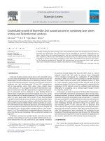
was analyzed by XRD. X-ray diffraction patterns of ZnO
nanorods with different growth temperature are shown in
Fig.1. All diffraction peaks well indexed to the standard
diffraction pattern of hexagonal ZnO phase except for
2θ=36o and 37o. In comparison with the standard XRD
pattern of ZnO, the much higher relative intensity of the
(002) diffraction peak provides further evidence that the
nanorods are preferentially oriented in the c-axis
direction. The strongest (002) peak of diffraction pattern
appears while the growth temperature is equal to 80 ℃.
Fig. 1. X-ray diffraction patterns of ZnO nanorods with
different growth temperature.
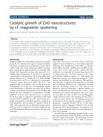
SEM was used to investigate the nanostructure of
ZnO nanorods. Figures 2 show the SEM images of ZnO
nanorods obtained under different growth temperatures.
They show that a dense array of hexagonal ZnO nanorods
having a diameter of from 30nm to 150nm are formed
under different growth temperatures, and the average
diameters of ZnO nanorods are listed in Table I. It is
noted that Fig. 2(d) shows the best nanostructure of ZnO
nanorods. From the results of Fig. 1 and Figs. 2, the best
growth temperature of ZnO nanorods is 80 ℃. The
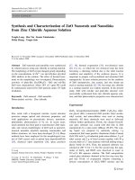
cross-section image of ZnO nanorods arrays grown at 80
℃ is shown in Fig. 3. It is found that all ZnO nanorods
grow almost vertically from the substrate, and the length
of nanorods is about 1.3um.
TABLE I
THE AVERAGE DIAMETER OF ZNO NANORODS
WITH DIFFERENT GROWTH TEMPERATURES
Fig. 2. SEM images of ZnO nanorods with different
growth temperatures:
(a)65
(d)80 ℃(e)85 ℃
618
℃(b)70 ℃(c)75 ℃
electrode of dye-sensitized solar cell to increase the
contact area between ZnO and dye, resulting in the
enhancement of efficiency for dye-sensitized solar cell.
ACKNOWLEDGEMENT
The research is supported by National Science
Council, R.O.C. under contract Nos. NSC 96-2622E-150-027-CC3 and NSC 96-2221-E-150-028.
REFERENCES
Fig. 3. A cross-section view of SEM image of ZnO
nanorods with growth temperature equal to 80 ℃.
Figure 4 shows the UV-Vis absorption spectra of ZnO
nanorods under different growth temperatures. The
absorption peaks appear at 330nm, 370nm and 390nm
while the growth temperature increases from 65 ℃ to 85
℃, and the strongest absorption peak at 390nm is
observed while the growth temperature is equal to 75 ℃.
It is indicated that the smallest average diameter of ZnO
nanorods has the best absorption for UV light. From the
results of XRD, SEM and UV-Vis analyses for ZnO
nanorods, we can apply these high quality ZnO nanorods
on the electrode of dye-sensitized solar cell to increase
the contact area between ZnO and dye, resulting in the
enhancement of efficiency for dye-sensitized solar cell.
Fig.4 The UV-Vis absorption spectra of ZnO under
different growth temperature from 65 ℃ to 85 ℃.
IV. CONCLUSION
In this study, we have successfully synthesized ZnO
nanorods on ITO glass substrate. From the results of
XRD and SEM, the best growth temperature of ZnO
nanorods is 80 ℃, at which the average diameter and
length of ZnO nanorods are about 70.4 nm and 1.3um.
The absorption peaks appear at 330nm, 370nm and
390nm while the growth temperature increases from 65
℃ to 85 ℃, and the strongest absorption peak at 390nm is
observed while the growth temperature is equal to 75 ℃.
These high quality ZnO nanorods can be applied on the
619
[1] M.H. Huang, S. Mao, H. Feick, H. Yan, Y. Wu,
H.Kind,E.Weber, R. Russo, P. Yang, “Room-temperature
ultraviolet nanowire nanolasers,” Science vol.292,p.1897,
2001.
[2] J. Zhong, A.H. Kitai, P. Mascher, W. Puff, “Effect of
substrate temperature on the growth and luminescence
properties of ZnO nanostructures,” J Electrochem. Soc.
vol.140,p.3644, 1993.
[3] N. Beermann, L. Vayssieres, S.-E. Lindquist, A.Hagfeldt,
“Photoelectrochemical studies of oriented nanorod thin
films of Hematite,”J. Electrochem. Soc. vol.147,p.2456,
2000.
[4] N. Yamazoe, “Photoelectrochemical studies of oriented
nanorod thin films of Hematite,”Sensors Actuators B
vol.5,p.7, 1991.
[5] G.S. Trivikrama Rao, D. Tarakarama Rao, “Study on
Sensitivity of Nano-Grain ZnO Gas Sensors,”Sensors
Actuators B vol.55, p.166, 1999.
[6] M. Law, L.E. Greene, J.C. Johnson, R. Saykally, P. Yang,
“Nanowire dye-sensitized solar cells,” Nat.Mater.
vol.4 ,p.455, 2005.
[7] T. Yoshida, K. Terada, D. Schlettwein, T. Oekermann, T.
Sugiura,H.
Minoura,
“Electrochemical
and
Photoelectrochemical
Properties
of
Organic
Semiconductors - Dye-Sensitization in Nanostructured
Hybrid Materials,” Adv. Mater. vol.12,p.1214, 2000.
[8] J.B. Baxter, E.S. Aydil, “Nanowire-based dye-sensitized
solar cells,”Appl. Phys. Lett. vol.86,p.53114, 2005 .
[9] E. Hosono, S. Fujihara, I. Honma, H. Zhou, “The
Fabrication of an Upright-Standing Zinc Oxide
Nanosheet for Use in Dye-Sensitized Solar Cells,” Adv.
Mater. vol.17,p.2091, 2005.
[10] L.E. Greene, M. Law, D.H.Tan, MMontano,J.Goldberger
,G. Somorjai, P. Yang, “ZnO Nanowire/p-GaN
Heterojunction LEDs,”Nano. Lett. vol.5, p.1231, 2005.
[11] W.U. Huynh, J.J. Dittmer, A.P. Alivisatos, “Investigation
of Properties of ZnO Nanorad Structures by Chemical
Vapor Deposition,” Science vol.295,p.2425, 2002.
[12] T. Stu binger, W. Bru tting, “Exciton diffusion and
optical interference in organic donor-acceptor photovotaic
cells,” J. Appl. Phys. vol.90,p.3632, 2001.
[13] C.J. Brabec, N.S. Sariciftci, J.C. Hummelen, “Origin of
the Open Circuit Voltage of Plastic Solar Cells,” Adv.
Funct. Mater. vol.11,p.15, 2001.
[14] B. Pradhan, A. Bandyopadhyay, A. J Pal, “Tuning
performance of donor-acceptor based self-assembled
photovoltaicdevices,”Appl. Phys.Lett.vol.85,p.633, 2004.
[15] M.H. Huang, Y.Wu, H. Feick, N. Tran, E.Weber, P.
Yang, “Catalytic growth of zinc oxide nanowires by
vapor transport,”Adv.Mater. vol.13,p.113, 2001.
[16] Y.C. Kong, D.P. Yu, B. Zhang,W. Fang, S.Q. Feng,
“Ultraviolet-emitting ZnO nanowires synthesized by a
[17]
[18]
[19]
[20]
[21]
physical vapor deposition approach,”Appl. Phys.Lett.
vol.78,p.407, 2001.
J.-J. Wu, S.-C. Liu, “Low-Temperature and Catalyst-Free
Synthesis of Well-Aligned ZnO Nanorods on Si (100),”
Adv. Mater. vol.14,p.215, 2002.
J.-J. Wu, S.-C. Liu, “Catalyst-Free Growth and
Characterization of ZnO Nanorods,”J. Phys. Chem. B
vol.106,p.9546, 2002.
J. Zhang, L. Sun, H. Pan, C. Liao, C. Yan, “ZnO
nanowires fabricated by a convenient route,” New J.
Chem. vol.26,p.33, 2002.
Y. Li, G.W. Meng, L.D. Zhang, F. Phillipp, “Ordered
semiconductor ZnO nanowire. arrays and their
photoluminescence properties,” Appl. Phys. Lett.
vol.76,p.2011, 2000.
L. Vayssieres, “Growth of arrayed nanorods and
nanowires of ZnO from aqueous solutions,” Adv. Mater.
620
vol.15,p.464, 2003.
[22] L. Vayssieres, K. Keis, A. Hagfeldt, S. Lindquist,
“Three-dimensional array of highly oriented crystalline
ZnO microtubes,”Chem. Mater. vol.13,p.4395, 2001.
[23] L.E. Greene,M. Law, J. Goldberger, F. Kim, J.C.
Johnson, Y. Zhang,R.J. Saykally, P. Yang, Angew.
“Low-temperature wafer-scale production of ZnO
nanowire arrays,” Chem. Int. Ed. vol.42 ,p.3031, 2003.
[24] J. Choy, E. Jang, J. Won, J. Chung, D. Jang, Y. Kim,
“Soft Solution Route to Directionally Grown ZnO
Nanorod Arrays on Si Wafer,”Adv. Mater. vol.15,p.1911,
2003.
[25] K. Govender, D. Boyle, P. Kenway, P. O’Brien,
“Understanding the factors that govern the deposition and
morphology of thin films of ZnO from aqueous
solution,”J. Mater. Chem. vol.14,p.2527, 2004.