Do plant exudates shape the root microbiome
Bạn đang xem bản rút gọn của tài liệu. Xem và tải ngay bản đầy đủ của tài liệu tại đây (2.88 MB, 18 trang )
Lawrence Berkeley National Laboratory
Recent Work
Title
Feed Your Friends: Do Plant Exudates Shape the Root Microbiome?
Permalink
/>
Journal
Trends in Plant Science, 23(1)
ISSN
1360-1385
Authors
Sasse, J
Martinoia, E
Northen, T
Publication Date
2018
DOI
10.1016/j.tplants.2017.09.003
Peer reviewed
eScholarship.org
Powered by the California Digital Library
University of California
TRPLSC 1597 No. of Pages 17
Feature Review
Feed Your Friends: Do Plant
Exudates Shape the Root
Microbiome?
Joelle Sasse,1 Enrico Martinoia,2 and Trent Northen1,3,*
Plant health in natural environments depends on interactions with complex and
dynamic communities comprising macro- and microorganisms. While many
studies have provided insights into the composition of rhizosphere microbiomes (rhizobiomes), little is known about whether plants shape their rhizobiomes. Here, we discuss physiological factors of plants that may govern plant–
microbe interactions, focusing on root physiology and the role of root exudates.
Given that only a few plant transport proteins are known to be involved in root
metabolite export, we suggest novel families putatively involved in this process.
Finally, building off of the features discussed in this review, and in analogy to
[580_TD$IF]well-known symbioses, we elaborate on a possible sequence of events governing rhizobiome assembly.
Trends
Recent advances in sequencing technology have been enabling highthroughput characterization of highly
complex plant-associated microbial
communities, which are relevant for
plant health.
Metagenomic approaches have been
identifying the metabolic potential of
microbes, and are starting to reveal
functional groups of microbes, reducing the overall complexity of
rhizobiomes.
Metabolomic studies are uncovering
differential root exudation in various
environments and plant developmental stages, as well as consumption of
specific exometabolites by microbes.
The Root Microbiome (Rhizobiome)
Plant growth and yield in natural environments depend on a plethora of interactions with
bacteria and fungi [1] (one example is discussed in Box 1). The microbial community associated
with roots was proposed to be assembled in two steps: first, the rhizosphere (see Glossary) is
colonized by a subset of the bulk soil community and, second, the rhizoplane and the
endosphere are colonized by a subset of the rhizosphere community [2,3]. Intriguingly, a
set of recurring plant-associated microbes has emerged (core microbiome) [581_TD$IF][2,4]. This review
focuses on how plants shape their rhizobiome. On the one hand, common factors among
plants likely lead to the assembly of the core microbiome. On the other hand, factors specific to
certain plants result in [582_TD$IF]an association with microbes that are not members of the core microbiome. Here, we discuss evidence that plant genetic factors, specifically root morphology and
root exudation, shape rhizobiomes.
Initial evidence for an influence of plant genotype on rhizobiome composition was that similar
rhizobiomes assembled in association with arabidopsis (Arabidopsis thaliana) and barley
(Hordeum vulgare) grown in the same experimental conditions, although they displayed
different relative abundances and some specific taxonomic groups [5]. A correlation between
phylogenetic host distance and rhizobiome clustering was described for Poaceae species [6],
distant relatives of arabidopsis [7], rice varieties [3], and maize lines (Zea mays) [6], but not for
closely related arabidopsis species and ecotypes [7]. Distinct rhizobiomes were also described
for domesticated plants, such as barley, maize, agave (Agave sp.), beet (Beta vulgaris), and
lettuce (Lactuca sp.), compared with their respective wild relatives [5,8–11]. Interestingly, not all
plants have a rhizobiome distinct from bulk soil: some species, such as maize and lotus (Lotus
japonicus) [12–15], have assembled a distinct rhizobiome, whereas other species, such as
arabidopsis and rice, assembled a rhizobiome similar to bulk soil [3,5,7]. The former species
display a strong, and the latter a weak rhizosphere effect (Figure 1, Key Figure). The cause of
Trends in Plant Science, Month Year, Vol. xx, No. yy
Genome-wide association studies and
other
comparative
genomic
approaches have been revealing a link
between plant genetic factors, exudation
profiles,
and
microbiome
compositions.
Molecular studies have been characterizing plant transport proteins necessary for interactions with specific
microbes, although the transport of
most nutrients and signaling molecules
remains unexplored.
1
Lawrence Berkeley National
Laboratory, Berkeley, CA 94720, USA
2
Department of Plant and Microbial
Biology, University of Zurich, Zurich
8008, Switzerland
3
Joint Genome Institute, Walnut
Creek, CA 94958, USA
*Correspondence:.
(T. Northen).
/>
1
TRPLSC 1597 No. of Pages 17
Box 1. Phytoremediation: Interplay of Exudates, Border Cells, and Rhizobiomes
Plants grown on soils contaminated with heavy metals and organic pollutants (phytoremediation) assemble a rhizobiome that is distinct from that of plants grown on non-contaminated rhizosphere, or bulk soils [50_TD$IF][111–113] supporting
plant growth [114,115] and higher heavy metal uptake [116]. Consequently, efforts have aimed at increasing the
phytoremediation potential of heavy metal accumulators by combining them with specific microbial communities.
However, due to limited understanding of the plant–microbe–environment interplay, these endeavors have had limited
success so far [51_TD$IF][111,112]. Below, we discuss both, the response of plants and microbes to contaminated soils.
Plants display distinct responses to contaminated soils: legumes exhibited a systemic response, [52_TD$IF]whereas grasses
exhibited a more local response [117]. Various wheat cultivars displayed varying degrees of heavy metal tolerance,
which were [53_TD$IF]associated not only with distinct rhizobiomes [118], but also with the ability to exude organic acids [54_TD$IF][75].
Tolerant lines increased the expression of specific genes, such as the malate transporter ALMT1 (see Box 2 and [5_TD$IF]Table 1
in the main text) and organic acid exporters of the MATE family [50_TD$IF][119]. Organic acid exudation has two main effects. [56_TD$IF]First,
(heavy) metal ions are chelated, and, second, they can act as anion exchanger and release tightly bound phosphate,
supporting plant growth [51_TD$IF][111,112]. Increased organic acid exudation could also lead to increased nutrient availability for
the microbial community, and have signaling functions. A second physiological response to protect the root from heavy
metals is an increased production and shedding of border cells accumulating heavy metals [57_TD$IF][120]. The interplay of border
cell production and differential exudation by alteration of transporter abundance not only determines [58_TD$IF]the performance of
the plant on contaminated soils, but also the environment for the microbial community.
Similar to plants, microbes in contaminated soils are under selective pressure from several sides: they have to tolerate
the toxic environment, grow on root exudates, and compete for niches and resources [59_TD$IF][111,114]. Thus, it is no surprise
that rhizobiomes of contaminated soils were found to be generally less diverse compared [560_TD$IF]with other environments [114].
The addition of a substrate mix or the supply of plants with distinct exudation patterns [561_TD$IF]to contaminated soils could
increase of the growth of specifically engineered or native microbes, leading to an increase [562_TD$IF]in the functional traits of the
rhizobiome, and the phytoremediation potential of the microbial community. In addition, a tritrophic bacteria–fungi–plant
interactions on contaminated soil was reported recently: microbes simultaneously increased arsenic tolerance in rice
and resistance against disease [563_TD$IF][115]. Overall, further investigations of the roles of exudates, transporters, border cells,
and bacterial and fungal communities will contribute to deciphering the effects of contaminated soils on plants, and lead
to more- efficient phytoremediation procedures.
this phenomenon is currently unknown. The strength of the rhizosphere effect varies with the
developmental stage of the plant [16–18]. Similarly, root exudation [16] and microbial communities were found to change with the age of the plant.
Furthermore, distinct rhizobiomes were associated with different developmental stages of
arabidopsis [16,19], rice [3], and Avena fatua grown during two consecutive seasons [20].
Pioneering studies demonstrated the ability of microbes to alter plant development[583_TD$IF]. Overall, it
appears evident that host genotype, domestication, and plant development influence the
composition of rhizobiomes. As an alternative to plant developmental stage, residence time
of plants in soil was discussed as a hypothesis for successive microbiomes [584_TD$IF][21]. These
contrasting results might be partially explained by differing environmental influences, host
plants, or soils, and additional work is needed to resolve these questions.
In this review, we discuss root morphology and root exudates as two genetic factors shaping
plant–microbiome interactions, and we examine the following aspects: (i) how root morphology
and border cells affect rhizobiomes; (ii) how plant exudates shape the rhizobiome; and (iii)
possible plant transport proteins involved in exudation. Figure 1 provides a general overview of
exometabolite networks in the rhizosphere, and Box 1 illustrates the interplay between root
exudates, border cells, and rhizobiomes in phytoremediation. We conclude by integrating
these ideas into a possible scenario of rhizobiome assembly.
Root Physiological Features Shape Rhizobiomes and Exudation
Rhizobiomes are influenced by their spatial orientation towards roots in two ways. First, the
radial proximity of microbial communities to roots defines community complexity and composition, as described in recent publications [58_TD$IF][3,19,22], and as outlined by the two-step model of
2
Trends in Plant Science, Month Year, Vol. xx, No. yy
Glossary
Antiporter: a transporter utilizing a
proton gradient to shuttle protons
and substrates in opposite directions
across a membrane.
Apoplasm: intercellular space
between plasma membranes of plant
tissue.
Border cells: root cap cells released
into the rhizosphere with a distinct
transcriptome, releasing mucilage,
distinct proteins, and extracellular
DNA.
Casparian strip: suberin-based
connection between endodermal root
cells, blocking the passive
apoplasmic flow of liquids and
compounds.
Endophyte: microbe living within
plant tissue in the endosphere.
Endosphere: all endophytes of a
plant. Relevant to this review are
microbes living in root tissues.
Epiphyte: microbe living on plant
tissue.
Exometabolites: compounds
released by roots or microbes into
the rhizosphere, often acting either
as nutrients or signaling molecules.
Isolate: a microbial strain isolated
from a natural environment, such as
the rhizosphere, to be used in a
laboratory setting.
Mucilage: matrix of high-molecularweight compounds released by
border cells and root tip.
Phytoremediation: accumulation of
heavy metals or organic pollutants of
contaminated soils in plant tissue,
with the aim to clean soils.
Rhizobiome: the rhizosphere
microbiome, comprising microbes
associated with plant roots.
Rhizoplane: root surface including
tightly adhered microbes.
Rhizosphere: 1–3-mm zone around
root shaped by roots, and exudates.
Rhizosphere effect: plants with a
strong rhizosphere effect have a
rhizosphere microbiome distinct from
bulk soil. Developmental stage and
the type of plant species both
influence the strength of the
rhizosphere effect.
Symporter: a transporter utilizing a
proton gradient to shuttle protons
and substrates in the same direction
across a membrane.
Uniporter: a transporter binding one
substrate molecule at a time,
facilitating diffusion across a
membrane.
TRPLSC 1597 No. of Pages 17
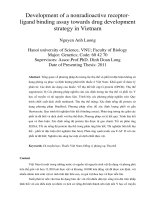
Key Figure
Plant and Microbial Exometabolite Networks
Figure 1. Plant roots and border cells (brown) and microbes (blue) synthesize metabolites and transporters (boxes), and
export certain metabolites into the rhizosphere. This network is depicted by broken arrows. Exometabolites can have
nutritional value and signaling functions ([49_TD$IF]unbroken arrows indicate direction; if the metabolite has only a nutrient or signaling
function, the role is specified in brackets). Some microbial epiphytes can migrate from the rhizosphere into the rhizoplane
and into the root, where they become endophytes. Plant–microbe and microbe–microbe exometabolite interactions are
displayed with numbers: (1) substrate competition between microbes, or between microbes and roots; (2) plant growth
promotion by microbial compounds; and (3) rhizosphere effect, likely influenced by the presence of exometabolites. Plant
and microbial exudates are displayed as gradients. Organisms and cells are not to scale.
microbial root colonization mentioned above [2]. Second, the lateral position of microbes along
a root shapes the community, as exemplified by early studies [586_TD$IF][23,24]. Importantly, recent
microbiome studies take into consideration the former, but not the latter aspect. In this section,
we discuss specific microbial associations with various root regions, and the role of spatially
distinct root exudation.
Root tips are the first tissues that make contact with bulk soil: root tips are associated with the
highest numbers of active bacteria compared with other root tissues, and likely select microbes
in an active manner [587_TD$IF][23]. The root elongation zone is specifically colonized by Bacillus subtilis,
which suggests a particular role of this zone in plant–microbe interactions [58_TD$IF][25]. Mature root
zones feature a microbial community distinct from root tips [58_TD$IF][25]. Their community includes
decomposers [589_TD$IF][11,24], which could be involved in the degradation of dead cells shedding from
old root parts [590_TD$IF][26]. Similarly, lateral roots are associated with distinct microbial communities,
differing between tips and bases, as well as between different types of lateral root [14].
One trait influencing the differential microbial colonization of root tissues could be the differential
exudation profiles of the distinct root parts. This is illustrated in the following example. Cluster
Trends in Plant Science, Month Year, Vol. xx, No. yy
3
TRPLSC 1597 No. of Pages 17
roots are densely packed lateral roots formed by some plants growing on extremely nutrient-poor
soils; these roots exude high amounts of organic acids and, in some cases, protons, to solubilize
phosphate [591_TD$IF][27]. The low pH and carboxylate-rich rhizosphere of cluster roots is associated with a
specialized rhizobiome, dominated by Burkholderia species that metabolize citrate and oxalate
[592_TD$IF][28]. Besides organic acids, mature cluster roots also exude isoflavonoids and fungal cell walldegrading enzymes, leading to a decrease in bacterial abundance, as well as fungal sporulation
[593_TD$IF][29]. Taken together, cluster root exudates not only solubilize phosphate, but also regulate
microbes in such a way that they do not interfere with phosphate uptake. Beyond this example,
spatial patterns of metabolite exudation are largely unexplored. We hypothesize that such patterns
exist in all root systems for the following reasons: (i) spatially distinct organic acid exudation is a trait
of all root systems (Table 1 [594_TD$IF]and Box 1); (ii) spatially distinct exudation was similarly detected for
strigolactones, amino acids, and sugars (Table 1) [59_TD$IF][30,31]; and (iii) root nutrient uptake, which is
sometimes coupled with proton transport, can also exhibit spatial patterns (Table 1). Overall,
spatially defined metabolite exudation by distinct root parts is likely an important factor in
structuring the rhizobiome. Future studies should aim at characterizing spatially distinct rhizobiomes and their functional traits, and at investigating spatially distinct root exudation.
Root Border Cells and Mucilage Shape Plant–Microbe Interactions
Root tips are not only associated with high numbers of bacteria ([148_TD$IF][24], see above), but also produce
border cells and mucilage (Figure 1), crucial for plant–microbe interactions. Depending on the
root meristem organization, border cells are released into the rhizosphere either as single cells or as
border-like cells (which remain attached to each other). Residence time in the soil is different for the
two types of border cell. Single maize border cells stayed alive in soil for months, likely due to the
presence of starch deposits [596_TD$IF][32], whereas arabidopsis border-like cells survived for only 2 weeks
[597_TD$IF][33]. Border cells have a transcriptional profile distinct from root tip wells, with overall lower primary
and higher secondary metabolism [596_TD$IF][32]. ABC transporters constitute a large fraction of differentially
expressed genes, which is consistent with transport of secondary metabolites [598_TD$IF][32,34]. Secondary
metabolites are likely central to the role of border cells in defense against pathogens [59_TD$IF][35–37].
Pathogen attack can result not only in higher border cell production and release [59_TD$IF][35–37], but
also in higher mucilage production by border cells and root tip cells. Mucilage contains proteins
with antimicrobial functions [60_TD$IF][35,38,39], as well as extracellular DNA involved in defense against
fungi [601_TD$IF][40] and certain bacteria [602_TD$IF][41]. Importantly, mucilage is also produced under nonpathogenic conditions, [603_TD$IF]serving as a lubricant for the root environment and stabilizing soil particles
[42]. Interestingly, mucilage also provides distinct carbon sources for microbes, thus influencing rhizobiome composition [604_TD$IF][43,44].
Border cells similarly interact with nonpathogenic microbes (Figure 2): they release flavonoids
that attract rhizobia, uncharacterized compounds that induce branching of mycorrhizal hyphae,
and arabinogalactans that trigger biofilm formation of specific beneficial bacteria (Box 2)
[605_TD$IF][32,33,45,46]. The full extent of how border cells and mucilage shape root–microbe interactions remains unclear. It is tempting to speculate that the specialized metabolism of the
border cells results in a distinct exudation profile of not only proteins and mucilage, but also lowmolecular-weight compounds that could serve as microbial nutrients or as signaling compounds. Further research should focus on the genetic and physiological differences between
border cells and border-like cells, as well as on the transport proteins involved in exudation of
low-molecular-weight compounds, DNA, and proteins.
How Microbial Communities Interact, and the Influence of Plant Exudates
Plant–microbe interactions are not only defined by plant root morphology and plant-derived
exudates, but also by microbe–microbe interactions (Figure 1). Thus, we focus further here on
4
Trends in Plant Science, Month Year, Vol. xx, No. yy
TRPLSC 1597 No. of Pages 17
Table 1. Transporters for Metabolite Uptake and Releasea,b,c
Transport mode
Metabolite examples
Transporter
family
Description and localization
Refs
Sugars (glucose, fructose, sucrose, arabinose, xylose, mannose, maltose, ribose, galactose, galactinol, glycerol)
Import
Export
MFS (SUC)
Sucrose: H+[51_TD$IF] [512_TD$IF]symporter, PM
[137]
+
MFS (STP, PMT)
Hexose: H symporter, PM
[81,82,138]
SWEETS
Mono- and disaccharides; indirect,
vacuolar
[514_TD$IF][83,84]
MFS (ESL)
Uniporter? Vacuolar
[51_TD$IF][139]
MFS family
Sugar: H [513_TD$IF] antiporter, sugar uniporter
[516_TD$IF][85]
+
Sugar alcohols (inositol, myo-inositol, threitol, xylitol, erythritol, ribitol)
Import
Export
MFS (INT)
Inositol: H+ symporter, PM
[138,140]
MFS (PMT)
Polyol: H+[517_TD$IF] symporter, PM
[518_TD$IF][138,141,142]
+
MFS (INT)
Inositol: H symporter, indirect, vacuolar
MFS family
H+[519_TD$IF] antiporter, uniporter
[138,142]
Sugar [520_TD$IF]phosphate (glucose-6-phosphate, glucose-1-phosphate)
Amino acids (glutamic acid, aspartic acid, alanine, threonine, serine, asparagine, glutamine, valine, glycine,
isoleucine, homoserine, histidine, lysine, arginine, leucine, proline, phenylalanine, 4-aminobutyric acid, methionine,
ornithine, tryptophan, tyrosine)
Import
Export
Neutral, acidic, basic amino acids, PM
[521_TD$IF][143–145]
GDU
Glutamine, vasculature, PM
[52_TD$IF][93]
BAT1
Bidirectional amino acids in yeast, PM
[523_TD$IF][146]
APC (LHT, AAP,
ProT, ANT)
SIAR2
Bidirectional amino acids in yeast, PM
[524_TD$IF][147]
CAT
Gamma-aminobutyric acid, bidirectional?
Vacuolar
[52_TD$IF][148]
UmamiT
Phloem, amino acid export, PM
[526_TD$IF][92]
Organic acids (succinic, malic, tartaric, lactic, formic, butyric, acetic, propionic, gluconic, oxalic, citric, pyruvic,
formic, malonic, a-ketoglutaric, fumaric, trans-aconitic, aspartic, benzoic, glyceric acid)
Export
ALMT
Malate, some aluminium, pathogen
activated, PM
[527_TD$IF][80,115,149]
MATE
Citrate, aluminium, iron activated. PM
[528_TD$IF][150,151]
Nucleotides (adenosine, guanosine, cytidine, thymine)
Import
Export
Heterocyclic
nitrogen
Allantoin: H+ symporter, PM
PUP
Purine: H+[529_TD$IF] symporter, PM
[95]
[530_TD$IF][94]
+
ENT
Nucleoside, nucleotide, some H
symporters, PM
[152]
P-type ATPase
Extracellular ATP degradation,
indirect, PM
[532_TD$IF][97]
ANT
Nucleotide transport, Escherichia
coli, PM
[53_TD$IF][153]
MDR (ABC)
Nucleotide transport, PM
[534_TD$IF][96,97]
OPT
Oligopeptide: H+[531_TD$IF] symporter, glutathione,
phytochelatins, PM
[53_TD$IF][154,155]
PTR
Di-, tripeptide transporter, PM
[536_TD$IF][132,155,156]
Peptides
Import
Trends in Plant Science, Month Year, Vol. xx, No. yy
5
TRPLSC 1597 No. of Pages 17
Table 1. (continued)
Transport mode
Export
Metabolite examples
Transporter
family
Description and localization
Refs
MDR (ABC)
Peptides, PM
[537_TD$IF][34]
Fatty acids (linoleic, oleic, palmitic, stearic)
Import
P4-ATPase (ALA)
ATP-dependent flippase, PM
[538_TD$IF][157]
Export
ABC (PDR, WBC)
Lipids for cutin, sterols, mycorrhizal
fungi, PM
[539_TD$IF][98,99,158–160]
Inorganics (nitrate, phosphate, sulfate, potassium)
Import
Export
NRT1, NRT2
NO3À/H+ symporter, high/low
affinity, PM
AMT
NH4+[540_TD$IF], PM?
[541_TD$IF][132]
MFS (PHT)
Phosphorus/H+ symporter, PM
[132,161]
SULTR
Sulfate, PM
[541_TD$IF][132]
KUP
Potassium, PM
[541_TD$IF][132]
ATPase
H [542_TD$IF], ATP dependent, PM
[543_TD$IF][162]
+
[155]
Secondary metabolites, hormones (Coumarins: esculetin, esculin, scopoletin, scopolin, 4-methylumbelliferone;
Sterols: campesterol, cholesterol, sitosterol, stigmasterol; Flavonoids: hormones, glucosinolates)
Import
Export
ABC (PDR)
Hormones, PM
[54_TD$IF][163,164]
AUX/LAX
Auxin, PM
[54_TD$IF][165]
NRT
Hormones, glucosinolates, PM
[546_TD$IF][163,166]
ABC (PDR, MRP,
MDR)
Hormones, heavy metals, ATP
dependent, PM
[547_TD$IF][31,34,101,103]
MATE
Flavonoids, anthocyanins, xenobiotics,
phenolics, PM
[548_TD$IF][134,167,168]
a
An overview of metabolite classes with examples frequently detected in root exudates, with transporter families involved in
metabolite import [549_TD$IF]or export. The transporter function is given in the description section, with localization of the family in
roots when not at the plasma membrane.
b
Text in italics: additional families likely involved in export of metabolites without experimental validation.
c
Abbreviation: PM, plasma membrane.
microbial communities. Specifically, we discuss: (i) how plant exudates influence microbial
diversity; (ii) how plant-responsive microbes are identified; (iii) how microbes interact and (iv)
how mycorrhizal fungi influence root–bacteria interactions.
The rhizosphere serves as carbon-rich niche for the establishment of microbial communities, in
contrast to bulk soil, which is rapidly depleted in carbon and other nutrients by heterotrophic
microbes. Given that the ability of microbes to metabolize plant-derived exometabolites might
determine their success in the microbial community, several studies have investigated whether
the diversity of plant exudates correlates with microbial diversity. Some studies found higher
plant diversity was associated with higher microbial diversity [60_TD$IF][47,48], and that the addition of a
diverse exudate mix to plant monocultures increased microbial diversity [607_TD$IF][49]. Interestingly,
isolates from soils with a diverse plant community consistently exhibited less-narrow niches
and displayed less resource competition than did isolates from low plant diversity environments
[608_TD$IF][50,51]. Although on a global scale, environmental factors had a larger impact on microbial
diversity than did plant diversity [609_TD$IF][48], we can conclude that, on a local scale, high plant diversity
likely promotes a diverse microbial community.
6
Trends in Plant Science, Month Year, Vol. xx, No. yy
TRPLSC 1597 No. of Pages 17
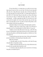
Figure 2. Metabolite Exchange Networks in the Rhizosphere. (A) Flavonoids are exuded, likely by an ABCG-type
transporter [50_TD$IF][122], and sensed by rhizobia
that in turn produce Nod factors. Rhizobacteria enter the root via root hairs or
cracks between epidermal cells [501_TD$IF][133]. (B)
Strigolactones are exuded by ABCGtype Petunia hybrida PDR1 localized in
the subepidermal layer of the root
maturation zone [502_TD$IF][31], and sensed by Glomeromycota that in turn produce Myc
factors. Chitin has a role in hyphal attachment to the root. (C) At Aluminium-Activated Malate Transporter 1 (ALMT1) is
located in the cortex of the elongation
zone, and is involved in malic acid exudation in Pseudomonas-infected Arabidopsis thaliana, which attracts Bacillus
subtilis [503_TD$IF][80]. B. subtilis forms biofilms
on roots, a process dependent on root
pectin and arabinogalactan [504_TD$IF][45]. (D)
ATPases exude protons altering rhizospheric pH, enabling proton-dependent
transport processes. Multidrug and Toxic
Compound Extrusion (MATE) transporters exude citrate [50_TD$IF][119], which can be
metabolized by microbes, and AtPDR9
transports phenolic compounds [506_TD$IF][134].
The signaling function and potential
crosstalk with microbes are currently
unknown. (E) Involvement of transporters
in metabolite exudation is generally
poorly understood [507_TD$IF][26,91,92]. Microbes
exude compounds that are utilized by
other microbes [508_TD$IF][59,135] and sensed by
plants. (F) Border cells produce mucilage
(red gradient), exude proteins, extracellular DNA, as well as metabolites, all of
which impact the microbial community
[509_TD$IF][33,40,136]. Currently, the mode of
transport of these compounds is not
characterized. Key: blue, microbial components; brown, plant components; red
transporters, characterized; orange
transporters, uncharacterized. Abbreviation: exDNA, extracellular DNA.
The large diversity of microbial communities is a current challenge for plant–microbe research,
because it is impractical to study questions such as how members of a community interact, and
what specific traits a microbial community has. Therefore, many studies currently aim at
identifying the subset of microbes responsive to plants. Strikingly, only 7% of bulk soil microbes
increased in abundance in the rhizosphere compared with bulk soil [148_TD$IF][24], which reduces the
Trends in Plant Science, Month Year, Vol. xx, No. yy
7
TRPLSC 1597 No. of Pages 17
number of taxa to investigate from thousands to hundreds. Other approaches to identifying
plant-responsive microbes have focused on transcriptional profiling. Compared with soilabundant microbes, plant-associated microbes exhibited distinct transcriptional responses
to plant exudates [610_TD$IF][52,53] and, intriguingly, displayed distinct phylogenetic clustering [61_TD$IF][18,52].
Network analyses further revealed that rhizosphere microbes displayed higher levels of interactions than did bulk soil microbes [612_TD$IF][54]. These studies illustrate the potential for the identification of a distinct set of plant-responsive microbes.
The above points highlight how plants influence microbial communities. However, the members of microbial communities also interact with each other. Compellingly, it is still unclear
whether microbe–microbe interactions are predominantly positive or negative. Network analyses reported predominantly positive intrakingdom interactions [613_TD$IF][54,55]. By contrast, laboratory
growth assays identified competition as the major factor in shaping isolate communities, and
cooperation could only be detected for 6–10% of the isolates [614_TD$IF][56–58]. One major difference
between the two experimental approaches is that the former investigates a natural system,
whereas the latter is based on the ability to culture microbes. Isolation of microbes introduces a
bias, since it can select against cooperators, precluding obligate syntrophs. Further evidence
that at least some microbes avoid competition was provided by co-cultivation experiments.
Environmental isolates: (i) displayed high substrate specialization [615_TD$IF][59]; (ii) did not necessarily
take up the compound with the highest energy [61_TD$IF][60]; and (iii) diverged in substrate use when
cultivated for several generations [617_TD$IF][50,57]. In addition, some metabolites exuded by microbes
could be metabolized by others [615_TD$IF][59], suggesting potential cross-feeding between community
members. The above findings suggest complex interactions of microbes. It remains to be
resolved in which situation competition or cooperation dominates communities. However, it is
evident that microbial interactions are based on altered gene expression. Microbes responded
to competing bacteria [618_TD$IF][61] or even close relatives [619_TD$IF][62] by differentially regulating genes involved
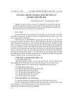
Box 2. Is there a Common Theme of Symbiosis?
The establishment of symbioses between plants and mycorrhiza or rhizobia is detailed in the literature, but the assembly
of plant-associated microbiomes remains unclear. Here, we present a hypothesis on for the assembly of a complex
microbial community in the rhizosphere that is [564_TD$IF]based on the mechanism reported for the aforementioned symbioses
(Figure I).
Plants induce symbioses with mycorrhiza and rhizobia in nutrient- poor soils. The symbionts are attracted by
strigolactones exported by an ABCG- type transporter located in a specific root zone, or by flavonoids likely exported
by a transporter of the same family (see Figure 2A,[56_TD$IF]B) [31,121,122]. Signaling molecules leading to the assembly of
rhizobiomes are largely uncharacterized, but one example illustrates a symbiotic interaction with a beneficial microbe
(see Figure 2C in the main text): pathogen-infected or elicitor- treated arabidopsis plants increased ALMT1 expression
and malic acid exudation (see Table 1 in the main text), which lead to specific attraction and root colonization of the
biocontrol agent Bacillus subtilis, [503_TD$IF][80]. Interestingly, B. subtilis root colonization was not malic acid dependent [503_TD$IF][80],
suggesting the presence of additional signaling compounds.
Signaling compounds are similarly exuded by mycorrhiza and rhizobia, and: lLipochitooligosaccharides (LCOSs) are
required for the induction of symbiosis [56_TD$IF][123,124]. Mycorrhiza further require plant-derived cutin to attach to the root
surface [567_TD$IF][125]. Some rhizosphere microbes produce N-acyl homoserine lactones (AHL) and volatile organic compounds
(VOCs) that are sensed by plants [568_TD$IF][64]. Biofilm-derived exopolysaccharides similarly elicit plant transcriptional responses
[569_TD$IF][126]. These compounds could be part of the plant – microbe crosstalk. Furthermore, plant-derived cell wall polysaccharides and other signals [504_TD$IF][45] were shown to initiate microbial root colonization and biofilm formation [570_TD$IF][22].
In a next step, plants respond to rhizobia and mycorrhiza by initiating the common symbiosis pathway (SYM), altering
gene expression and root morphology [571_TD$IF][127]. The response of the immune system is distinct, with mycorrhiza eliciting,
and rhizobia suppressing, an initial pathogen response [572_TD$IF][128,129]. The immune system is also important in microbiome
establishment (see Figure 1 in the main text): for example, the phytohormone salicylic acid is not only involved in
responses to microbial pathogens, but is also required for assembly of a typical microbiome [573_TD$IF][130]. Also, the genetic
network for phosphate starvation signaling was found to influence the structure of microbiomes [574_TD$IF][131]. Nevertheless, the
exact mechanism of how the plant immune system shapes microbiome formation remains to be determined.
8
Trends in Plant Science, Month Year, Vol. xx, No. yy
TRPLSC 1597 No. of Pages 17
After the [57_TD$IF]successful establishment of symbiosis with rhizobia and mycorrhiza, specifically expressed transporters
translocate nutrients between the partners [541_TD$IF][132]. Plants deliver sugars, organic acids, and lipids, and, in return, receive
phosphate, nitrogen, and other nutrients provided by the microbes [576_TD$IF][99,127]. Compound exchange between plants and
rhizobiomes, remains uncharacterized, and we propose plant transporters that could be involved in the process (see
Table 1 and the discussion in the [57_TD$IF]main text).
Figure I. Comparison of Symbiosis Establishment in Mycorrhization and Nodulation with Root Microbiome Formation.
in metabolite exudation and transport processes [620_TD$IF][61,63], making the study of microbial
transporters a compelling topic for future studies. Thus, metabolite uptake, release, and
sensing are important factors in shaping microbial communities.
Metabolite turnover in soil is influenced not only by plants, but also by functionally diverse
bacteria, fungi, and animals [568_TD$IF][64]. Plant–fungal and plant–animal interactions in the rhizosphere
go beyond the scope of this review, and are discussed elsewhere [621_TD$IF][64–66]. Here, we provide a
few brief examples focusing on the impacts of mycorrhiza on rhizobiomes and exometabolite
turnover. Endomycorrhizal fungi receive a significant fraction of the carbon fixed by plants (Box
2). Interestingly, these fungi also exude sugars [62_TD$IF][67], shaping a distinct bacterial community
[623_TD$IF][67,68]. Likewise, Ectomycorrhiza receive carbon from plants, and form a dynamic bacterial
community [624_TD$IF][69]; they even participate in plant-to-plant carbon transport [625_TD$IF][70]. The field of fungal
microbiomes is nascent: if and how fungi control exudation, whether fungal microbiomes have
beneficial functions, and how plant and fungal microbiomes influence each other are all
unknowns. Although many questions remain, these recent findings already suggest that a
holistic view of rhizosphere nutrient cycling and signaling exchange via exometabolites requires
a whole-community approach including all domains of life.
Trends in Plant Science, Month Year, Vol. xx, No. yy
9
TRPLSC 1597 No. of Pages 17
Exudates Are Diverse and Dynamic
Plant exudates shape microbial communities. Overall, plants exude up to 20% of fixed carbon
and 15% of nitrogen [62_TD$IF][65,71], which includes an array of simple molecules, such as sugars,
organic acids, and secondary metabolites, as well as complex polymers, such as mucilage
(Table 1 [627_TD$IF]and Figures 1 and 2). Although every plant produces exudates, the amount and
composition of root exudates varies. First, exudation is defined by the genotype of the host, as
observed in the distinct exudation patterns of 19 arabidopsis accessions [628_TD$IF][72]. Strikingly, the
amount of variation between the accessions depended on the metabolite class investigated.
Glucosinolates displayed most, flavonoids medium, and phenylpropanoids low variability [629_TD$IF][73].
Second, exudation changes with plant developmental stage: with increasing age, arabidopsis
sugar exudation decreased, and amino acid and phenolic exudation increased [16]. Third,
exudation is modulated by abiotic stresses: the amounts of exuded amino acids, sugars, and
organic acids changed in maize grown in phosphate-, iron-, nitrogen-, or potassium-deficient
conditions [630_TD$IF][53]. In addition, phosphate-deficient arabidopsis plants increased coumarin and
oligolignol exudation [631_TD$IF][74], heavy metal-treated poplar (Populus tremula) induced organic acid
exudation [54_TD$IF][75], and zinc-deficient wheat increased phytosiderophore exudation [632_TD$IF][76]. Differential exudation is a plausible mechanism by which plants could modulate their interaction with
microbes, as exemplified by the correlation between exudation patterns and rhizobiome
variation reported for eight arabidopsis accessions [63_TD$IF][77]. Differential exudation modulated by
transport proteins is discussed below.
Characterized and Putative Plant Transporters for Exudation
Plant-derived exometabolites need to cross at least one membrane to transit from the cytoplasm of root cells into the rhizosphere. There is considerable discussion as to what degree
plants are able to regulate this transport. In general, different modes of transport could be
envisioned. First, small, hydrophilic compounds could diffuse from the root into the rhizosphere,
driven by the large concentration gradient [634_TD$IF][26,78]. Second, channel proteins could facilitate
such diffusion. Third, active (ATP-driven) or secondary active (proton gradient driven) transporters could shuttle compounds across membranes against a concentration gradient. Diffusion of compounds can only be relevant in young root tissue, which is still devoid of Casparian
strips or suberized endodermis that both block apoplasmic flow in adult tissues. Transport
proteins involved in exudation are mostly elusive. From a conceptual point of view, plasma
membrane-localized exporters likely have a direct, and vacuolar transporters an indirect effect
on exudation. The vacuole is a major storage organelle for many metabolites detected in
exudates, such as sugars, organic acids, and secondary metabolites [635_TD$IF][79]. Alteration of vacuolar
transporter levels impacts vacuolar and cytosolic concentrations and, thus, can influence
metabolite exudation into the rhizosphere.
The few characterized transporters involved in exudation are essential for the transport of
specific compounds (Figure 2 [63_TD$IF]and Box 2) [31,80], and are presented in Table 1. Since only a few
transporters involved in exudation have been characterized, we suggest additional families that
might be involved in the process. To complete the picture of metabolite exchange between
roots and soil, Table 1 additionally contains a few important plasma membrane-localized
metabolite uptake systems. Below, we discuss the evidence for transport processes involved
in the import and exudation of compounds detected in root exudates, such as sugars, organic
acids, and secondary metabolites.
Sugars
Sugars constitute a significant fraction of exudates, and are a main carbon source for microbes
[637_TD$IF][14,42]. Interestingly, many more sugar uptake than release systems have been described.
Sugar Transport Proteins (STPs) utilize high extracellular proton levels to import sugars, and
mutation of STPs leads to higher external sugar levels [638_TD$IF][81,82]. Sugars Will Eventually Be
10
Trends in Plant Science, Month Year, Vol. xx, No. yy
TRPLSC 1597 No. of Pages 17
Exported Transporters (SWEETs) are sugar uniporters, and all root-expressed members
localize to the vacuole [514_TD$IF][83,84]. Due to an alteration of root sugar homeostasis, SWEET mutant
plants exhibited higher sugar export from roots compared with wild-type plants, and were more
susceptible to disease [639_TD$IF][85]. Intriguingly, no transporters directly exporting sugars into the
rhizosphere have been characterized so far, and it is debated whether sugar exudation is a
transport-driven process at all [590_TD$IF][26]. Potential evidence for passive sugar efflux was supported
by the observation of higher sucrose concentrations around young, permeable root tissue than
around older, less-permeable root tissue [640_TD$IF][30]. However, because sugars are synthesized in
leaves, they still need to be unloaded either from phloem or from root cells to be exuded into the
rhizosphere, a process likely depending on transporters due to the hydrophilic nature of sugars.
A further indication of the presence of elusive transporters is the differential sugar exudation in
various environments, as shown, for example, for maize grown in potassium-, phosphate-, or
iron-deficient conditions [641_TD$IF][85–88].
Sugar Alcohols and Phosphates
Sugar alcohols are imported by secondary active proteins with broad substrate specificity
(Table 1), whereas the modes of export are enigmatic. Sugar phosphates are involved in
intracellular carbohydrate metabolism, and plastid-localized sugar–phosphate co-transporters
have been reported in several species [642_TD$IF][89]. Although sugar phosphates are detected in
exudates, neither import nor export mechanisms are currently characterized.
Amino Acids
Amino acids are recognized by microbial chemoreceptors crucial for the early steps of root
colonization [643_TD$IF][90], making amino acids an important fraction of exudates. Modulation of amino
acid transport could be either a means of communication with microbes, or a response to
microbial presence. Amino acid uptake is mediated by several transporter families with broad
substrate specificity (Table 1) [64_TD$IF][91]. Amino acid exudation is affected by several transporters
expressed in vascular tissue: mutation of phloem-localized UmamiTs resulted in lower amino
acid exudation [645_TD$IF][92], whereas mutation of xylem-localized Glutamine Dumpers (GDUs) caused
increased exudation [52_TD$IF][93]. Although no plasma membrane-localized amino acid exporters have
been characterized so far, several lines of evidence suggest their presence. First, higher
tryptophan exudation from older root zones than younger parts [640_TD$IF][30] suggests the involvement
of transport proteins in exudation, due to the fully formed Casparian strips and thick cell walls in
mature root parts interfering with diffusion. Second, concentration differences between amino
acids in root exudates and root extracts are not the same for all the amino acids [64_TD$IF][91],
suggesting the selective transport of at least some amino acids. Third, various transporter
families exhibit bidirectional amino acid transport characteristics in heterologous systems
(Table 1), and could be involved in amino acid exudation.
Organic Acids
Organic acids constitute a large fraction of exudates, and are microbial nutrients. No importers
have been characterized so far, but the release of malate and citrate by Aluminium-Activated
Malate Transporters (ALMT) and Multidrug and Toxic Compound Extrusion (MATE) families are
among the few well-understood examples of transporters involved in exudation (Table 1 [64_TD$IF]and
Figure 2). Activity of members of both families is often modulated by metal ions (Box 1) and
microbes (Box 2). Uncharacterized ALMT and MATE family members are primary candidates
for exporters of other organic acids due to their similarity to already-characterized members,
their plasma membrane localization, and their function as proton antiporters.
Nucleotides and Peptides
Nucleotides are imported by secondary active transporters, but their exudation remains elusive
(Table 1) [647_TD$IF][94,95]. It is well established that extracellular ATP has a signaling function, and ABC
Trends in Plant Science, Month Year, Vol. xx, No. yy
11
TRPLSC 1597 No. of Pages 17
transporters were proposed to mediate cellular export [97,98]. Peptide uptake is transporter
mediated in heterologous systems, and a role of ABC transporters in peptide exudation has
been suggested (Table 1).
Fatty Acid
Fatty acid transport is necessary for mycorrhizal symbiosis: mycorrhizal fungi depended on
their hosts for the synthesis of certain fatty acids [648_TD$IF][98], and the current model includes transport
of lipids by ABCG proteins in the symbiotic membrane [649_TD$IF][98,99]. One ABCG member, STR, was
previously shown to be required for mycorrhization [650_TD$IF][100]. Interestingly, arabidopsis ABCG
transporters were similarly shown to export fatty acids for cutin synthesis in aboveground
tissues (Table 1). Lipid transport was required not only for symbiotic interactions, but also for
pathogen colonization [648_TD$IF][98]. Fatty acids are detected in root exudates (Table 1), but the mode of
lipid exudation into the rhizosphere has yet to be discovered. A role in lipid exudation could be
envisioned for root-expressed ABCG members (Table 1 [64_TD$IF]and Figure 2).
Secondary Metabolites
Secondary metabolites are ubiquitous in root exudates, and ABC transporters are likely
candidates for specialized metabolite transport into the rhizosphere. A distinct exudation
profile was described for seven ABC mutants [651_TD$IF][101], and one mutant line displayed an altered
microbial community [652_TD$IF][102]. Although the causal metabolites could not be identified, the authors
noted transport of the same compound by various proteins, and possible broad substrate
specificity for some transporters [651_TD$IF][101]. In a later study, exudates of arabidopsis ABCG37/PDR9
mutant lines were found to be deficient in several phenylpropanoids [653_TD$IF][103] (Figure 2D). Arabidopsis PDR9 was previously characterized as auxin precursor transporter [654_TD$IF][104], which suggests a broad substrate specificity for PDR9. Interestingly, a PDR9 homolog was highly
expressed in cluster roots of white lupin devoid of phosphate [65_TD$IF][105], illustrating PDR9 involvement in response to various abiotic stresses. These studies illustrate the potential for the
discovery of novel transporter functions in the ABC family, an excellent target for future studies
investigating root exudation. In addition, MATE proteins transport secondary metabolites into
the vacuole, and plasma membrane-localized members could also be involved in secondary
metabolite exudation.
In summary, more transport proteins involved in metabolite import into roots than in export from
roots have been reported so far (Table 1). The characterization of additional transport families
involved in exudation will enable the generation of mutant lines that are devoid of the exudation
of specific metabolites. Such lines could be used to investigate the correlation of exudation
profiles and microbial communities.
How Do Rhizobiomes Assemble?
Plant-derived transporters and exometabolites are intrinsic to plant–mycorrhizal and Àrhizobial
symbioses (Box 2). We speculate that, although there is paucity of evidence, plants analogously
select for a beneficial rhizobiome. Given that plants evolved in the presence of microbes, a
subset of which benefits plant growth, we hypothesize that, over millennia, plant exudation via
active transport processes evolved with the substrate specificity of plant-associated bacteria.
In Box 2, we discuss exudates and other steps involved in root microbiome assembly,
analogously to the establishment of plant–mycorrhizal and Àrhizobial symbioses. However,
intense future research is needed to reveal the precise mechanisms governing plant microbiome assembly, and the possible beneficial functions of the microbial community.
The major mechanisms by which plants are thought to modulate microbial interactions
currently include: (i) modulation of their exudate profiles (alteration of biosynthesis and/or
transport of microbial substrates and signaling molecules); (ii) root morphology (number
12
Trends in Plant Science, Month Year, Vol. xx, No. yy
TRPLSC 1597 No. of Pages 17
and length of roots, and root surface); and (iii) regulation of immune system activities (tolerance
or avoidance). In turn, mechanisms for successful rhizosphere colonization by soil microbes
require that they: (i) are metabolically active (catabolism of exudates); (ii) sense the plant
(receptors for exudates); (iii) move towards the root (chemotaxis and [65_TD$IF]mobility) and (iv) successfully compete with other microbes for root niches (physical colonization, substrate competition,
and defense against toxins). In addition, for successful colonization of the rhizoplane or root
tissue, microbes must be able to (v) attach to the surface (cell wall sensing or biofilm formation)
or (vi) enter root tissue (evasion and/or manipulation of immune system).
Despite apparent parallels between plant microbiomes and the aforementioned symbioses,
plant microbiomes have some specific characteristics. First, microbiomes are detected in all
environmental conditions, whereas mycorrhizal and rhizobial symbioses are induced in specific
circumstances. Second, microbiomes occur on various tissues, whereas rhizobia and mycorrhiza interface with roots only. Third, microbiomes comprise many members, whereas the
aforementioned symbioses persist between two predominant partners. Fourth, although most
members of the microbiome originate from the environment [657_TD$IF][2,4,106] similar to rhizobia and
mycorrhiza, there is evidence that some endophytes may be vertically transmitted via seeds
[658_TD$IF][107–110]. Future research should focus on the factors involved in microbiome assembly, the
relative contribution of epi- and endophytes to microbiomes, and the signaling crosstalk
between plants and microbial communities.
Concluding Remarks and Future Directions
Rhizobiome assembly and the involvement of the plant in this process are currently enigmatic.
Here, we have discussed multiple factors shaping the rhizobiome, including host genotype and
development, root morphology, border cells and mucilage, and root exudates. Root exudation
is a dynamic process, likely dependent on a plethora or transporters that are mostly uncharacterized. Spatially defined exudation likely results in distinct microbial communities that are
observed to be associated with specific root parts. The success of microbial colonization of the
rhizosphere depends on several aspects, such as chemotaxis, substrate specificity, competitiveness, and cooperativeness. Furthermore, endophytes likely form biofilms on the root
surface, [659_TD$IF]and encounter the plant immune system. Although some factors shaping root microbiomes emerge, many open questions [60_TD$IF]remain (see Outstanding Questions).
One major challenge will be to analyze root exudation in natural settings. Due to the chemical
complexity of soil, exudation is traditionally analyzed in hydroponic culture [61_TD$IF][14,16,72,88], an
environment distant from the more natural settings of plant microbiome studies. Furthermore,
novel technologies enabling high-throughput screening of putative transporters against possible substrates are needed to reveal the impact of the respective substrates on the rhizobiome
and, in turn, on plant health. An increased understanding of root morphology, exudation, and
involved transporters will likely enable the engineering or breeding of plants with altered abilities
to interact with specific beneficial microbes or pathogens. This needs to be complemented with
an improved understanding of the substrate preferences of plant-associated microbes, their
interactions, and the mechanisms through which they benefit the plant. A holistic understanding of the functions of a healthy plant rhizobiome would enable the directed design of
customized microbial communities. With this, specific plants in a given environment could
be tailored to a specific purpose, such as phytoremediation, stress resistance, altered plant
development, or increased yield[62_TD$IF].
Outstanding Questions
Can we define standardized laboratory
plant–soil–rhizobiome relationships to
decipher the mechanisms of plant and
microbial nutrient exchange and other
beneficial activities by controlling confounding environmental variables?
How and to what degree do plants
control exudation to specifically interact with microbes, shaping the rhizobiome? Special attention should be
given to transporter families expressed
in the root epidermis and root tip.
How can novel, high-throughput techniques be utilized to identify key
nutrients and signaling molecules
exchanged between plants and
microbes, as well as the transport proteins involved in the process?
What are the distinct aspects of the
microbial communities associated with
root tips, lateral roots, mature root
parts, and border cells? How are these
communities influenced by exometabolites and nutrient uptake by the
plant?
Are there functional classes and
potentially crucial taxa common to
plant rhizobiomes that can be used
to design customized rhizobiomes
persisting in a given environment, supporting host plant growth?
How are bacterial communities associated with mycorrhizal hyphae
assembled, and how do they interact
with rhizobiomes? Are microbes transferred between roots and hyphae?
How do reciprocal interactions
between the rhizobiome and plant alter
the trajectory of plant development,
stress responses, as well as microbial
community succession and activities?
How can we alter the host genotype to
more efficiently select a beneficial rhizobiome that increases plant health
and yield?
Acknowledgments
We thank Estelle Couradeau, Kateryna Zhalnina, and Pascal Schläpfer for critical reading of the manuscript J.S. is
supported by the National Science Foundation (NSF Proposal 1617020), and work in the laboratory of E.M. is supported
Trends in Plant Science, Month Year, Vol. xx, No. yy
13
TRPLSC 1597 No. of Pages 17
by the Swiss National Foundation. [63_TD$IF]The work conducted by the U.S. Department of Energy Joint Genome Institute, a DOE
Office of Science User Facility, is supported under Contract No. DE-AC02-05CH11231.
References
1.
Schmidt, J.E. et al. (2016) Using ancient traits to convert soil
health into crop yield: impact of selection on maize root and
rhizosphere function. Front. Plant Sci. 7, 351–11
23. DeAngelis, K.M. et al. (2005) Two novel bacterial biosensors for
detection of nitrate availability in the rhizosphere. Appl. Environ.
Microbiol. 71, 8537–8547
2.
Bulgarelli, D. et al. (2013) Structure and functions of the bacterial
microbiota of plants. Annu. Rev. Plant Biol. 64, 807–838
24. DeAngelis, K.M. et al. (2008) Selective progressive response of
soil microbial community to wild oat roots. ISME J. 3, 168–178
3.
Edwards, J. et al. (2015) Structure, variation, and assembly of
the root-associated microbiomes of rice. Proc. Natl. Acad. Sci.
U. S. A. 112, E911–E920
25. Massalha, H. et al. (2017) Live imaging of root–bacteria interactions in a microfluidics setup. Proc. Natl. Acad. Sci. U. S. A.
114, 4549–4554
4.
Müller, D.B. et al. (2016) The plant microbiota: systems-level
insights and perspectives. Annu. Rev. Genet. 50, 211–234
26. Jones, D.L. et al. (2009) Carbon flow in the rhizosphere: carbon
trading at the soil–root interface. Plant Soil 321, 5–33
5.
Bulgarelli, D. et al. (2015) Structure and function of the bacterial
root microbiota in wild and domesticated barley. Cell Host
Microbe 17, 392–403
27. Neumann, G. and Martinoia, E. (2002) Cluster roots–an underground adaptation for survival in extreme environments. Trends
Plant Sci. 7, 162–167
6.
Bouffaud, M.-L. et al. (2014) Root microbiome relates to plant
host evolution in maize and other Poaceae. Environ. Microbiol.
16, 2804–2814
28. Weisskopf, L. et al. (2011) Burkholderia species are major
inhabitants of white lupin cluster roots. Appl. Environ. Microbiol.
77, 7715–7720
7.
Schlaeppi, K. et al. (2014) Quantitative divergence of the bacterial root microbiota in Arabidopsis thaliana relatives. Proc. Natl.
Acad. Sci. U. S. A. 111, 585–592
8.
Szoboszlay, M. et al. (2015) Comparison of root system architecture and rhizosphere microbial communities of Balsas teosinte and domesticated corn cultivars. Soil Biol. Biochem. 80,
34–44
29. Weisskopf, L. et al. (2006) White lupin has developed a complex
strategy to limit microbial degradation of secreted citrate
required for phosphate acquisition. Plant Cell Environ. 29,
919–927
30. Jaeger, C.H. et al. (1999) Mapping of sugar and amino acid
availability in soil around roots with bacterial sensors of sucrose
and tryptophan. Appl. Environ. Microbiol. 65, 2685–2690
Coleman-Derr, D. et al. (2015) Plant compartment and biogeography affect microbiome composition in cultivated and native
Agave species. New Phytol. 209, 798–811
31. Kretzschmar, T. et al. (2012) A petunia ABC protein controls
strigolactone-dependent symbiotic signalling and branching.
Nature 483, 341–344
10. Zachow, C. et al. (2014) Differences between the rhizosphere
microbiome of Beta vulgaris ssp maritima – ancestor of all beet
crops – and modern sugar beets. Front. Microbiol. 5, 1–13
32. Watson, B.S. et al. (2015) Integrated metabolomics and transcriptomics reveal enhanced specialized metabolism in Medicago truncatula root border cells. Plant Physiol. 167, 1699–
1716
9.
11. Cardinale, M. et al. (2014) Bacterial networks and co-occurrence relationships in the lettuce root microbiota. Environ.
Microbiol. 17, 239–252
12. Zhu, S. et al. (2016) Nitrogen fertilizer rate affects root exudation,
the rhizosphere microbiome and nitrogen-use-efficiency of
maize. Appl. Soil Ecol. 107, 324–333
13. Peiffer, J.A. et al. (2013) Diversity and heritability of the maize
rhizosphere microbiome under field conditions. Proc. Natl.
Acad. Sci. U. S. A. 110, 6548–6553
14. Kawasaki, A. et al. (2016) Microbiome and exudates of the root
and rhizosphere of Brachypodium distachyon, a model for
wheat. PLoS One 11, e0164533
15. Zgadzaj, R. et al. (2016) Root nodule symbiosis in Lotus japonicus drives the establishment of distinctive rhizosphere, root,
and nodule bacterial communities. Proc. Natl. Acad. Sci. U. S.
A. 113, E7996–E8005
16. Chaparro, J.M. et al. (2013) Rhizosphere microbiome assemblage is affected by plant development. ISME J. 8, 790–803
17. Schreiter, S. et al. (2014) Effect of the soil type on the microbiome in the rhizosphere of field-grown lettuce. Front. Microbiol.
5, 1–13
18. Shi, S. et al. (2015) Successional trajectories of rhizosphere
bacterial communities over consecutive seasons. mBio 6,
e00746–15
19. Lundberg, D.S. et al. (2012) Defining the core Arabidopsis
thaliana root microbiome. Nature 488, 86–90
20. Dombrowski, N. et al. (2016) Root microbiota dynamics of
perennial Arabis alpina are dependent on soil residence time
but independent of flowering time. ISME J. 11, 43–55
21. Panke-Buisse, K. et al. (2017) Cultivated sub-populations of soil
microbiomes retain early flowering plant trait. Microb. Ecol. 73,
1–10
22. Bulgarelli, D. et al. (2012) Revealing structure and assembly
cues for Arabidopsis root-inhabiting bacterial microbiota. Nature
488, 91–95
14
Trends in Plant Science, Month Year, Vol. xx, No. yy
33. Vicre, M. et al. (2005) Root border-like cells of Arabidopsis.
Microscopical characterization and role in the interaction with
rhizobacteria. Plant Physiol. 138, 998–1008
34. Kang, J. et al. (2011) Plant ABC transporters. Arabidopsis Book
9, e0153
35. Koroney, A.S. et al. (2016) Root exudate of Solanum tuberosumis enriched in galactose-containing molecules and impacts
the growth of Pectobacterium atrosepticum. Ann. Bot. 118,
797–808
36. Curlango-Rivera, G. et al. (2010) Transient exposure of root tips
to primary and secondary metabolites: impact on root growth
and production of border cells. Plant Soil 332, 267–275
37. Cannesan, M.A. et al. (2011) Association between border cell
responses and localized root infection by pathogenic Aphanomyces euteiches. Ann. Bot. 108, 459–469
38. Weiller, F. et al. (2016) The Brassicaceae species Heliophila
coronopifolia produces root border-like cells that protect the
root tip and secrete defensin peptides. Ann. Bot. 119, 803–813
39. Basu, U. et al. (2006) Extracellular proteomes of Arabidopsis
thaliana and Brassica napus roots: analysis and comparison by
MudPIT and LC-MS/MS. Plant Soil 286, 357–376
40. Wen, F. et al. (2009) Extracellular DNA is required for root tip
resistance to fungal infection. Plant Physiol. 151, 820–829
41. Minh Tran, T. et al. (2016) Extracellular DNases of Ralstonia
solanacearum modulate biofilms and facilitate bacterial wilt virulence. Environ. Microbiol. 18, 4103–4117
42. Traoré, O. and Renaud, V.G. (2000) Effect of root mucilage and
modelled root exudates on soil structure. Eur. J. Soil Sci. 51,
575–581
43. Knee, E.M. et al. (2001) Root mucilage from pea and its utilization by rhizosphere bacteria as a sole carbon source. MPMI 14,
775–784
44. Benizri, E. et al. (2007) Additions of maize root mucilage to soil
changed the structure of the bacterial community. Soil Biol.
Biochem. 39, 1230–1233
TRPLSC 1597 No. of Pages 17
45. Beauregard, P.B. et al. (2013) Bacillus subtilis biofilm induction
by plant polysaccharides. Proc. Natl. Acad. Sci. U. S. A. 110,
E1621–E1630
46. Nagahashi, G. and Douds, D.D., Jr (2004) Isolated root caps,
border cells, and mucilage from host roots stimulate hyphal
branching of the arbuscular mycorrhizal fungus, Gigaspora
gigantea. Mycol. Res. 108, 1079–1088
47. Eisenhauer, N. et al. (2013) Plant diversity effects on soil food
webs are stronger than those of elevated CO2 and N deposition
in a long-term grassland experiment. Proc. Natl. Acad. Sci. U. S.
A. 110, 6889–6894
48. Prober, S.M. et al. (2014) Plant diversity predicts beta but not
alpha diversity of soil microbes across grasslands worldwide.
Ecol. Lett. 18, 85–95
selected by Pinus sylvestris roots colonized by different ectomycorrhizal fungi. Environ. Microbiol. 18, 1470–1483
70. Klein, T. et al. (2016) Belowground carbon trade among tall trees
in a temperate forest. Science 352, 342–344
71. el Zahar Haichar, F. et al. (2016) Stable isotope probing of
carbon flow in the plant holobiont. Curr. Opin. Biotechnol. 41,
9–13
72. Mönchgesang, S. et al. (2016) Natural variation of root exudates
in Arabidopsis thaliana-linking metabolomic and genomic data.
Sci. Rep. 6, 1–11
73. Mönchgesang, S. et al. (2016) Plant-to-plant variability in root
metabolite profiles of 19 Arabidopsis thaliana accessions is
substance-class-dependent. IJMS 17, 1565–1569
49. Steinauer, K. et al. (2016) Root exudate cocktails: the link
between plant diversity and soil microorganisms? Ecol. Evol.
6, 7387–7396
74. Ziegler, J. et al. (2016) Non-targeted profiling of semi-polar
metabolites in Arabidopsis root exudates uncovers a role for
coumarin secretion and lignification during the local response to
phosphate limitation. J. Exp. Bot. 67, 1421–1432
50. Essarioui, A. et al. (2017) Nutrient use preferences among soil
Streptomyces suggest greater resource competition in monoculture than polyculture plant communities. Plant Soil 409, 1–15
75. Qin, R. et al. (2007) Exudation of organic acid anions from poplar
roots after exposure to Al, Cu and Zn. Tree Physiol. 27, 313–320
51. Essarioui, A. et al. (2017) Plant community richness mediates
inhibitory interactions and resource competition between Streptomyces and Fusarium populations in the rhizosphere. Microb.
Ecol. 6, 1–11
52. Zhang, N. et al. (2016) Comparative genomic analysis of Bacillus
amyloliquefaciens and Bacillus subtilis reveals evolutional traits
for adaptation to plant-associated habitats. Front. Microbiol. 7,
332–14
53. Carvalhais, L.C. et al. (2013) Linking plant nutritional status to
plant-microbe interactions. PLoS One 8, e68555
54. Shi, S. et al. (2016) The interconnected rhizosphere: high network complexity dominates rhizosphere assemblages. Ecol.
Lett. 19, 926–936
55. Agler, M.T. et al. (2016) Microbial hub taxa link host and abiotic
factors to plant microbiome variation. PLoS Biol. 14
56. Foster, K.R. and Bell, T. (2012) Competition, not cooperation,
dominates interactions among culturable microbial species.
Curr. Biol. 22, 1845–1850
57. Lawrence, D. et al. (2012) Species interactions alter evolutionary
responses to a novel environment. PLoS Biol. 10, e1001330–11
58. Coyte, K.Z. et al. (2015) The ecology of the microbiome: Networks, competition, and stability. Science 350, 663–666
59. Baran, R. et al. (2015) Exometabolite niche partitioning among
sympatric soil bacteria. Nat. Commun. 6, 1–9
60. Erbilgin, O. et al. (2017) Dynamic substrate preferences predict
metabolic properties of a simple microbial consortium. BMC
Bioinformatics 18, 1–12
61. Garbeva, P. et al. (2011) Transcriptional and antagonistic
responses of Pseudomonas fluorescens Pf0-1 to phylogenetically different bacterial competitors. ISME J. 5, 973–985
62. González-Torres, P. et al. (2015) Interactions between closely
related bacterial strains are revealed by deep transcriptome
sequencing. Appl. Environ. Microbiol. 81, 8445–8456
63. Baran, R. et al. (2011) Untargeted metabolic footprinting reveals
a surprising breadth of metabolite uptake and release by Synechococcus sp. PCC 7002. Mol. BioSyst. 7, 3200–3207
76. Rengel, Z. and Romheld, V. (2000) Root exudation and Fe
uptake and transport in wheat genotypes differing in tolerance
to Zn deficiency. Plant Soil 222, 25–34
77. Micallef, S.A. et al. (2009) Influence of Arabidopsis thaliana
accessions on rhizobacterial communities and natural variation
in root exudates. J. Exp. Bot. 60, 1729–1742
78. Farrar, J. et al. (2003) How roots control the flux of carbon to the
rhizosphere. Ecology 84, 827–837
79. Martinoia, E. et al. (2012) Vacuolar transporters in their physiological context. Annu. Rev. Plant Biol. 63, 183–213
80. Rudrappa, T. et al. (2008) Root-secreted malic acid recruits
beneficial soil bacteria. Plant Physiol. 148, 1547–1556
81. Truernit, E. et al. (1996) The sink-specific and stress-regulated
Arabidopsis STP4 gene: enhanced expression of a gene encoding a monosaccharide transporter by wounding, elicitors, and
pathogen challenge. Plant Cell 8, 2169–2182
82. Yamada, K. et al. (2011) Monosaccharide absorption activity of
Arabidopsis roots depends on expression profiles of transporter
genes under high salinity conditions. J. Biol. Chem. 286, 43577–
43586
83. Chen, H.Y. et al. (2015) The Arabidopsis vacuolar sugar transporter SWEET2 limits carbon sequestration from roots and
restricts Pythium infection. Plant J. 83, 1046–1058
84. Guo, W.J. et al. (2014) SWEET17, a facilitative transporter,
mediates fructose transport across the tonoplast of Arabidopsis
roots and leaves. Plant Physiol. 164, 777–789
85. Chaudhuri, B. et al. (2011) Dynamic imaging of glucose flux
impedance using FRET sensors in wild-type Arabidopsis plants.
J. Exp. Bot. 62, 2411–2417
86. Chaparro, J.M. et al. (2013) Root exudation of phytochemicals in
Arabidopsis follows specific patterns that are developmentally
programmed and correlate with soil microbial functions. PLoS
One 8, 1–10
87. Chen, L.-Q. et al. (2015) Transport of sugars. Annu. Rev. Biochem. 84, 865–894
64. Leach, J.E. et al. (2017) Communication in the phytobiome. Cell
169, 587–596
88. Carvalhais, L.C. et al. (2011) Root exudation of sugars, amino
acids, and organic acids by maize as affected by nitrogen,
phosphorus, potassium, and iron deficiency. J. Plant Nutr. Soil
Sci. 174, 3–11
65. Venturi, V. and Keel, C. (2016) Signaling in the rhizosphere.
Trends Plant Sci. 21, 187–198
89. Flugge, U.I. et al. (2011) The role of transporters in supplying
energy to plant plastids. J. Exp. Bot. 62, 2381–2392
66. van Dam, N.M. and Bouwmeester, H.J. (2016) Metabolomics in
the rhizosphere: tapping into belowground chemical communication. Trends Plant Sci. 21, 256–265
90. Allard-Massicotte, R. et al. (2016) Bacillus subtilis early colonization of Arabidopsis thaliana roots involves multiple chemotaxis
receptors. mBio 7, e01664–16–10
67. Kaiser, C. et al. (2014) Exploring the transfer of recent plant
photosynthates to soil microbes: mycorrhizal pathway vs direct
root exudation. New Phytol. 205, 1537–1551
91. Moe, L.A. (2013) Amino acids in the rhizosphere: from plants to
microbes. Am. J. Bot. 100, 1692–1705
68. Toljander, J.F. et al. (2017) Influence of arbuscular mycorrhizal
mycelial exudates on soil bacterial growth and community structure. FEMS Microbiol. Ecol. 61, 295–304
69. Marupakula, S. et al. (2016) Analysis of single root tip microbiomes suggests that distinctive bacterial communities are
92. Besnard, J. et al. (2016) UMAMIT14 is an amino acid exporter
involved in phloem unloading in Arabidopsis roots. J. Exp. Bot.
67, 6385–6397
93. Pratelli, R. et al. (2010) Stimulation of nonselective amino acid
export by glutamine dumper proteins. Plant Physiol. 152, 762–
773
Trends in Plant Science, Month Year, Vol. xx, No. yy
15
TRPLSC 1597 No. of Pages 17
94. Gillissen, B. et al. (2000) A new family of high-affinity transporters
for adenine, cytosine, and purine derivatives in arabidopsis.
Plant Cell 12, 291–300
95. Desimone, M. (2002) A novel superfamily of transporters for
allantoin and other oxo derivatives of nitrogen heterocyclic compounds in Arabidopsis. Plant Cell 14, 847–856
96. Roux, S.J. and Steinebrunner, I. (2007) Extracellular ATP: an
unexpected role as a signaler in plants. Trends Plant Sci. 12,
522–527
97. Thomas, C. et al. (2000) A role for ectophosphatase in xenobiotic resistance. Plant Cell 12, 519–533
98. Jiang, Y. et al. (2017) Plants transfer lipids to sustain colonization
by mutualistic mycorrhizal and parasitic fungi. Science 356,
1172–1175
99. Luginbuehl, L.H. et al. (2017) Fatty acids in arbuscular mycorrhizal fungi are synthesized by the host plant. Science 356,
1175–1178
100. Zhang, Q. et al. (2010) Two Medicago truncatula half-ABC
transporters are essential for arbuscule development in arbuscular mycorrhizal symbiosis. Plant Cell 22, 1483–1497
101. Badri, D.V. et al. (2008) Altered profile of secondary metabolites
in the root exudates of Arabidopsis ATP-binding cassette transporter mutants. Plant Physiol. 146, 762–771
102. Badri, D.V. et al. (2009) An ABC transporter mutation alters root
exudation of phytochemicals that provoke an overhaul of natural
soil microbiota. Plant Physiol. 151, 2006–2017
103. Fourcroy, P. et al. (2013) Involvement of the ABCG37 transporter in secretion of scopoletin and derivatives by Arabidopsis
roots in response to iron deficiency. New Phytol. 201, 155–167
104. Ruzicka, K. et al. (2010) Arabidopsis PIS1 encodes the ABCG37
transporter of auxinic compounds including the auxin precursor
indole-3-butyric acid. Proc. Natl. Acad. Sci. U. S. A. 107,
10749–10753
105. Wang, Z. et al. (2014) The regulatory network of cluster-root
function and development in phosphate-deficient white lupin
(Lupinus albus) identified by transcriptome sequencing. Physiol.
Plant. 151, 323–338
106. Hodgson, S. et al. (2014) Vertical transmission of fungal endophytes is widespread in forbs. Ecol. Evol. 4, 1199–1208
107. Hardoim, P.R. et al. (2012) Dynamics of seed-borne rice endophytes on early plant growth stages. PLoS One 7, e30438–13
108. Barret, M. et al. (2015) Emergence shapes the structure of the
seed microbiota. Appl. Environ. Microbiol. 81, 1257–1266
109. Truyens, S. et al. (2014) Bacterial seed endophytes: genera,
vertical transmission and interaction with plants. Environ. Microbiol. Rep. 7, 40–50
110. Johnston-Monje, D. and Raizada, M.N. (2011) Conservation
and diversity of seed associated endophytes in Zea across
boundaries of evolution, ethnography and ecology. PLoS One
6, e20396–22
111. Thijs, S. et al. (2016) Phytoremediation: state-of-the-art and a
key role for the plant microbiome in future trends and research
prospects. Int. J. Phytoremediation 19, 23–38
112. Thijs, S. et al. (2016) Towards an enhanced understanding of
plant–microbiome interactions to improve phytoremediation:
engineering the metaorganism. Front. Microbiol. 7, 416–15
113. Deng, Z. and Cao, L. (2017) Fungal endophytes and their
interactions with plants in phytoremediation: a review. Chemosphere 168, 1100–1106
114. Yergeau, E. et al. (2015) Transplanting soil microbiomes leads to
lasting effects on willow growth, but not on the rhizosphere
microbiome. Front. Microbiol. 6, 921–14
115. Lakshmanan, V. et al. (2016) Killing two birds with one stone:
natural rice rhizospheric microbes reduce arsenic uptake and
blast infections in rice. Front. Plant Sci. 7, 962–12
116. Muehe, E.M. et al. (2015) Rhizosphere microbial community
composition affects cadmium and zinc uptake by the metalhyperaccumulating plant Arabidopsis halleri. Appl. Environ.
Microbiol. 81, 2173–2181
117. Kawasaki, A. et al. (2011) Indirect effects of polycyclic aromatic
hydrocarbon contamination on microbial communities in
legume and grass rhizospheres. Plant Soil 358, 169–182
16
Trends in Plant Science, Month Year, Vol. xx, No. yy
118. Mahoney, A.K. et al. (2017) Community structure, species variation, and potential functions of rhizosphere-associated bacteria of different winter wheat (Triticum aestivum) cultivars. Front.
Plant Sci. 8, 2276–14
119. Sharma, S. et al. (2016) Halotolerant Rhizobacteria promote
growth and enhance salinity tolerance in peanut. Front. Microbiol. 7, 523–11
120. Hawes, M. et al. (2016) Extracellular trapping of soil contaminants by root border cells: new insights into plant defense.
Agronomy 6, 5–9
121. Peters, N.K. and Long, S.R. (1988) Alfalfa root exudates and
compounds which promote or inhibit induction of Rhizobium
meliloti nodulation genes. Plant Physiol. 88, 396–400
122. Banasiak, J. et al. (2013) A Medicago truncatula ABC transporter belonging to subfamily G modulates the level of isoflavonoids. J. Exp. Bot. 64, 1005–1015
123. Maillet, F. et al. (2011) Fungal lipochitooligosaccharide symbiotic
signals in arbuscular mycorrhiza. Nature 469, 58–63
124. Denarie, J. et al. (1996) Rhizobium lipo-chitooligosaccharide
nodulation factors: signaling molecules mediating recognition
and morphogenesis. Annu. Rev. Biochem. 65, 503–535
125. Murray, J.D. et al. (2013) Signaling at the root surface: the role of
cutin monomers in mycorrhization. Mol. Plant 6, 1381–1383
126. Zipfel, C. and Oldroyd, G.E.D. (2017) Plant signalling in symbiosis and immunity. Nature 543, 328–336
127. Oldroyd, G.E.D. (2013) Speak, friend, and enter: signalling systems that promote beneficial symbiotic associations in plants.
Nat. Rev. Microbiol. 11, 252–263
128. Mithofer, A. (2002) Suppression of plant defence in rhizobialegume symbiosis. Trends Plant Sci. 7, 440–444
129. Pozo, M.J. and Azcon-Aguilar, C. (2007) Unraveling mycorrhizainduced resistance. Curr. Opin. Plant Biol. 10, 393–398
130. Lebeis, S.L. et al. (2015) Salicylic acid modulates colonization of
the root microbiome by specific bacterial taxa. Science 349,
860–864
131. Castrillo, G. et al. (2017) Root microbiota drive direct integration
of phosphate stress and immunity. Nature 543, 1–22
132. Garcia, K. et al. (2016) Take a trip through the plant and fungal
transportome of mycorrhiza. Trends Plant Sci. 21, 937–950
133. Madsen, L.H. et al. (2010) The molecular network governing
nodule organogenesis and infection in the model legume Lotus
japonicus. Nat. Commun. 1, 10–12
134. Baetz, U. and Martinoia, E. (2014) Root exudates: the hidden
part of plant defense. Trends Plant Sci. 19, 90–98
135. Bezemer, T.M. et al. (2010) Divergent composition but similar
function of soil food webs of individual plants: plant species and
community effects. Ecology 91, 3027–3036
136. Ma, W. et al. (2010) The mucilage proteome of maize (Zea mays
L.) primary roots. J. Proteome Res. 9, 2968–2976
137. Doidy, J. et al. (2012) Sugar transporters in plants and in their
interactions with fungi. Trends Plant Sci. 17, 413–422
138. Büttner, M. (2007) The monosaccharide transporter(-like) gene
family in Arabidopsis. FEBS Lett. 581, 2318–2324
139. Yamada, K. et al. (2010) Functional analysis of an Arabidopsis
thaliana abiotic stress-inducible facilitated diffusion transporter
for monosaccharides. J. Biol. Chem. 285, 1138–1146
140. Schneider, S. (2006) Arabidopsis INOSITOL TRANSPORTER4
mediates high-affinity H+ symport of myoinositol across the
plasma membrane. Plant Physiol. 141, 565–577
141. Klepek, Y.-S. et al. (2010) Arabidopsis thaliana POLYOL/
MONOSACCHARIDE TRANSPORTERS 1 and 2: fructose
and xylitol/H+ symporters in pollen and young xylem cells. J.
Exp. Bot. 61, 537–550
142. Schneider, S. et al. (2008) Functional and physiological characterization of Arabidopsis INOSITOL TRANSPORTER1, a novel
tonoplast-localized transporter for myo-inositol. Plant Cell 20,
1073–1087
143. Svennerstam, H. et al. (2008) Root uptake of cationic amino
acids by Arabidopsis depends on functional expression of
amino acid permease 5. New Phytol. 180, 620–630
TRPLSC 1597 No. of Pages 17
144. Hirner, A. (2006) Arabidopsis LHT1 is a high-affinity transporter
for cellular amino acid uptake in both root epidermis and leaf
mesophyll. Plant Cell 18, 1931–1946
145. Tegeder, M. and Ward, J.M. (2012) Molecular evolution of plant
AAP and LHT amino acid transporters. Front. Plant Sci. 3, 1–11
146. Dündar, E. and Bush, D.R. (2009) BAT1, a bidirectional amino
acid transporter in Arabidopsis. Planta 229, 1047–1056
147. Ladwig, F. et al. (2012) Siliques Are Red1 from Arabidopsis Acts
as a bidirectional amino acid transporter that is crucial for the
amino acid homeostasis of siliques. Plant Physiol. 158, 1643–
1655
148. Snowden, C.J. et al. (2015) A tonoplast Glu/Asp/GABA
exchanger that affects tomato fruit amino acid composition.
Plant J. 81, 651–660
157. Poulsen, L.R. et al. (2015) A phospholipid uptake system in the
model plant Arabidopsis thaliana. Nat. Commun. 6, 1–14
158. Pighin, J.A. et al. (2004) Plant cuticular lipid export requires an
ABC transporter. Science 306, 702–704
159. Bird, D. et al. (2007) Characterization of Arabidopsis ABCG11/
WBC11, an ATP binding cassette (ABC) transporter that is
required for cuticular lipid secretion. Plant J. 52, 485–498
160. Hwang, J.-U. et al. (2016) Plant ABC transporters enable many
unique aspects of a terrestrial plant’s lifestyle. Mol. Plant 9, 338–
355
161. Mudge, S.R. et al. (2002) Expression analysis suggests novel
roles for members of the Pht1 family of phosphate transporters
in Arabidopsis. Plant J. 31, 341–353
162. Falhof, J. et al. (2016) Plasma membrane H+-ATPase regulation
in the center of plant physiology. Mol. Plant 9, 323–337
149. Sharma, T. et al. (2016) The ALMT family of organic acid transporters in plants and their involvement in detoxification and
nutrient security. Front. Plant Sci. 7, 663–12
163. Boursiac, Y. et al. (2013) ABA transport and transporters.
Trends Plant Sci. 18, 325–333
150. Furukawa, J. et al. (2007) An aluminum-activated citrate transporter in barley. Plant Cell Physiol. 48, 1081–1091
164. Borghi, L. et al. (2015) The role of ABCG-type ABC transporters
in phytohormone transport. Biochem. Soc. Trans. 43, 924–930
151. Magalhaes, J.V. et al. (2007) A gene in the multidrug and toxic
compound extrusion (MATE) family confers aluminum tolerance
in sorghum. Nat. Genet. 39, 1156–1161
165. Luschnig, C. (2002) Auxin transport: ABC proteins join the club.
Trends Plant Sci. 7, 329–332
152. Wormit, A. et al. (2004) Characterization of three novel members
of the Arabidopsis thaliana equilibrative nucleoside transporter
(ENT) family. Biochem. J. 383, 19–26
153. Rieder, B. and Neuhaus, H.E. (2012) Identification of an Arabidopsis plasma membrane-located ATP transporter important
for anther development. Plant Cell 23, 1932–1944
154. Koh, S. et al. (2002) An oligopeptide transporter gene family in
Arabidopsis. Plant Physiol. 128, 21–29
155. Tsay, Y.-F. et al. (2007) Nitrate transporters and peptide transporters. FEBS Lett. 581, 2290–2300
166. Nour-Eldin, H.H. et al. (2012) NRT/PTR transporters are essential for translocation of glucosinolate defence compounds to
seeds. Nature 488, 531–534
167. Gomez, C. et al. (2009) Grapevine MATE-type proteins act as
vacuolar H+-dependent acylated anthocyanin transporters.
Plant Physiol. 150, 402–415
168. Marinova, K. et al. (2007) The Arabidopsis MATE transporter
TT12 acts as a vacuolar flavonoid/H+-antiporter active in proanthocyanidin-accumulating cells of the seed coat. Plant Cell 19,
2023–2038
156. Léran, S. et al. (2014) A unified nomenclature of NITRATE
TRANSPORTER 1/PEPTIDE TRANSPORTER family members
in plants. Trends Plant Sci. 19, 5–9
Trends in Plant Science, Month Year, Vol. xx, No. yy
17