12 APAPARI 2017 lung function testing en
Bạn đang xem bản rút gọn của tài liệu. Xem và tải ngay bản đầy đủ của tài liệu tại đây (1.17 MB, 50 trang )
APAPARI Workshop Hanoi 2017
Lung function tests
Dr. Michael Lim
Division of Paediatric Pulmonary and Sleep
Khoo Teck Puat - National University Childrens Medical
Institute
(KTP-NUCMI)
National University Hospital Singapore 29th April 2017
Overview
•
•
•
•
Spirometry
Body plethysmography
Helium dilution
Carbon monoxide
transfer
Introduction
• Spirometry
– Measure dynamic lung volumes and flow rates during
forced ventilatory manoeuvres
• Plethysmography
– Measure static lung volumes (TLC, RV). Effort
independent measures of airway obstruction may also be
generated
• Gas diffusion techniques
– To measure static lung volumes, and to determine the
efficiency of gas exchange
Indications
• Diagnosis
– Characterise impairment in physiological function
– Quantification of impairment in physiological function
• Monitoring of chronic disease
– Asthma
– Neuromuscular disease
• Establishing the effectiveness of therapeutic
intervention
– Asthma
– Bronchiectasis
• Assessing risk of an intervention
– Chemotherapy
– Anaesthetic
Spirometry
• Uses forced ventilatory manoeuvres to assess
maximal flow rates and dynamic lung
volumes
• Flow and time measured
• Volumes derived from these
• Flow measured using pneumotachometer
(measures pressure change across a fixed
resistance) or speed of rotating fan
• Two curves:
– Flow volume curve
– Volume time curve
Flow depends on:
•
•
•
•
Elastic recoil of the lung
Dimensions of the airway
Stiffness of the airway
Lung volume (airway supported open in
inflated lungs, but narrows down as the
lung empties)
• (Density and viscosity of the gas)
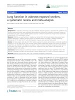
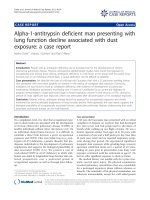
Physiology behind forced expiratory
manoeuvres
• Flow limitation theory
– Dynamic compression of the
airways
– Wave speed theory
spirxpert
P mo=pressure at mouth
P br=pressure inside the
airway P pl=intrapleural
pressure
P alv=intra alveolar pressure P
L.el=elastic recoil pressure of
Wave speed theory
• Flow in elastic tubes limited by the ability of elastic
tubes to propagate pressure waves
• Bulk flow cannot occur at speeds above which
pressures driving the flow can be propagated along
the tube (tube wave speed)
• At tube wave speed – choke point
• Increasing driving pressure above choke point does
not lead to increased flow
• Max flows proportional to density of gas, airway
wall
compliance, and surface area of lumen
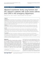
Wave speed theory (2)
• As lung volume diminishes,
total small airways crosssectional area decreases,
peripheral airway resistance
increases, EPP moves
towards alveoli
• Proportion of airways
dynamically compressed
increases
• Reduction in surface area
increases number of airways
that are choked or flow
limited exponentially
• Gives rise to expiratory flowvolume loop
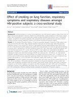
After medication, without predicted value
Flow [l/s]
Vol [l]
Spirometry – measures changes in
flow and volume
• Non-invasive
• Cheap, easy, quick to do
• Widely available (but not always with
graphical display)
• Highly reproducible when airway function
is
stable
• Wide range of predicted values
•
What can we learn from forced
flow- volume measurements?
(1) blow out? –
How much air can the subject
can be reduced in restrictive disorders, or if
there is airway narrowing precipitating early
airway closure (e.g. asthma or CF)
• How fast is the air expelled? – can be
reduced
with airway narrowing.
• Pattern of change in flow-volume curve (insp
& exp) can indicate site of obstruction
What can we learn from forced
flow- volume measurements?
(2)
• RespoŶse to treatŵeŶt ;e.g. β2agonist)
• Change with age or growth
• Progression of disease
͚͚Spiroŵetry is aŶ effort-dependent
manoeuvre that requires
understanding, co-ordination, and
co-operation by the
subject/patient, ǁho ŵust ďe
The person
making
the recordings is
Đarefully
iŶstruĐtedd
every bit as important as the
spirometer!
How do we get from a
spirogram (volume-time
graph) to a flow-volume
curve?
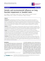
Static lung volumes and capacities
based on a volume–time spirogram
IVC: inspiratory vital capacity
IRV: inspiratory reserve volume VT: tidal
volume (TV)
ERV: expiratory reserve volume RV:
residual volume
IC: inspiratory capacity
FRC: functional residual capacity TLC:
total lung capacity
•
•
•
•
•
•
•
Forced Vital Capacity (FVC)
Forced Expired Volume in 1 second (FEV1) (can do FEV0.5 or FEV0.75 in small children)
FEV1/FVC
(Inspiratory flows/volumes)
Maximum expiratory flow when x% of the FVC has been exhaled (FEFx%) or x% of the FVC
remain to be exhaled (MEFx%, now deprecated) – 25% 50% 75%
Flows at 25% of FVC exhaled = FEF25 or MEF75
Maximal mid-expiratory flow (MMEF) - average expiratory flow over the middle half of the FVC –
may be more sensitive index of obstructive small airways disease as it reflects flow rates once
the dynamic compression-wave has reached the small diseased airways
Quality control and practical aspects
(1)
• Demonstration and careful instruction
• Observe the subject
• Inspect raw data – timebase and flowvolume
• Minimum 3 attempts, maximum of 8 – but
may need more, especially in preschool
children
Quality control and practical aspects
(2)
• Noseclips, Yes or No?
• Filters may be used
• Posture, seated or
standing?
• Use of incentive spirometry