Nghiên cứu mối liên quan giữa kháng thể kháng nucleosome và c1q với mức độ hoạt động của bệnh và tổn thương thận trong lupus ban đỏ hệ thống trẻ em copy tt tiếng anh
Bạn đang xem bản rút gọn của tài liệu. Xem và tải ngay bản đầy đủ của tài liệu tại đây (205.13 KB, 27 trang )
MINISTRY OF EDUCATION & TRAINING
MINISTRY OF HEALTH
HANOI MEDICAL UNIVERSITY
BUI SONG HUONG
STUDY ON THE RELATIONSHIP BETWEEN
ANTINUCLEOSOME AND ANTI-C1q
ANTIBODIES WITH DISEASE ACTIVITY AND
RENAL DAMAGE IN SYSTEMIC LUPUS
ERYTHEMATOSUS IN CHILDREN
Specialized : Pediatrics
Code
: 62720135
SUMMARY OF DOCTORAL THESIS
HANOI - 2019
Research completed in
HANOI MEDICAL UNIVERSITY
Scientific supervisors
Assoc. Prof. Ph.D Le Thi Minh Huong
Ph.D Tran Thị Chi Mai
Scientific reviewer 1:
Scientific reviewer 2:
Scientific reviewer 3:
The thesis will be defended in front of The
Council for Philosophy Doctor in Medicine at
Hanoi Medical University
At………………………….2019.
The thesis can be founf at:
− The National Library
−
Hanoi Medical University Library
−
National Children’s Hospital Library
LIST OF PUBLISHED PAPERS RELATIVE
TO THIS DISSERTATION
1. Apply SLICC 2012 classification criteria in systemic
lupus erythematosus in children, 2017. Pediatric
Journal, 10 (6), 60-64.
2. Relation between anti-nucleosome antibodies and the
level of disease activity in systemic lupus erythematosus
in children, 2017. Medicine Ho Chi Minh city,
Appendix 21, 6, 263-266.
3. Correlations between anti-dsDNA, anti-nucleosome and
anti-C1q antibodies with the disease activity in pediatric
systematic lupus erythematosus, 2019. Journal of Pediatric
Research and Practice, 1, 9-15.
1
ABBREVIATIONS
AC1qAb
Anti-C1q antibody
Anti-dsDNA Anti-double stranded DNA antibody
AnuAb
Anti-nucleosome antibodies
AUC
Area under the ROC curve
GFR
Glomerular filtration rate
LN
Lupus nephritis
PCU
Protein/creatinin urinary ratio
Pos
Positive
SLE
Systemic Lupus Erythematosus
SLEDAI
Systemic Lupus Erythematosus Disease Activity Index
SLICC
Systemic Lupus International Collaborating Clinics
INTRODUCTION
1. Urgency of topics
Systemic Lupus Erythematosus (SLE) is a common systemic
autoimmune disease and more frequent in women. The appearance of
a series of pathological autoantibodies against the antigens that are
part of the body's tissues is a speciality of SLE. Some autoantibodies
have an important role in pathogenesis, diagnosis, assessment of
disease activity level, damage organs especially kidneys and
prognosis for SLE. Anti-double stranded DNA antibody (AntidsDNA) has been a high worth in assessment of disease activity
level, renal damage but revealed limitations actually, so it is
necessary to seek alternative immunological markers. Assessing the
disease activity level by scales is also complicated, time consuming
and sometimes difficult especially in children. Renal biopsy is the
2
gold standard for accurately assessing renal histological lesions
however there are contraindications and limitations.
Finding an autoantibody that can show disease activity and
kidney damage is extremely significant by practical value,
convenience and safe. The anti-nucleosome antibody (AnuAb) and
anti-C1q antibody (AC1qAb) are currently focused on by researchers
about the value in assessing disease activity and renal damage, which
may be superior to anti-dsDNA. However, the values of these two
autoantibodies are not yet confirmed and need further study on
different subjects, different geographical regions. Research on the
SLE in children is limited, especially in Vietnam, so this issue needs
to be further explored to improve the assessment and monitoring of
SLE activity even so effectiveness of treatment.
To having a better understanding of the characteristics of
AnuAb and AC1qAb in assessing disease activity and kidney damage
in SLE, we decide to reseach the topic: “Study on the relationship
between antinucleosome and C1q antibodies with disease activity
and kidney damage in pediatric systemic lupus erythematosus” for
the following purposes:
1. Describe some clinical and laboratory characteristics of
systemic Lupus erythematosus in children.
2. Analysis the association between antinucleosome and antiC1q antibodies with disease activity of systemic lupus
erythematosus according to SLEDAI score.
3. Evaluate the association between antinucleosome and antiC1q antibodies with kidney damage in systemic lupus
erythematosus.
3
2. New contributions of the thesis
AnuAb and AC1qAb concentrations were studied and
quantified at the first time in systemic Lupus erythematosus in
Vietnamese children. This study can be used to compare with
regional and world studies.
AnuAb and AC1qAb have been recorded having corrilation
with disease activity level so they can be used to monitor SLE in
children. AC1qAb suggests diagnosis of lupus nephritis in SLE. This
helps clinicians to early perform a kidney biopsy, choose appropriate
treatment regimens, improve treatment effectiveness and reduce the
risk of death.
3. Layout of the thesis
The thesis consists of 104 pages including: Introduction (2
pages), Chapter 1- Overview (35 pages), Chapter 2- Objects and
Methods (16 pages), Chapter 3- Results (20 pages), Chapter 4Discussion (28 pages), Conclusions (2 pages) and Recommendations
(1 page).
The thesis has 25 tables, 4 pictures, 10 charts and 165
references (including 13 Vietnamese documents, 152 English
documents).
Chapter 1. OVERVIEW
1.1. Pathogenesis mechanism
The cause of SLE is unclear but is complicated by genetic
factors, immune, sex hormones and environment, causing damage
immune system, thereby producing immune response to form
4
autoantibodies against endogenous antigens. There are three main
immune pathways in Lupus which are disorders of programmed cell
death, reducing ability to clean up dead cells and activated T and B
lymphocyte abnormalities, thereby producing autoantibodies. Lupus
pathogenesis is related to many cells and molecules as well as
congenital and acquired immune responses. Autoantibodies may
appear for many years before the onset of clinics. Recently, some
autoantibodies have found that play a major role in SLE
pathophysiology.
Nucleosomes are the basic units of chromosomes playing an
important role in SLE. Programmed cell death releases nuclear
fragments that increase circulating nucleosomes which are altered
and escaped from the normal cleaning process, so leading to increase
expression of nucleosomes to the immune system. Modified
nucleosomes activate nucleosome-specific self-reactive T cells then
stimulate B cells produce AnuAb. The immune complex nucleosomeAnuAb attaches to molecules in the basal membrane of the skin and
kidneys such as heparin sulphate, lamin, collagen 4 or AnuAb is
carried directly to the cross-reactive molecule in the basement
membrane as the alkaline-actinin to organize pathological injury.
C1q is the first component in the complementary activating
chain, stimulates phagocytosis cleaning dead cells, prevents T cell
proliferation, inhibits activation of plasmacytoid dendritic cells,
prevents the production of IFN and inflammatory cytokines. That
plays protecting and inhibiting the immune response against Lupus.
AC1qAb can alter the physiological role of C1q by occupying
important positions associated with C1q receptors, prevent the
5
cleaning process of programmed cells and immun complex leading to
exis immun complex, fixe in the organization and cause organ
damage leading to extensive clinical manifestations of the disease.
SLE usually begins involving several organs and gradually affects
many organs, most commonly kidney damage.
1.2. Diagnostic criteria
We use the classification criteria of the International Clinical
Association Lupus-SLICC 2012 (The Systemic Lupus International
Collaborating Clinics) covering all areas of articulation, dermatology,
neurology, nephrology and immunology which is simple to assess
and easy to use. Patients who have 4 of 17 criterias are diagnosis of
SLE.
1.3. Assess the disease activity of SLE
We use the SLEDAI index of Bombardier, 1992. The
SLEDAI is an overall index of disease activity in the previous 10
days. It consists of 24 weighted clinical and laboratory variables of
nine organ systems. The scores of the descriptors range from 1 to 8,
and the total possible score for all 24 descriptors is 105.
SLEDAI has disadvantage is that it does not catch the
progression, is less sensitive to change than other tools and
does not include the severity of an organ system. SLEDAI
has advantage which is an easy-to-use and validated for use
in children. We use the SLEDAI scale because its sensitivity
to changes in assessment results is estimated to be the
smallest compared to other scales, less fluctuating indicator
among reviewers. Most studies on children use SLEDAI to
6
assess disease activity.
Evaluating disease activity by scales give us a specific
number, but sometimes is difficult and time-consuming.
Therefore, scientists still try to find new immunological
markers related to the disease activity to identify more
accurately, more sensitive and quickly.
1.4. Lupus nephritis
In SLE, kidneys are the most common, early and severe
organ, especially in children accounting for 37-82%. Lupus nephritis
(LN) may appear in the first year but usually occur in the first 5 years
after diagnosis of SLE. The gold standard is kidney biopsy that
indicates glomerulonephritis mediating by immune complex. Early
diagnosis and treatment of LN is very important to improve survival
in LN patients so it is necessary to identify biomarkers that can
predict the development of LN in SLE.
1.5. Role of anti-nucleosome and anti-C1q antibodies in Lupus
Many autoantibodies have been found in SLE patients but
only some of them have clinical significance. No biological marker
accurately measures the SLE disease activity. Anti-dsDNA has been
widely used in diagnosis, monitoring of disease activity and
assessment of kidney damage during the past time but also revealed
limitations.
Although there have been many reports of AnuAb and aCqA
over the past time, most studies in adults, moreover results are
conflict due to heterogeneous clinical characteristics of SLE. Many
studies have shown that AnuAb is valuable in SLE diagnosis and
related to disease activity level, even the author has suggested using
7
AnuA instead in case of negative Anti-dsDNA. To avoid repeated
kidney biopsies, biomarkers are used to assess kidney damages.
AC1qAb has been shown to play an important role in LN
pathogenesis and is closely related to disease activity as well as
the appearance of nephritis. The final conclusion about the values of
AnuAb and AC1qAb in SLE still needs time to prove. So we perform
this research to evaluate the values of AnuAb and AC1qAb in disease
activity for pediatric SLE and particulaly in LN.
CHAPTER 2. SUBJECTS AND METHODS
2.1. Study subjects
Subjects of the study included 125 children who were
diagnosed with SLE were examined and treated at National
Children’s Hospital in Vietnam from January 2015 to December
2017.
Criteria to select patients:
- Patients are eligible for diagnosis of SLE according to SLICC
2012 classification standards.
- Children aged over 1 month and under 16 years old.
- Family of patients and children agree to participate in the study.
Exclusion criteria
SLE Patients coordinate with other autoimmune diseases
(such as rheumatoid arthritis, polyarthritis, Sharp Syndrome,
scleroderma, antiphospholipid syndrome) and drug-induce Lupus.
2.2. Research Methods
Case series descriptive study.
2.3. Research process
8
- Eligible patients are invited to participate in the study.
- The patient was evaluated for
disease history, clinical
manifestations, assessment of disease activity on the SLEDAI scale
for the first time (T0) admission to hospital and was diagnosed SLE
and taken to study, the second time (T3) about 3 months and the third
time (T6) about 6 months after the first time.
- Laboratory was evaluated 3 times at T0, T3, T6 and at the same
time SLEDAI score for hematological tests (full blood count, urinary
sediment), biochemical tests (ure, creatinine, AST, ALT, protein,
albumin, C3, C4 serum concentrations, urine protein and creatinine
levels), quantification of antinuclear antibody, Anti-dsDNA, AnuAb,
AC1qAb.
- Collect data, assess and discuss symptoms with renal and
immuno experts.
2.4. Location and time of study
- SLE patients were examined and treated at the Kidney-Dialysis
Department and Immunology-Allergy-Arthritis Department, National
Children’s Hospital from January 2015 to December 2017.
- Research tests: blood formula tests, biochemical tests,
quantitative antibody tests (AnuAb and AC1qAb) are made in
Hematology and Biochemistry Department in Vietnam National
Children’s Hospital. These laboratories have been accredited with
ISO standards.
2.5. Data processing
The data were processed by STATA 14 software.
2.6. Ethics Research
This is a descriptive, non-intervention study. The research subjects
9
voluntarily participate. Collected data are only for research and
patient care, not for other purposes.
CHAPTER 3. RESULTS
During the period from January 2015 to December 2016, we
collected 125 SLE patients who met the research criterias.
3.1. Clinical and subclinical characteristics
Mean age of SLE onset is 10,52 ± 2,91 age (N=125).
Femal/male ratio=7,9/1.
Bảng 3.1. Distribution of patients according to age group
Age group
(year)
n
%
>10
79
63,2%
5 – 10
<5
Total
41
32,8%
5
4%
125
100%
The most common are children over 10 years old (63,2%),
children under 5 years of age are rare (4%).
Table 3.2: Clinical characteristics according to LN and
non-LN groups
Clinical characteristics
Butterfly rash
Discoid
Photosensitivity
Oral ulcer
Alopecia
Arthritis
Fever
LN
Non-LN
n = 99 (100%) n = 26 (100%)
57
13
(57,6)
(50)
3
3
(3)
(11,5)
27
6
(27,3)
(23,1)
20
7
(20,2)
(26,9)
18
9
(18,2)
(34,6)
47
12
(47,5)
(46,2)
40
17
(40,4)
(65,4)
p
0,49
0,2
0,67
0,46
0,07
0,90
0,03
10
21
2
0,16
(21,2)
(7,7)
7
4
Neurologic disorder
0,24
(7)
(15,4)
Common clinical symptoms in both LN and non-LN groups
are butterfly rash, arthritis and fever. The rate of LN in SLE is
99/125, accounting for 79,2%. The non-LN group had a higher rate
of fever than LN group, the difference was statistically significant
with p < 0,05.
Table 3.3: Clinical characteristics of Lupus nephritis group
Serositis
Clinical characteristics
N (n = 99)
% (100 %)
Edema
58
58,6
Hypertention
37
37,4
Oliguria
32
32,3
Macroscopic hematuria
18
18,2
In LN group, edema is the most common clinical symptom,
accounting for 58,6%, followed by hypertension 37,4% and oliguria
32,3%.
Table 3.4: Paraclinical characteristics of Lupus nephritis group
Characteristics
N (n=99)
% (100%)
Increased serum creatinine
60
60,6
Increased serum ure
34
34,3
Decreased serum protein
43
43,4
Decreased serum Albumin
48
48,5
Urinary red blood cells
60
60,6
Urinary white blood cells
68
68,7
Urinary casts
18
18,2
PCU> 200 mg/mmol
72
72,7
Nephrotic syndrom
44
44,4
GFR < 90
40
40,4
The common paraclinical disorders are increased serum
11
creatinine 60,6%, urinary red blood cell 60,6%, white blood cell
68,7%, PCU (protein/creatinine ratio) > 200 mg/mmol 72,7%,
nephrotic syndrome 44,4%, decreased GFR (glomerular filtration
rate)<90 ml/min/1,73m2 40,4%.
3.2. Relationship between antibodies and disease activity
Table 3.5: Relationship between the positive antibodies and
SLEDAI level
Antibody
T0, T3, T6 ≤ 10
AnuAb
16
Pos
AC1qA
6
Pos
Anti15
dsDNA Pos
SLEDAI
T3
≤ 10 >10 P2
T0
>10
P1
T6
≤ 10 >10
98
0,008
44
12
0,032
39
16
0,016
78
0,0000
28
8
0,216
14
10
0,005
88
0,148
40
10
0,315
39
15
0,056
P3
Positive AnuAb and AC1qA rate are related significantly to
SLEDAI level. The positive anti-dsDNA rate was not associated with
SLEDAI level.
12
Relation between antibody concentrations and SLEDAI level
Table 3.6: Relationship between antibody concentrations and
SLEDAI level at T0 (n = 125)
Antibody T0
AnuAb
AC1qAb
Anti-dsDNA
SLEDAI T0
SLEDAI ≤ 10
SLEDAI > 10
60,3
334,1
(7,5-6888,1)
5,3
(5,7-8200)
16,6
(1,7-19)
70,1
(0,2-992,2)
189,45
p
0,014
<0,01
0,034
(5,3-1200)
(0,1-9143,4)
The median concentration of all antibodies at the time of T0
in the group of patients with SLEDAI> 10 (strong and very strong
SLE) was higher than that of patients with SLEDAI ≤ 10 (mild,
moderate or no activity SLE), p <0.05.
Table 3.7: Relationship between the antibody concentration and
SLEDAI level at T3 (n = 75)
SLEDAI T3
p
SLEDAI ≤ 10
SLEDAI > 10
59
159,8
AnuAb
0,023
0,6 - 4200
30,9 – 1689,2
8,4
15,35
AC1qAb
0,155
0,8 – 85,2
1,1 – 83,2
33,25
113,55
Anti-dsDNA
0,053
0,1 – 4200,2
12,5 – 799,5
At the time of T3, only median concentrations of AnuAb in
patients with SLEDAI> 10 were higher than those with SLEDAI ≤
10, the difference was statistically significant with p<0,05. .
Table 3.8: Relationship between the concentration of antibodies
Antibody T3
and SLEDAI level at T6 (n = 72)
13
Kháng thể T6
AnuAb
SLEDAI T6
SLEDAI ≤ 10
SLEDAI > 10
35,5
333,95
AC1qAb
Anti-dsDNA
2,6 – 4391,4
5,6
32,4 – 5494,4
25,25
0,8 – 233,7
43,15
3,6 – 138,6
326,15
2,1 – 2012,4
21,1 – 4762,2
p
0,000
0,000
0,000
The median concentration of all autoantibodies at the time of
T6 in the group of patients with SLEDAI>10 was higher than the
group with SLEDAI≤10, p <0.001.
Correlation between antibody concentration and SLEDAI score
Table 3.9: Correlation between antibody concentration and
SLEDAI score
Antibody
concentration
T0, T3, T6
AnuAb
AC1qA
Anti-dsDNA
T0
r
p
0,281
0,417
0,289
0,002
0,000
0,001
SLEDAI
T3
r
p
0,328
0,262
0,31
0,004
0,023
0,007
T6
r
p
0,372
0,429
0,507
0,001
0,000
0,000
The concentration of all antibodies correlated positively with
SLEDAI score at different levels.
14
3.3. Relationship between antibody and kidney damage
Table 3.10: Relationship between the changes antibodies rate and
nephritis
LN
Non-LN
n=99 (%)
n=26 (%)
AnuAb Pos
90 (90,91)
24 (92,31)
0,59
AC1qAb Pos
71 (71,72)
13 (50)
0,001
Anti-dsDNA Pos
83(83,84)
20(76,92)
0,41
Dicreased C3
94 (94,95)
19 (73,08)
0,0008
Dicreased C4
92 (92,93)
21 (80,77)
0,061
Immunology marker
P
The positive rate of AC1qAb and the dicreased rate of C3
were associated with lupus nephritis, p = 0,001 and p <0,001,
respectively.
15
Table 3.11: Relationship between antibody concentration and
nephritis
Immunology marker
AnuAb
AC1qAb
Anti-dsDNA
C3
C4
LN (n=99)
Non-LN (n=26)
75,3
52,9
(4-5494,4)
(2,6-4391,4)
7,4
5
(0,8-233,7)
(1,9-12,4)
54,2
89,8
(2,1-4762,2)
(3,8-422,3)
0,92
1
(0,14-1,82)
(0,563-1,66)
0,15
0,213
(0,003-0,772)
(0,03-0,57)
p
0,652
0,011
0,113
0,000
0,014
Median concentration of AC1qAb, C3 and C4 was associated
with nephritis with p <0,05; p <0,001 and 0,05 respectively.
16
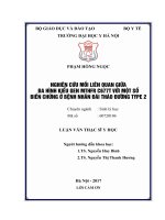
AUC = 0,663
Cut point = 21,1 U/ml
sensitivity=44,4%
specificity=84,6%
Figure 3.1: Area under the ROC curve of AC1qAb
The area under the curve (AUC) of AC1qAb is 0,663, so
there is a value to diagnosis of nephritis with the cut point =21,1
U/ml, sensitivity of 44,4% and a specificity of 84,6%.
Table 3.12: Subclinical manifestations of kidney damage
group III and IV
Manifestation
Group III
Group IV
n=22
n=28
p
(100%)
(100%)
Increased serum creatinine
5(22,7)
18(64,3)
0,003
GFR < 90
6(27,3)
19(69,7)
0,004
PCU >200 mg/mmol
16(72,7)
27(96,4)
0,023
Urinary red blood cells
10(45,5)
25(89,3)
0,001
Urinary red blood cells
11(50)
24(85,7)
0,007
The rate of subclinical disorders of kidney damage in group
IV was higher than that of group III, p <0,01 (except PCU p <0,05).
Table 3.13: Relationship between antibody concentration and
kidney damage Group III and IV
17
Antibody
AnuAb
AC1qAb
Group III
Group IV
(n=22)
(n=28)
200
184
(19,3-8200)
18,9
(5,7-1200)
14
(2,4-992,2)
(0,2-600)
150,5
157,35
Anti-dsDNA
(0,1-4200)
p
0,092
0,39
(0,4-
0,784
5153,7)
Median concentrations of antibodies of LN group III and IV
do not different significantly.
Table 3.14: Correlation between antibody concentrations and
chronic and active points of kidney damage
Antibody
A
C
r
p
r
p
AnuAb
-0,02
0,89
-0,11
0,46
AC1qAb
0,07
0,63
-0,25
0,09
Anti-dsDNA
0,09
0,56
-0,01
0,94
Antibody concentrations are not correlated with the chronic
and active points of kidney damage.
18
CHAPTER 4: DISCUSSION
4.1. Clinical and subclinical characteristics of children SLE
The mean age of disease onset is: 10,52 ± 2,91 years, the
most common group is over 10 years old (63,2%) (Bảng 3.1). The
disease is predominant in female accounting for 88,8% with female /
male ratio = 7,9/1. This result is appropriate with many domestic and
foreign researches. It shows common disease in puberty girls.
SLE is a chronic autoimmune disease with a diverse clinical
phenotype, varying among individuals, also stage of disease. The
common clinical symptoms of SLE in this study were butterfly rash,
arthritis and fever. Assessing general clinical difference between LN
and non-LN group shows a higher rate of fever in the non-LN group
than in LN group, the difference was statistically significant with
p <0,05 (Table 3.2). The non-LN group has higher rate of fever which
is also the reason for hospitalization of this group in our study, so the
rate seems to be higher. Non-LN patients have milder clinical
symptoms and often outpatient treatment. We encountered 99 patients
(79,2%) of LN, in which edema accounted for 58,6%, hypertension
37,4%, oliguria 32,3%, macroscopic hematuria 18,2% (Table 3.3).
The paraclinical characteristics of LN group are increased serum
creatinine 60,6%, decreased serum albumin at the nephrotic syndrom
threshold 48,5%, urinary red blood cells 60,6%, urinary white blood
cells without infection 68,7%, nephrotic syndrome 44,4%, decreased
GFR 40,4%, PCU of nephrotic syndrome >200 mg/mmol 72,7%
(Table 3.4).
Each author has different rates may be due to not only
different groups of patients, different disease stages when selecting
the study sample, but also the complex clinical properties of SLE.
19
4.2. Relationship between antibody and disease activity
Lupus disease activity is expressed through clinical and
paraclinical symptoms that change clearly during the period of active
disease or flare.
Relation between positive antibody rate and SLEDAI level
Positive AnuAb rate is always related to the SLEDAI level (≤
10 or > 10) in all 3 times with p <0.05. The positive AC1qAb rate is
associated with the SLEDAI level at the first and third time with p
<0.01 while the positive Anti-dsDNA ratio was not associated with
the SLEDAI level at all three times ( Table 3.5).
Relation between antibody concentration and SLEDAI level
At the first time (T0), the median concentration of all
antibodies in the group of patients with strong and very strong
disease activity (SLEDAI> 10) was higher than the group of patients
with mild and moderate disease activity (SLEDAI ≤ 10), the
difference was statistically significant with p <0.05
(Table 3.6). At the second time (T3), only median concentration of
AnuAb in patients with SLEDAI> 10 was higher than that of patients
with SLEDAI ≤ 10, the difference was statistically significant with p
<0, 05. AC1qAb and Anti-dsDNA did not differ between these two
groups of patients (Table 3.7). At the third time (T6), the median
concentration of all antibodies in the group of patients with
SLEDAI> 10 was higher than that of patients with SLEDAI ≤ 10, the
difference was statistically significant with p <0.001 (Table 3.8).
Correlation between antibody concentrations and SLEDAI score
The concentration of all antibodies at the test times correlated
with SLEDAI at different levels. The correlation level was
20
statistically significant with AnuAb at the second time (r = 0.328, p
<0.01) and the third time (r = 0.372, p = 0.001), AC1qAb at the first
time (r = 0.417, p <0.001 ) and the third time (r = 0.429, p <0.001),
Anti-dsDNA was the second time (r = 0.31, p <0.01) and the third
time (r = 0.507, p <0.001) (Table 3.9). Thus, according to our
research, antibodies are associated with different levels of disease
activity. AnuAb and AC1qAb are both associated with disease
activity at both positive antibody rates and differences in antibody
concentrations between SLEDAI levels and correlations between
antibody concentrations and SLEDAI scores. We only found that
Anti-dsDNA median concentration was higher in group of patients
with SLEDAI>10 than groupe with SLEDAI≤10 and the AntidsDNA concentration correlated with SLEDAI score. Meanwhile, the
positive Anti-dsDNA rate is not related to the SLEDAI level.
Immunologic markers showing the activity of SLE are associated
with SLEDAI scores at different levels similar to researches in the
world. Monitoring of these immunologic markers allows assessment
of the SLE and response to treatment.
4.3. Relationship between antibody and kidney damage
LN is a serious complication of SLE. The SLE diagnosis at
younger ages is related to poor prognosis, affecting life more than
any other organ system involvement.
4.3.1. Relationship between immunology markers and nephritis
Very few antibodies shown in the pathogenesis of SLE have
been associated simultaneously with disease activity as well as with
nephritis development. This study showed that the positive rate of
AC1qAb and C3 in the LN group was higher than the non-LN group
21
with p≤0,001 (Table 3.10). Median concentration of AC1qAb in LN
group was higher than non-LN group with p <0.05. The median
concentration of C3 and C4 in the LN group was lower than the nonLN group with statistical significance with p <0.001 and p <0.05
respectively (Table 3.11). Thus, the positive rate as well as the
reduction of AC1qAb
concen tration complementary levels are
related to LN. This result is similar to many studies in the world.
Nephritis diagnostic value of antibodies
We found the ability to predict LN of AnuAb, AC1qAb and
Anti-dsDNA antibodies by ROC curve analysis. The ROC analysis
showed that the area under the curve (AUC) for AnuAb was 0.471,
the AUC for Anti-dsDNA was 0.60, the AUC for AC1qAb was 0.663
(Figure 3.1). Thus, only AC1qAb has value to suggest diagnosis of
LN in SLE. The optimal threshold value for AC1qAb to predict LN is
21.1 U / ml with 44.4% sensitivity and 84.6% specificity. The
majority of authors suggest that aC1q is a useful marker, highly
specific, non-invasive biomarker to diagnose nephritis. Studies with
different results may be due to different samples, serum tests of
different manufacturers. Our diagnostic value of AC1qAb is not high
may be due to the research sample. It is necessary to have a research
design that is more suitable for this purpose.
4.3.2. Relationship between antibody and kidney lesion
In our study, group III and group IV of kidney lesions were
differed in subclinical symptoms of kidney damage. The rate of
increased serum creatinine, decreased GFR, PCU > 200 mg/mmol,
Urinary red blood cells, urinary white blood cells in group IV was
higher than that of group III, the difference was statistically
22
significant p <0.01, (PCU p <0.05 except) (Table 3.12). We found
that kidney lesions associated with classification of renal lesions. The
rate of increased serum creatinine, decreased GFR, PCU > 200
mg/mmol, urinary red blood cells, urinary white blood cells in kidney
damage group IV was highest. Most children with LN have normal
renal function despite the presence of active LN, even if there is
kidney damage. Renal biopsy provides information about the level of
inflammation and injury accumulated in LN. Other studies also found
different associations of histopathology with the most common
manifestations of kidney damage in group IV, similar to our results.
Relationship between antibodies and kidney damage
The concentration of antibodies (AnuAb, AC1qAb, AntidsDNA) between the two groups of lesions of group III and IV
kidney disease surgery was not statistically significant (Table 3.13).
The concentration of antibodies was not correlated with the activity
and chronic index scores of lesions of kidney disease surgery (Table
3.14). Many authors also suggest that antibodies are not relevant or
predictive values of kidney damage in SLE. AC1qAb may be a good
serum marker predicting the development of nephritis in SLE
patients, thus closely monitoring kidney disease activity. Antibodies
levels are potential markers to assess disease activity and early
suggestions related to nephritis. Renal biopsy is indispensable in LN
management to specifically assess kidney damage.