Fundamentals of anatomy and physiology 9th ed f martini, j nath, e bartholomew (pearson, 2012) 1
Bạn đang xem bản rút gọn của tài liệu. Xem và tải ngay bản đầy đủ của tài liệu tại đây (10.24 MB, 80 trang )
Quick Reference
Table of Contents
UNIT 1: LEVELS OF ORGANIZATION
1.
An Introduction to Anatomy and Physiology
1
2.
The Chemical Level of Organization
26
3.
The Cellular Level of Organization
62
4.
The Tissue Level of Organization
108
UNIT 2: SUPPORT AND MOVEMENT
5.
The Integumentary System
144
6.
Osseous Tissue and Bone Structure
169
7.
The Axial Skeleton
197
8.
The Appendicular Skeleton
232
9.
Articulations
253
10. Muscle Tissue
279
11. The Muscular System
322
UNIT 3: CONTROL AND REGULATION
12. Neural Tissue
374
13. The Spinal Cord, Spinal Nerves, and
Spinal Reflexes
416
14. The Brain and Cranial Nerves
448
15. Neural Integration I: Sensory Pathways
and the Somatic Nervous System
494
16. Neural Integration II: The Autonomic Nervous
System and Higher-Order Functions
516
17. The Special Senses
548
18. The Endocrine System
593
UNIT 4: FLUIDS AND TRANSPORT
19. Blood
638
20. The Heart
669
21. Blood Vessels and Circulation
707
22. The Lymphatic System and Immunity
764
UNIT 5: ENVIRONMENTAL EXCHANGE
23. The Respiratory System
813
24. The Digestive System
862
25. Metabolism and Energetics
916
26. The Urinary System
953
27. Fluid, Electrolyte, and Acid–Base Balance
997
UNIT 6: CONTINUITY OF LIFE
28. The Reproductive System
1031
29. Development and Inheritance
1076
“I’m glad I didn’t
sell my book
back!…I still use
it today!”
Alissa Lawrence, RN, BSN
Clearwater, Florida
Your A&P textbook is a valuable
investment in your future—an
investment you will want to keep!
“I’m glad I kept my A&P textbook because I used it as a reference in
graduate school, and I still use it occasionally to help explain issues to
patients. It is important to have access to texts that help make the topic
understandable and that approach the topic in a meaningful way. I feel
that being able to integrate the information in the text with actual
practice is critical for learning and practice.”
Meg Portwood, RN, MS, FNP
Lincoln City, Oregon
“I still have the text and used it several times throughout Physician Assistant
school. My Martini/Nath Fundamentals of A&P text was definitely a valuable text
throughout my PA program because of the constant learning process. As I
went through topics such as pharmacology it was often imperative to review
specific physiology and occasionally anatomy in order to fully understand how
medications, etc. affect the various body systems in order to achieve the
desired result.”
Aaron McCloud, PA
San Francisco, California
“I still have my A&P textbook! As a Registered Nurse, I find my A&P
textbook extremely valuable. The study of anatomy and physiology will
provide you with the building blocks of knowledge in understanding
the complexities of the human body and its functions.”
Cynthia Pronze, RN, MSN
Ann Arbor, Michigan
F U N D A M E N TA L S O F
Anatomy &
Physiology
Ninth Edition
Frederic H. Martini, Ph.D.
University of Hawaii at Manoa
Judi L. Nath, Ph.D.
Lourdes College
Edwin F. Bartholomew, M.S.
William C. Ober, M.D.
Art Coordinator and Illustrator
Claire W. Garrison, R.N.
Illustrator
Kathleen Welch, M.D.
Clinical Consultant
Ralph T. Hutchings
Biomedical Photographer
Executive Editor: Leslie Berriman
Project Editor: Robin Pille
Director of Development: Barbara Yien
Development Editor: Anne A. Reid
Editorial Assistant: Nicole McFadden
Senior Managing Editor: Deborah Cogan
Production Project Manager: Caroline Ayres
Copyeditor: Michael Rossa
Production Management and Compositor: S4Carlisle
Publishing Services, Inc.
Cover Photo Credit: Mike Powell/Getty Images
Director of Media Development: Lauren Fogel
Media Producer: Aimee Pavy
Design Manager: Marilyn Perry
Interior and Cover Designer: tani hasegawa
Contributing Illustrators: imagineeringart.com
Senior Photo Editor: Donna Kalal
Photo Researcher: Maureen Spuhler
Senior Manufacturing Buyer: Stacey Weinberger
Marketing Manager: Derek Perrigo
Notice: Our knowledge in clinical sciences is constantly changing. The authors and the publisher of
this volume have taken care that the information contained herein is accurate and compatible with
the standards generally accepted at the time of the publication. Nevertheless, it is difficult to ensure
that all information given is entirely accurate for all circumstances. The authors and the publisher
disclaim any liability, loss, or damage incurred as a consequence, directly or indirectly, of the use
and application of any of the contents of this volume.
Copyright © 2012 by Frederic H. Martini, Inc., Judi L. Nath, LLC, and Edwin F. Bartholomew, Inc.
Published by Pearson Education, Inc., publishing as Pearson Benjamin Cummings, 1301 Sansome
St., San Francisco, CA 94111. All rights reserved. Manufactured in the United States of America. This
publication is protected by Copyright and permission should be obtained from the publisher prior
to any prohibited reproduction, storage in a retrieval system, or transmission in any form or by any
means, electronic, mechanical, photocopying, recording, or likewise. To obtain permission(s) to
use material from this work, please submit a written request to Pearson Education, Inc.,
Permissions Department, 1900 E. Lake Ave., Glenview, IL 60025. For information regarding
permissions, call (847) 486-2635.
Many of the designations used by manufacturers and sellers to distinguish their products are
claimed as trademarks. Where those designations appear in this book, and the publisher was aware
of a trademark claim, the designations have been printed in initial caps or all caps.
MasteringA&P™, A&P Flix™, Practice Anatomy Lab™ (PAL™), and Interactive Physiology® are
trademarks, in the U.S. and/or other countries, of Pearson Education, Inc. or its affiliates.
Library of Congress Cataloging-in-Publication Data
Martini, Frederic.
Fundamentals of anatomy & physiology/Frederic H. Martini, Judi L. Nath, Edwin F. Bartholomew;
with William C. Ober, art coordinator and illustrator; Claire W. Garrison, illustrator; Kathleen
Welch, clinical consultant; Ralph T. Hutchings, biomedical photographer. — 9th ed. p.; cm.
Includes bibliographical references and index.
ISBN-13: 978-0-321-70933-2 (student edition : alk. paper)
ISBN-10: 0-321-70933-0 (student edition : alk. paper) 1. Human physiology—Textbooks. 2.
Human anatomy—Textbooks.
I. Nath, Judi Lindsley. II. Bartholomew, Edwin F. III. Title. IV. Title: Fundamentals of anatomy and
physiology.
[DNLM: 1. Anatomy. 2. Physiology. QS 4]
QP34.5.M27 2012
612—dc22
2010043347
0-321-70933-0 (Student edition)
978-0321-70933-2 (Student edition)
0-321-76625-3 (Exam Copy)
978-0321-76625-0 (Exam Copy)
1 2 3 4 5 6 7 8 9 10—DOW—14 13 12 11 10
Text and Illustration Team
Frederic (Ric) H. Martini, Ph.D.
Judi L. Nath, Ph.D.
Author
Author
Dr. Martini received his Ph.D. from Cornell University in comparative and functional anatomy for work on the
pathophysiology of stress. In addition to
professional publications that include
journal articles and contributed chapters, technical reports, and
magazine articles, he is the lead author of nine undergraduate
texts on anatomy and physiology or anatomy. Dr. Martini is
currently affiliated with the University of Hawaii at Manoa and
has a long-standing bond with the Shoals Marine Laboratory, a
joint venture between Cornell University and the University of
New Hampshire. He has been active in the Human Anatomy
and Physiology Society (HAPS) for 18 years and was a member
of the committee that established the course curriculum guidelines for A&P. He is now a President Emeritus of HAPS after
serving as President-Elect, President, and Past-President over
2005–2007. Dr. Martini is also a member of the American Physiological Society, the American Association of Anatomists, the
Society for Integrative and Comparative Biology, the Australia/New Zealand Association of Clinical Anatomists, the
Hawaii Academy of Science, the American Association for the
Advancement of Science, and the International Society of Vertebrate Morphologists.
Dr. Nath is a biology professor at Lourdes College, where she teaches anatomy
and physiology, pathophysiology, medical terminology, and pharmacology.
She received her Bachelor’s and Master’s
degrees from Bowling Green State University and her Ph.D.
from the University of Toledo. Dr. Nath is devoted to her students and strives to convey the intricacies of science in a captivating way that students find meaningful, interactive, and
exciting. She is a multiple recipient of the Faculty Excellence
Award, granted by the college to recognize her effective teaching, scholarship, and community service. She is active in many
professional organizations, notably the Human Anatomy and
Physiology Society (HAPS), where she has served several terms
on the board of directors. On a personal note, Dr. Nath enjoys
family life with her husband, Mike, and their three dogs. Piano
playing and cycling are welcome diversions from authoring,
and her favorite charities include the local Humane Society, the
Cystic Fibrosis Foundation, and Real Partners Uganda.
Edwin F. Bartholomew, M.S.
William C. Ober, M.D.
Author
Art Coordinator and Illustrator
Edwin F. Bartholomew received his undergraduate degree from Bowling Green
State University in Ohio and his M.S.
from the University of Hawaii. Mr.
Bartholomew has taught human
anatomy and physiology at both the secondary and undergraduate levels and a wide variety of other science courses (from
botany to zoology) at Maui Community College and at historic
Lahainaluna High School, the oldest high school west of the
Rockies. Working with Dr. Martini, he coauthored Essentials of
Anatomy & Physiology, Structure and Function of the Human Body,
and The Human Body in Health and Disease (all published by
Pearson Benjamin Cummings). Mr. Bartholomew is a member
of the Human Anatomy and Physiology Society (HAPS), the
National Association of Biology Teachers, the National Science
Teachers Association, the Hawaii Science Teachers Association,
and the American Association for the Advancement of Science.
Dr. Ober received his undergraduate degree from Washington and Lee University and his M.D. from the University of
Virginia. He also studied in the Department of Art as Applied to Medicine at
Johns Hopkins University. After graduation, Dr. Ober completed a residency in Family Practice and later was on the faculty
at the University of Virginia in the Department of Family Medicine and in the Department of Sports Medicine. He also served
as Chief of Medicine of Martha Jefferson Hospital in Charlottesville, VA. He is currently a Visiting Professor of Biology at
Washington and Lee University, where he has taught several
courses and led student trips to the Galápagos Islands. He is on
the Core Faculty at Shoals Marine Laboratory, where he teaches
Biological Illustration every summer. Dr. Ober has collaborated
with Dr. Martini on all of his textbooks in every edition.
iii
iv Text and Illustration Team
Claire W. Garrison, R.N.
Ralph T. Hutchings
Illustrator
Biomedical Photographer
Claire W. Garrison, R.N., B.A., practiced
pediatric and obstetric nursing before
turning to medical illustration as a fulltime career. She returned to school at
Mary Baldwin College, where she received her degree with distinction in studio art. Following a
five-year apprenticeship, she has worked as Dr. Ober’s partner
in Medical & Scientific Illustration since 1986. She is on the
Core Faculty at Shoals Marine Laboratory and co-teaches the
Biological Illustration course with Dr. Ober every summer. The
textbooks illustrated by Medical & Scientific Illustration have
won numerous design and illustration awards.
Mr. Hutchings was associated with
Royal College of Surgeons for 20 years.
An engineer by training, he has focused
for years on photographing the structure
of the human body. The result has been
a series of color atlases, including the Color Atlas of Human
Anatomy, the Color Atlas of Surface Anatomy, and The Human
Skeleton (all published by Mosby-Yearbook Publishing). For his
anatomical portrayal of the human body, the International
Photographers Association has chosen Mr. Hutchings as the
best photographer of humans in the twentieth century. He lives
in North London, where he tries to balance the demands of his
photographic assignments with his hobbies of early motor cars
and airplanes.
Kathleen Welch, M.D.
Clinical Consultant
Dr. Welch received her M.D. from the
University of Washington in Seattle and
did her residency in Family Practice at
the University of North Carolina in
Chapel Hill. For two years, she served as
Director of Maternal and Child Health at the LBJ Tropical Medical Center in American Samoa and subsequently was a member of the Department of Family Practice at the Kaiser
Permanente Clinic in Lahaina, Hawaii. She has been in private
practice since 1987 and is licensed to practice in Hawaii, Washington, and New Zealand. Dr. Welch is a Fellow of the American Academy of Family Practice and a member of the Hawaii
Medical Association and the Human Anatomy and Physiology
Society (HAPS). With Dr. Martini, she has coauthored both a
textbook on anatomy and physiology and the A&P Applications
Manual. She and Dr. Martini were married in 1979, and they
have one son, PK.
Preface
The Ninth Edition of Fundamentals of Anatomy & Physiology is a
comprehensive textbook that fulfills the needs of today’s students while addressing the concerns of their professors. This
edition was shaped by the collaboration among three experienced instructors, authors Ric Martini, Judi Nath, and Ed
Bartholomew. The Martini/Nath/Bartholomew team focused
their attention on the question “How can we best make this information meaningful, manageable, and comprehensible?”
During the revision process, we drew upon our content knowledge, research skills, artistic talents, and a collective 75 years of
classroom experience to make this edition the best yet.
The broad changes to this edition are presented in the New
to the Ninth Edition section below. Also below are the sections Terminology Changes in the Ninth Edition, Learning
Outcomes, and Chapter-by-Chapter Changes in the Ninth
Edition. A visual tour of the book follows in the remaining
pages of the Preface.
◗ New to the Ninth Edition
In addition to the many technical changes in this edition, such
as updated statistics and anatomy and physiology descriptions,
we have simplified the presentations to make the narrative easier to read. We have also focused on improving the integration
of illustrations with the narrative. These are the key changes in
this new edition:
• Easier narrative uses simpler, shorter, more active
sentences and a quantifiably lower reading level to make
reading and studying easier for students.
• “Spotlight” figures combine text and art to communicate
key topics in visually effective single-page or two-page
presentations.
• Improved text-art integration throughout the illustration
program enhances the readability of figures. Part captions
are now integrated into the figures so that the relevant text
is located immediately next to each part of a figure.
• More visual Clinical Notes draw students’ attention to
clinical information and scenarios they might encounter in
their future careers.
• New System Integrator figures for each body system
replace the “Systems in Perspective” figures from previous
editions. These “build-a-body” figures reinforce the
mechanisms of system integration by gradually increasing
in complexity as each new system is examined.
• Easier-to-read tables have been redesigned and
simplified, and references to them within the narrative are
now in color to make them easier to find.
• Updated Related Clinical Terms sections at the end of
each chapter have been revised to include the most current
relevant clinical terms and procedures.
• MasteringA&P™ (www.masteringaandp.com) is an online
learning and assessment system designed to help
instructors teach more efficiently and proven to help
students learn. Instructors can assign homework from
proven media programs such as Practice Anatomy Lab™
(PAL™), Interactive Physiology®, and A&P Flix™—all
organized by chapter—and have assignments automatically
graded. There are also abundant assessments from each
chapter’s content, including Reading Quizzes. All
assessments are organized by the chapter Learning
Outcomes. In the MasteringA&P Study Area, students can
access a full suite of self-study tools, listed in detail at the
very end of each textbook chapter.
◗ Terminology Changes in the Ninth
Edition
We have revised terminology in selected cases to match the most
common usage in medical specialties. We used Terminologia
Anatomica and Terminologia Histologica as our reference for
anatomical and tissue terms. Furthermore, possessive forms of
diseases are now used when the proposed alternative has not
been widely accepted, e.g., Parkinson disease is now Parkinson’s
disease. In addition, several terms that were primary in the
Eighth Edition have become secondary terms in the Ninth Edition. The changes, which affect virtually all of the chapters in the
text, are detailed in the table on the following page.
v
vi Preface
Eighth Edition Primary Term
Ninth Edition Primary Term
acrosomal cap
acrosome
adenohypophysis
anterior lobe of the pituitary gland
aqueduct of midbrain
cerebral aqueduct
awake-asleep cycle
sleep-wake cycle
basal lamina
basement membrane
canal of Schlemm
scleral venous sinus
creatine phosphokinase
creatine kinase (CK)
diaphragma sellae
sellar diaphragm
fibrous cartilage
fibrocartilage
fibrous tunic, vascular tunic, and
neural tunic
fibrous layer, vascular layer, and
inner layer
induced immunity
artificially induced immunity
infundibulopelvic ligament
suspensory ligament
inner ear
internal ear
intercellular cement
proteoglycans
lymphoid system
lymphatic system
macula adherens
desmosome
macula lutea
macula
mesencephalon
midbrain
neurohypophysis
posterior lobe of the pituitary gland
nonspecific defenses
innate (nonspecific) defenses
occluding junction
tight junction
organ of Corti
spiral organ
specific defenses
adaptive (specific) defenses
stratum germinativum
stratum basale
subcutaneous layer
hypodermis
suprarenal
adrenal
tympanic duct
scala tympani
vestibular duct
scala vestibuli
◗ Learning Outcomes
The chapters of the Ninth Edition are organized around concrete Learning Outcomes that indicate what students should be
able to do after studying the chapter.
• Learning Outcomes on the chapter-opening page are
correlated by number with the chapter headings in the
textbook. The Learning Outcomes are also correlated to the
test items in MasteringA&P™ (www.masteringaandp.com)
and to the test items in the Test Bank, making it possible
for instructors to organize the course material and assess
student learning based on specific Learning Outcomes. The
Learning Outcomes are derived from the Learning
Outcomes recommended by the Human Anatomy and
Physiology Society (HAPS).
• Full-sentence section headings, correlated by number with
the Learning Outcomes, state a core fact or concept to help
students readily see and learn the chapter content. There is a
one-to-one correspondence between the Learning Outcomes
and the full-sentence section headings in every chapter.
• Checkpoints are located at the close of each section and
ask students to pause and check their understanding of
facts and concepts. The Checkpoints reinforce the Learning
Outcomes presented on the chapter-opening page,
resulting in a systematic integration of the Learning
Outcomes over the course of the chapter. Answers are
located in the blue Answers tab at the back of the book.
All assessments in MasteringA&P are organized by the Learning Outcomes, making it easy for instructors to organize their
courses and demonstrate results against departmental goals for
student achievement.
◗ Chapter-by-Chapter Changes in the
Ninth Edition
This annotated Table of Contents provides select examples of
revision highlights in each chapter of the Ninth Edition.
Chapter 1: An Introduction to Anatomy and Physiology
• New Spotlight Figure 1–1 Levels of Organization
• New Figure 1–4 Positive Feedback: Blood Clotting
• Figure 1–5 Anatomical Landmarks revised
• Figure 1–7 Directional References revised
• Figure 1–8 Sectional Planes revised
• Figure 1–9 Relationships among the Subdivisions of the Ventral
Body Cavity revised
• Clinical Note: The Visible Human Project revised
• Clinical Note: Fatty Acids and Health revised
Chapter 2: The Chemical Level of Organization
• Figure 2–3 The Formation of Ionic Bonds revised
• New Spotlight Figure 2–7 Chemical Notation
• Figure 2–10 pH and Hydrogen Ion Concentration revised
• Figure 2–19 Amino Acids revised
• Figure 2–22 A Simplified View of Enzyme Structure and Function
revised
• Clinical Note: Solute Concentrations revised
Chapter 3: The Cellular Level of Organization
• Old Table 3–1 incorporated into new Spotlight Figure 3–1
Anatomy of a Model Cell
• Old Figure 3–7 incorporated into new Spotlight Figure 3–7
Protein Synthesis
• Figure 3–10 The Nucleus revised to include new figure of nuclear
pore
• Figure 3–12 mRNA Transcription revised
• Figure 3–17 Osmotic Flow across a Plasma Membrane revised
• Old Figure 3–23 incorporated into new Spotlight Figure 3–24
Stages of a Cell’s Life Cycle
• Old Figure 3–25 incorporated into new Spotlight Figure 3–24
Stages of a Cell’s Life Cycle
• Table 3–1 Examples of the Triplet Code switched order of template
strand with coding strand to show that the coding strand sequence
is the same as the mRNA sequence except for T and U
• Table 3–2 Template Strand and Coding Strand switched for clarity
• Clinical Note: Parkinson’s Disease revised
Preface vii
Chapter 4: The Tissue Level of Organization
• Reordered connective tissue proper cell populations in text under
Components of Connective Tissue Proper
• New Figure 4–1 The Polarity of Epithelial Cells
• New Figure 4–2 Cell Junctions
• Figure 4–4 Cuboidal and Transitional Epithelia, Transitional
Epithelium part revised
• Figure 4–5 Columnar Epithelia revised to include anatomical
location within human figure
• Figure 4–6 Modes of Glandular Secretion revised
• Figure 4–12 Formed Elements of the Blood revised
• Old Figure 4–20 incorporated into new Spotlight Figure 4–20
Tissue Repair
• Clinical Note: Problems with Serous Membranes revised
Chapter 5: The Integumentary System
• Figure 5–1 The Components of the Integumentary System revised
• Figure 5–10 Hair Follicles and Hairs changed order and revised
• Figure 5–14 Repair of Injury to the Integument revised
• Clinical Note: Skin Cancer revised
• Clinical Note: Burns and Grafts revised
• New Figure 5–17 System Integrator
Chapter 6: Osseous Tissue and Bone Structure
• Figure 6–1 A Classification of Bones by Shape revised
• Figure 6–3 Types of Bone Cells revised
• Figure 6–10 Endochondral Ossification revised
• Figure 6–15 A Chemical Analysis of Bone revised
• Figure 6–16 Factors That Alter the Concentration of Calcium Ions
in Body Fluids revised
• Old Figures 6–17 and 6–18 incorporated into new Spotlight
Figure 6–17 Types of Fractures and Steps in Repair
• Figure 6–18 The Effects of Osteoporosis on Spongy Bone revised
• Clinical Note: Heterotopic Bone Formation revised
• Clinical Note: Abnormal Bone Development revised
Chapter 7: The Axial Skeleton
• Figure 7–1 The Axial Skeleton revised and combined into a onepage figure
• Figure 7–2 Cranial and Facial Subdivisions of the Skull revised so
that the chart is above and connections between the chart and
the art are clearly apparent
• Figure 7–7 The Temporal Bones revised by switching positions of
(a) and (b) to show which part is the source of the dissected
mastoid air cells
• Figure 7–16 The Vertebral Column revised
• Clinical Note: Kyphosis, Lordosis, and Scoliosis revised
Chapter 8: The Appendicular Skeleton
• Figure 8–1 The Appendicular Skeleton revised
• Figure 8–4 The Humerus added views of the elbow joint
• Figure 8–5 The Radius and Ulna revised to show the interosseous
membrane and added a lateral view of the trochlear notch
• Figure 8–12 The Right Patella revised and added inferior view of
right femur and patella
• Figure 8–13 The Tibia and Fibula revised and added cross section
of tibia and fibula
• Figure 8–14 Bones of the Ankle and Foot revised
Chapter 9: Articulations
• Reorganized section on synovial joints for improved flow
• Included discussion and art on vertebral end plates
• Reorganized old Tables 9–1 and 9–2 into one simpler Table 9–1
Functional and Structural Classifications of Articulations
• New Spotlight Figure 9–6 Synovial Joints
• Figure 9–7 Intervertebral Articulations revised
• New Figure 9–13 System Integrator
• Clinical Note: Knee Injuries revised
Chapter 10: Muscle Tissue
• Moved Table 10–1 Steps Involved in Skeletal Muscle Contraction
and Relaxation to the end of Section 10-4 to better serve as a
summary of the topics
• Figure 10–1 The Organization of Skeletal Muscles revised
• New Figure 10–9 An Overview of Skeletal Muscle Contraction
• New Spotlight Figure 10–11 Skeletal Muscle Innervation
• New Spotlight Figure 10–12 The Contraction Cycle
• Figure 10–13 Shortening during a Contraction revised
• Figure 10–14 The Effect of Sarcomere Length on Active Tension
revised
• Figure 10–18 Concentric, Eccentric, and Isometric Contractions
revised and added new eccentric contractions part to figure
• Figure 10–21 Fast versus Slow Fibers revised
• Figure 10–24 Smooth Muscle Tissue revised
• Clinical Note: Tetanus revised
• Clinical Note: Delayed-Onset Muscle Soreness revised
Chapter 11: The Muscular System
• Nearly all figures in this chapter are now presented in the
anterior view first and the posterior view second
• New Figure 11–3 An Overview of the Major Skeletal Muscles
• New Figure 11–10 Muscles of the Vertebral Column
• New Figure 11–11 Oblique and Rectus Muscles and the
Diaphragm revised and new part (a) added
• Figure 11–13 An Overview of the Appendicular Muscles of the
Trunk revised
• Figure 11–14 Muscles That Position the Pectoral Girdle revised
• Figure 11–15 Muscles That Move the Arm revised
• Figure 11–17 Muscles That Move the Hand and Fingers revised
• Figure 11–18 Intrinsic Muscles of the Hand revised
• Table 11–15 Intrinsic Muscles of the Hand reorganized
• Figure 11–19 Muscles That Move the Thigh revised
• Figure 11–20 Muscles That Move the Leg revised
• New Figure 11–21 Extrinsic Muscles That Move the Foot and Toes
• Figure 11–22 Intrinsic Muscles of the Foot revised
• Table 11–19 Intrinsic Muscles of the Foot reorganized
• New Figure 11–23 System Integrator
• Clinical Note: Hernia revised
• Clinical Note: Intramuscular Injections revised
Chapter 12: Neural Tissue
• New Figure 12–3 A Structural Classification of Neurons
• New Figure 12–4 An Introduction to Neuroglia
• Figure 12–7 Peripheral Nerve Regeneration after Injury revised
• Figure 12–8 An Overview of Neural Activities revised
• New Figure 12–9 The Resting Potential Is the Transmembrane
Potential of an Undisturbed Cell
• New Figure 12–10 Electrochemical Gradients for Potassium and
Sodium Ions
• Old Figure 12–14 combined with old Table 12–3 for a new
Spotlight Figure 12–14 Generation of an Action Potential
• New Figure 12–16 Saltatory Propagation along a Myelinated
Axon
• Table 12–4 Synaptic Activity revised
• New Figure 12–17 Events in the Functioning of a Cholinergic Synapse
• New Figure 12–19 Temporal and Spatial Summation
• Table 12–4 Synaptic Activity Revised
• Clinical Note: Demyelination revised
Chapter 13: The Spinal Cord, Spinal Nerves, and Spinal Reflexes
• Figure 13–1 An Overview of Chapters 13 and 14 revised
• Figure 13–6 A Peripheral Nerve revised
viii Preface
• New Spotlight Figure 13–7 Peripheral Distribution of Spinal Nerves
• New Figure 13–14 Neural Circuits: The Organization of
Neuronal Pools
• New Figure 13–16 The Classification of Reflexes
• Figure 13–21 The Babinski Reflex revised
• Clinical Note: Spinal Anesthesia revised
Chapter 14: The Brain and Cranial Nerves
• New Table 14–1 Development of the Brain
• Figure 14–5 The Diencephalon and Brain Stem revised
• New Figure 14–7 The Cerebellum
• New Figure 14–12 The Brain in Lateral View
• Figure 14–16 Hemispheric Lateralization revised
• New Figure 14–17 Brain Waves
• Clinical Note: Epidural and Subdural Hemorrhages revised
• Clinical Note: Aphasia and Dyslexia revised
Chapter 15: Neural Integration I: Sensory Pathways and the Somatic
Nervous System
• Reorganized Section 15–4 Separate pathways carry somatic
sensory and visceral sensory information
• Figure 15–1 An Overview of Neural Integration revised
• Figure 15–3 Tactile Receptors in the Skin revised
• New Spotlight Figure 15–5 Somatic Sensory Pathways
• Clinical Note: Assessment of Tactile Sensitivities revised
Chapter 16: Neural Integration II: The Autonomic Nervous System and
Higher-Order Functions
• Enhanced Section 16–9 Neurotransmitters influence brain
chemistry and behavior
• Figure 16–10 The Autonomic Plexuses and Ganglia revised
• Figure 16–12 A Comparison of Somatic and Autonomic Function
revised
• New Figure 16–14 Levels of Sleep
• New Figure 16–16 System Integrator
• Clinical Note: Amnesia revised
• Clinical Note: Alzheimer’s Disease revised
Chapter 17: The Special Senses
• Figure 17–1 The Olfactory Organs revised
• New Spotlight Figure 17–2 Olfactory and Gustatory Receptors
• Figure 17–6 The Pupillary Muscles revised
• Figure 17–11 Accommodation revised
• New Spotlight Figure 17–13 Accommodation Problems
• New Spotlight Figure 17–17 Photoreception
• New Figure 17–18 Bleaching and Regeneration of Visual Pigments
• Figure 17–20 The Visual Pathways revised
• Figure 17–21 The Anatomy of the Ear revised
• Figure 17–30 Sound and Hearing revised
• New Figure 17–32 Pathways for Auditory Sensations
• Clinical Note: Glaucoma revised
• Clinical Note: Motion Sickness revised
Chapter 18: The Endocrine System
• Figure 18–1 Organs and Tissues of the Endocrine System revised
• New Spotlight Figure 18–2 Structural Classification of Hormones
• Figure 18–3 G Proteins and Hormone Activity revised
• Figure 18–13 The Homeostatic Regulation of Calcium Ion
Concentrations revised
• New Figure 18–15 The Pineal Gland
• Figure 18–17 The Regulation of Blood Glucose Concentrations
revised
• New Spotlight Figure 18–18 Diabetes Mellitus
• Figure 18–19 Endocrine Functions of the Kidneys revised
• New Spotlight Figure 18–20 The General Adaptation Syndrome
• New Figure 18–21 System Integrator
• Clinical Note: Hormones and Athletic Performance revised
Chapter 19: Blood
• Added information on aspirin as an anticoagulant
• New Spotlight Figure 19–1 The Composition of Whole Blood
• Figure 19–5 Recycling of Red Blood Cell Components revised
• Figure 19–7 Blood Types and Cross-Reactions revised
• Figure 19–8 Blood Type Testing revised
• New Spotlight Figure 19–9 Hemolytic Disease of the Newborn
• Figure 19–11 The Origins and Differentiation of Formed
Elements revised
• New Figure 19–12 The Vascular, Platelet, and Coagulation Phases
of Hemostasis and Clot Retraction
• Clinical Note: Plasma Expanders revised
• Clinical Note: Abnormal Hemoglobin revised
Chapter 20: The Heart
• Figure 20–4 The Heart Wall revised
• Figure 20–8 Valves of the Heart revised
• New Spotlight Figure 20–10 Heart Disease and Heart Attacks
• Figure 20–12 Impulse Conduction through the Heart revised
• Figure 20–13 An Electrocardiogram revised
• New Spotlight Figure 20–14 Cardiac Arrhythmias
• Figure 20–15 The Action Potential in Skeletal and Cardiac Muscle
revised
• New Figure 20–16 Phases of the Cardiac Cycle
• New Figure 20–19 A Simple Model of Stroke Volume
• New Figure 20–20 Factors Affecting Cardiac Output
• Figure 20–21 Autonomic Innervation of the Heart revised
• New Figure 20–23 Factors Affecting Stroke Volume
• Figure 20–24 A Summary of the Factors Affecting Cardiac Output
revised
Chapter 21: Blood Vessels and Circulation
• Figure 21–2 Histological Structure of Blood Vessels revised
• New Figure 21–4 Capillary Structure
• New Figure 21–6 Valves in the Venous System
• New Figure 21–9 Factors Affecting Friction and Vascular Resistance
• Figure 21–10 Relationships among Vessel Diameter, CrossSectional Area, Blood Pressure, and Blood Velocity revised
• New Figure 21–14 Short-Term and Long-Term Cardiovascular
Responses
• New Figure 21–15 Baroreceptor Reflexes of the Carotid and
Aortic Sinuses
• New Figure 21–16 The Chemoreceptor Reflexes
• New Figure 21–17 Hormonal Regulation of Blood Pressure and
Blood Volume
• New Figure 21–18 Cardiovascular Responses to Hemorrhaging
and Blood Loss
• Figure 21–19 A Schematic Overview of the Pattern of Circulation
revised
• New Figure 21–24 Arteries of the Brain
• Figure 21–25 Major Arteries of the Trunk revised
• Figure 21–26 Arteries Supplying the Abdominopelvic Organs
revised
• New Figure 21–29 Major Veins of the Head, Neck, and Brain
• Figure 21–33 The Hepatic Portal System revised
• New Spotlight Figure 21–35 Congenital Heart Problems
• New Figure 21–36 System Integrator
Chapter 22: The Lymphatic System and Immunity
• New Figure 22–1 An Overview of the Lymphatic System: The
Lymphatic Vessels, Lymphoid Tissues, and Lymphoid Organs
• New Figure 22–5 Classes of Lymphocytes
• Figure 22–11 Innate Defenses revised
• Figure 22–12 How Natural Killer Cells Kill Cellular Targets revised
• New Figure 22–13 Interferons
• New Figure 22–14 Pathways of Complement Activation
Preface ix
•
•
•
•
•
•
•
•
•
•
New Figure 22–15 Inflammation and the Steps in Tissue Repair
Figure 22–16 Forms of Immunity revised
New Figure 22–17 An Overview of the Immune Response
New Figure 22–18 Antigens and MHC Proteins
New Figure 22–19 Antigen Recognition by and Activation of
Cytotoxic T Cells
New Figure 22–20 Antigen Recognition and Activation of Helper
T Cells
Figure 22–22 The Sensitization and Activation of B Cells
New Figure 22–26 An Integrated Summary of the Immune Response
New Spotlight Figure 22–28 Cytokines of the Immune System
New Figure 22–30 System Integrator
Chapter 23: The Respiratory System
• Included information on spirometry
• Figure 23–7 The Gross Anatomy of the Lungs revised
• New Figure 23–12 An Overview of the Key Steps in External
Respiration
• Figure 23–13 Gas Pressure and Volume Relationships revised
• Figure 23–16 The Respiratory Muscles revised
• Figure 23–17 Pulmonary Volumes and Capacities revised
• Figure 23–18 Henry’s Law and the Relationship between
Solubility and Pressure revised
• Figure 23–23 Carbon Dioxide Transport in Blood revised
• Figure 23–34 A Summary of the Primary Gas Transport
Mechanisms revised
• Figure 23–25 Basic Regulatory Patterns of Respiration revised
• New Spotlight Figure 23–26 Control of Respiration
• New Figure 23–27 The Chemoreceptor Response to Changes in PCO2
• New Figure 23–29 System Integrator
Chapter 24: The Digestive System
• Included information on vomiting
• Reorganized the section Control of Digestive Functions
• New Figure 24–1 The Components of the Digestive System
• Figure 24–4 Peristalsis revised
• New Figure 24–5 The Regulation of Digestive Activities
• Figure 24–8 Teeth revised
• Figure 24–11 The Swallowing Process revised
• Figure 24–13 The Stomach Lining revised
• New Figure 24–14 The Secretion of Hydrochloric Acid
• New Spotlight Figure 24–15 Regulation of Gastric Activity
• Figure 24–16 Segments of the Intestine revised
• New Figure 24–21 The Anatomy and Physiology of the
Gallbladder and Bile Ducts
• New Figure 24–22 Major Duodenal Hormones
• New Figure 24–23 The Activities of Major Digestive Tract Hormones
• New Figure 24–26 The Defecation Reflex
• New Spotlight Figure 24–27 Chemical Events in Digestion
• New Figure 24–28 Digestive Secretion and Absorption of Water
• New Figure 24–29 System Integrator
Chapter 25: Metabolism and Energetics
• Included information on exercise as a mechanism for lowering
cholesterol
• New Figure 25–2 Nutrient Use in Cellular Metabolism
• New Figure 25–8 Beta-Oxidation
• New Figure 25–9 Lipid Transport and Utilization
• New Figure 25–10 Amino Acid Catabolism and Synthesis
• New Spotlight Figure 25–11 Absorptive and Postabsorptive States
• New Figure 25–13 Caloric Expenditures for Various Activities
• New Figure 25–14 Mechanisms of Heat Transfer
Chapter 26: The Urinary System
• Included information on the myogenic mechanism
• Figure 26–2 The Position of the Kidneys revised
•
•
•
•
•
•
•
•
New Figure 26–9 An Overview of Urine Formation
New Figure 26–10 Glomerular Filtration
New Figure 26–11 The Response to a Reduction in the GFR
Figure 26–14 Tubular Secretion and Solute Reabsorption at the
DCT revised
New Figure 26–15 The Effects of ADH on the DCT and Collecting
Duct
New Spotlight Figure 26–16 Summary of Renal Function
New Figure 26–20 The Micturition Reflex
New Figure 26–21 System Integrator
Chapter 27: Fluid, Electrolyte, and Acid–Base Balance
• New Figure 27–4 Fluid Shifts between the ICF and ECF
• New Figure 27–5 The Homeostatic Regulation of Normal
Sodium Ion Concentrations in Body Fluids
• New Figure 27–6 The Integration of Fluid Volume Regulation
and Sodium Ion Concentrations in Body Fluids
• New Figure 27–7 Major Factors Involved in Disturbances of
Potassium Balance
• New Figure 27–8 Three Classes of Acids that Can Threaten pH
Balance
• New Figure 27–9 The Basic Relationship between PCO2 and
Plasma pH
• New Figure 27–10 Buffer Systems in Body Fluids
• New Figure 27–12 The Carbonic Acid–Bicarbonate Buffer System
• New Figure 27–13 Kidney Tubules and pH Regulation
• New Figure 27–14 Interactions among the Carbonic
Acid–Bicarbonate Buffer System and Compensatory Mechanisms
in the Regulation of Plasma pH
• New Figure 27–15 Respiratory Acid–Base Regulation
• New Figure 27–16 Responses to Metabolic Acidosis
• New Figure 27–17 Metabolic Alkalosis
• Figure 27–18 A Diagnostic Chart for Suspected Acid–Base
Disorders revised
Chapter 28: The Reproductive System
• Added information on straight tubules
• Figure 28–7 Spermatogenesis revised
• New Spotlight Figure 28–12 Regulation of Male Reproduction
• Figure 28–15 Oogenesis revised
• Figure 28–16 The Ovarian Cycle revised
• New Figure 28–21 The Histology of the Vagina
• Figure 28–22 The Female External Genitalia revised to include
vestibular bulb and vestibular gland
• Figure 28–24 Pathways of Steroid Hormone Synthesis in Males
and Females revised
• New Spotlight Figure 28–25 Regulation of Female Reproduction
• New Figure 28–26 System Integrator
Chapter 29: Development and Inheritance
• Added information on Apgar score
• Figure 29–1 Fertilization revised
• New Figure 29–4 The Inner Cell Mass and Gastrulation
• Figure 29–5 Extraembryonic Membranes and Placenta Formation
revised
• New Figure 29–10 Factors Involved in the Initiation of Labor and
Delivery
• New Figure 29–12 The Milk Let-Down Reflex
• New Figure 29–13 Growth and Changes in Body Form and
Proportion
• Figure 29–15 Major Patterns of Inheritance revised
• New Figure 29–16 Predicting Phenotypic Characters by Using
Punnett Squares
• Figure 29–17 Crossing Over and Translocation revised
• New Figure 29–18 Inheritance of an X-Linked Trait
SPOTLIGHT ON
Text-art integration
Spotlight
NEW
Figure 10–11
Skeletal Muscle Innervation
292 UNIT 1 Levels of Organization
Spotlight figures are one-
Motor neuron
A single axon may branch to control more than one skeletal
muscle fiber, but each muscle fiber has only one neuromuscular junction (NMJ). At the NMJ, the synaptic terminal of
the neuron lies near the motor end plate
of the muscle fiber.
or two-page presentations that
combine text and art to communicate
physiological, organizational, or clinical
information in a visually effective format.
Path of electrical impulse
(action potential)
Axon
Neuromuscular
junction
Synaptic
terminal
SEE BELOW
Sarcoplasmic
reticulum
Motor
end plate
Myofibril
1
Vesicles
The synaptic cleft, a
narrow space, separates the synaptic
terminal of the neuron
from the opposing
motor end plate.
Spotlight
1
Contraction Cycle Begins
The contraction cycle, which
involves a series of interrelated
steps, begins with the arrival of
calcium ions within the zone of
overlap.
2
Calcium ions bind to troponin,
weakening the bond between
actin and the troponin–tropomyosin
complex. The troponin molecule
then changes position, rolling the
tropomyosin molecule away from
the active sites on actin and
allowing interaction with the
energized myosin heads.
Arriving action
potential
ACh
Spotlight
Figure 10–12
3
The stimulus for ACh release
is the arrival of an electrical
impulse, or action potential,
at the synaptic terminal. An
action potential is a sudden
change in the transmembrane
potential that travels along the
length of the axon.
Junctional AChE
fold of
motor end plate
The Contraction Cycle
Active-Site Exposure
2
The cytoplasm of the synaptic
terminal contains vesicles filled
with molecules of acetylcholine,
or ACh. Acetylcholine is a
neurotransmitter, a chemical
released by a neuron to change
the permeability or other
properties of another cell’s
plasma membrane. The synaptic
cleft and the motor end plate
contain molecules of the enzyme
acetylcholinesterase (AChE),
which breaks down ACh.
Clear steps—
combining text and art—
guide students through
complex processes.
More examples of
text-art integration:
Motor end plate
Cross-Bridge Formation
Once the active sites are
exposed, the energized
myosin heads bind to them,
forming cross-bridges.
4
Myosin Head Pivoting
5
After cross-bridge formation,
the energy that was stored in the
resting state is released as the
myosin head pivots toward the M
line. This action is called the
power stroke; when it occurs,
the bound ADP and phosphate
group are released.
Cross-Bridge Detachment
When another ATP binds to the
myosin head, the link between
the myosin head and the active
site on the actin molecule is
broken. The active site is now
exposed and able to form
another cross-bridge.
6
Figure 9–6
Synovial Joints
Synovial joints are described as gliding, hinge, pivot, condylar, saddle,
or ball-and-socket on the basis of the shapes of the articulating
surfaces. Each type permits a different range and type of motion.
Myosin Reactivation
Myosin reactivation occurs
when the free myosin head
splits ATP into ADP and P.
The energy released is used
to recock the myosin head.
Gliding joint
Movement: slight nonaxial or multiaxial
Clavicle
Examples:
• Acromioclavicular and claviculosternal joints
• Intercarpal and intertarsal joints
• Vertebrocostal joints
• Sacro-iliac joints
Manubrium
Hinge joint
Movement: monaxial
ADP
+
P
Ca2+
Myosin head
ADP
+ P
Ca2+
ADP
+
P
Ca2+
ADP
+
P
Ca2+
Ca2+
Ca2+
Actin
ADP + P
Sarcoplasm
Troponin
Tropomyosin
Active
site
ATP
P
Ca2+
Ca2+
ADP + P
Humerus
Ca2+
Ca2+
Ca2+
Ca2+
ADP
+
ADP
P+
ADP
+
P
ATP
Ulna
ADP
P+
Examples:
• Elbow joint
• Knee joint
• Ankle joint
• Interphalangeal joint
Pivot joint
Movement: monaxial (rotation)
Atlas
Examples:
• Atlanto-axial joint
• Proximal radio-ulnar joint
Axis
Resting Sarcomere
Contracted Sarcomere
In the resting sarcomere, each
myosin head is already
“energized”—charged with the
energy that will be used to
power a contraction. Each
myosin head points away from
the M line. In this position, the
myosin head is “cocked” like
the spring in a mousetrap.
Cocking the myosin head
requires energy, which is
obtained by breaking down
ATP; in doing so, the myosin
head functions as ATPase, an
enzyme that breaks down ATP.
At the start of the contraction
cycle, the breakdown products,
ADP and phosphate (often
represented as P), remain
bound to the myosin head.
The entire cycle is repeated several
times each second, as long as Ca2+
concentrations remain elevated and ATP
reserves are sufficient. Calcium ion
levels will remain elevated only as long
as action potentials continue to pass
along the T tubules and stimulate the
terminal cisternae. Once that stimulus is
removed, the calcium channels in the
SR close and calcium ion pumps pull
Ca2+ from the sarcoplasm and store it
within the terminal cisternae. Troponin
molecules then shift position, swinging
the tropomyosin strands
over the active sites and
preventing further
cross-bridge formation.
Condylar joint
Zone of Overlap
(shown in sequence above)
Movement: biaxial
Scaphoid
bone
Radius
Examples:
• Radiocarpal joint
• Metacarpophalangeal joints 2–5
• Metatarsophalangeal joints
Ulna
Saddle joint
Movement: biaxial
III
II
Examples:
• First carpometacarpal joint
Metacarpal
bone of thumb
Trapezium
Ball-and-socket joint
Movement: triaxial
Scapula
Examples:
• Shoulder joint
• Hip joint
Humerus
294
x
263
295
The Contraction Cycle
Synovial Joints
Chapter 10, pages 294–295
Chapter 9, page 263
Muscle Fiber
When the action potential
reaches the neuron's synaptic
terminal, permeability changes in
the membrane trigger the
exocytosis of ACh into the
synaptic cleft. Exocytosis occurs
as vesicles fuse with the neuron's
plasma membrane.
4
ACh molecules diffuse across the
synaptic cleft and bind to ACh
receptors on the surface of the
motor end plate. ACh binding
alters the membrane’s permeability to sodium ions. Because the
extracellular fluid contains a high
concentration of sodium ions, and
sodium ion concentration inside
the cell is very low, sodium ions
rush into the sarcoplasm.
Motor
end plate
5
The explanation is built
directly into the illustration
for efficient and effective
learning.
The sudden inrush of sodium
ions results in the generation
of an action potential in the
sarcolemma. AChE quickly
breaks down the ACh on the
motor end plate and in the
synaptic cleft, thus inactivating
the ACh receptor sites.
Action
potential
The all-in-one-place
presentation means no
flipping back and forth
between narrative and
illustration to get the
full story.
Na+
Na+
ACh
receptor site
Na+
AChE
Spotlight
Figure 12-14
Generation of an Action Potential
Sodium channels close, voltagegated potassium channels open, and
potassium ions move out of the cell.
Repolarization begins.
Each neuron receives information in the form of graded
potentials on its dendrites and cell body, and graded
potentials at the synaptic terminals trigger the release
of neurotransmitters. However, the two ends of the
neuron may be a meter apart, and even the largest
graded potentials affect only a tiny area. Such relatively
long-range communication requires
a different mechanism—the action potential.
Action potentials are propagated changes in the
transmembrane potential that, once initiated,
affect an entire excitable membrane.
Whereas the resting potential depends
on leak channels and the graded potential
we considered depends on chemically
gated channels, action potentials depend on
voltage-gated channels.
D E P O L A R I Z AT I O N
Axon hillock
2
Resting
potential
Threshold
–60
1
–70
A graded depolarization brings an area of
excitable membrane to
threshold (–60 mV).
+
+
+–
–
+
–
–
+
+
+ +
–
+
+
+
+
The axolemma contains both voltagegated sodium channels and voltage-gated
potassium channels that are closed when
the membrane is at the resting potential.
KEY
+
+
=
Sodium ion
=
Potassium ion
RELATIVE REFRACTORY PERIOD
During the relative refractory
period, the membrane can
respond only to a largerthan-normal stimulus.
During the absolute refractory
period, the membrane cannot
respond to further stimulation.
0
2
Depolarization to Threshold
+
–60 mV
+
+
Local
current
+
–
+
+
+
+
+
+
+
–
4
ABSOLUTE REFRACTORY PERIOD
1
–70 mV
Potassium channels
close, and both sodium
and potassium channels
return to their
normal states.
Voltage-gated sodium
channels open and
sodium ions move into
the cell. The
transmembrane
potential rises to +30
mV.
–40
Initial segment
–
R E P O L A R I Z AT I O N
0
Steps in the formation of an action potential
at the initial segment of an axon. The first
step is a graded depolarizaton caused by the
opening of chemically gated sodium ion
channels, usually at the axon hillock. Note that
when illustrating action potentials, we can
ignore both the leak channels and the
chemically gated channels, because their
properties do not change.
Resting
Potential
– +
+ –
3
Activation of Sodium
Channels and Rapid
Depolarization
+
+10 mV
+
+
–
+
+ –
The stimulus that initiates an action potential
is a graded depolarization large enough to
open voltage-gated sodium channels. The
opening of the channels occurs at a
transmembrane potential known as the
threshold.
+ +
+
–
+
+
+
+
–
+
+
+
+
When the sodium channel activation gates
open, the plasma membrane becomes
much more permeable to Na+. Driven by
the large electrochemical gradient, sodium
ions rush into the cytoplasm, and rapid
depolarization occurs. The inner
membrane surface now contains more
positive ions than negative ones, and the
transmembrane potential has changed
from –60 mV to a positive value.
+
+
+
+
+
+
+
+
–90 mV
+
+
+
+
2
1
Closing of Potassium Channels
Resting
Potential
+
+
+
+
+
Time (msec)
4
Inactivation of Sodium
Channels and Activation
of Potassium Channels
+30 mV
+
+
Changes in the
transmembrane
potential at one
location during the
generation of an
action potential. The
circled numbers in
the graph correspond
to the steps
illustrated below.
3
+30
Transmembrane potential (mV)
3
The action potential
generated at the motor
end plate now sweeps
across the entire membrane
surface. The effects are almost
immediate because an action
potential is an electrical event that
flashes like a spark across the
sarcolemmal surface. The effects are
brief because the ACh has been
removed, and no further stimulus
acts upon the motor end plate until
another action potential arrives at
the synaptic terminal.
+
As the transmembrane potential
approaches +30 mV, the inactivation gates
of the voltage-gated sodium channels
close. This step is known as sodium
channel inactivation, and it coincides with
the opening of voltage-gated potassium
channels. Positively charged potassium
ions move out of the cytosol, shifting the
transmembrane potential back toward
resting levels. Repolarization now begins.
+
+
+
– – –
– +
+
+
–70 mV
+
+
– +
+
+
+– –
–
–
+
The voltage-gated sodium channels remain
inactivated until the membrane has repolarized to near threshold levels. At this time,
they regain their normal status: closed but
capable of opening. The voltage-gated
potassium channels begin closing as the
membrane reaches the normal resting
potential (about –70 mV). Until all of these
potassium channels have closed, potassium
ions continue to leave the cell. This produces
a brief hyperpolarization.
+
–
+–
–
+
–
–
+
+
+ +
–
+
+
+
–
+
+
As the voltage-gated potassium
channels close, the transmembrane
potential returns to normal resting
levels. The action potential is now
over, and the membrane is once
again at the resting potential.
396
397
Diabetes Mellitus
Generation of an Action Potential
Chapter 18, page 623
Chapter 12, pages 396–397
xi
SPOTLIGHT ON
Easy Readability
Topic headings are full
sentences so students can learn
◗ A skeletal muscle contains
muscle tissue, connective tissues,
blood vessels, and nerves
10-2
something about new topics just by
reading the headings.
Figure 10–1 illustrates the organization of a representative
Topic headings correlate by number
with HAPS-based Learning Outcomes on the
chapter-opening page for easy assessment. The Learning
Outcomes are derived from those recommended by the Human
Anatomy and Physiology Society (HAPS). The Learning
Outcomes are also tied directly to assessment in MasteringA&P
(www.masteringaandp.com) and the Test Bank.
skeletal muscle. Here we consider how connective tissues are
organized in skeletal muscle, and how skeletal muscles are supplied with blood vessels and nerves. In the next section we examine skeletal muscle tissue in detail.
Organization of Connective Tissues
As you can see in Figure 10–1, each muscle has three layers of
connective tissue: (1) an epimysium, (2) a perimysium, and
(3) an endomysium.
The epimysium (ep-i-MIZ-e-um; epi-, on ϩ mys, muscle) is
a dense layer of collagen fibers that surrounds the entire muscle. It separates the muscle from nearby tissues and organs. It is
connected to the deep fascia, a dense connective tissue layer.
The perimysium (per-i-MIZ-e-um; peri-, around) divides
the skeletal muscle into a series of compartments. Each compartment contains a bundle of muscle fibers called a fascicle
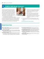
Clinical Note
More visual
Clinical Notes draw
Giants and dwarfs
—it all comes
down to bones and
cartilage
students’ attention to clinical
information they will need
in their future careers.
A variety of endocrine or metabolic problems can result in
characteristic skeletal changes. In pituitary dwarfism
(Figure 6–14a), inadequate production of growth hormone
leads to reduced epiphyseal cartilage activity and abnormally
short bones. This condition is becoming increasingly rare in the
United States, because children can be treated with synthetic
human growth hormone.
Gigantism results from an overproduction of growth
hormone before puberty. (The world record for height is 272 cm,
or 8 ft, 11 in., reached by Robert Wadlow, of Alton, Illinois, who
died at age 22 in 1940. Wadlow weighed 216 kg, or 475 lb.) If
growth hormone levels rise abnormally after epiphyseal
cartilages close, the skeleton does not grow longer, but bones
get thicker, especially those in the face, jaw, and hands. Cartilage
growth and alterations in soft-tissue structure lead to changes in
physical features, such as the contours of the face. These physical
changes occur in the disorder called acromegaly.
Several inherited metabolic conditions that affect many
systems influence the growth and development of the skeletal
system. These conditions produce characteristic variations in
body proportions. For example, many individuals with Marfan’s
Figure 6–14 Examples of Abnormal Bone Development.
a Pituitary dwarfism
b Marfan’s syndrome
I N T E G R A T O R
Transports calcium and phosphate for
bone deposition; delivers EPO to red
bone marrow, parathyroid hormone, and
calcitonin to osteoblasts and osteoclasts
Skeletal muscle contractions assist in
moving blood through veins; protects
superficial blood vessels, especially in neck
and limbs
Delivers oxygen and nutrients, removes
carbon dioxide, lactic acid, and heat during
skeletal muscle activity
Controls patterns of circulation in
peripheral tissues; modifies heart rate and
regulates blood pressure; releases ADH
Endothelial cells maintain blood–brain
barrier; helps generate CSF
Erythropoietin regulates production of
RBCs; several hormones elevate blood
pressure; epinephrine stimulates cardiac
muscle, elevating heart rate and
contractile force
Distributes hormones throughout the
body; heart secretes ANP and BNP
Skeletal
Page 000
Provides calcium needed for normal
cardiac muscle contraction; protects blood
cells developing in red bone marrow
Integumentary
Page 000
Body System
Muscular
Page 000
Cardiovascular System
Delivers immune system cells to injury
sites; clotting response seals breaks in
skin surface; carries away toxins from
sites of infection; provides heat
Nervous
Page 000
Integumentary
Skeletal
Muscular
Nervous
Endocrine
Other examples of
easy-to-read features:
Cardiovascular System
Stimulation of mast cells produces
localized changes in blood flow and
capillary permeability
syndrome are very tall and have long, slender limbs
(Figure 6–14b), due to excessive cartilage formation at the
epiphyseal cartilages. Although this is an obvious physical
distinction, the characteristic body proportions are not in
themselves dangerous. However, the underlying mutation, which
affects the structure of connective tissue throughout the body,
commonly causes life-threatening cardiovascular problems.
Endocrine
Page 000
S Y S T E M
Body System
Abnormal Bone Development
Respiratory
Page 000
Urinary
Page 000
The most extensive communication occurs
between the cardiovascular and lymphatic
systems. Not only are the two systems
physically interconnected, but cells of the
lymphatic system also move from one part of
the body to another within the vessels of the
cardiovascular system. We examine the
lymphatic system in detail, including its role in
Digestive
Page 000
The section on vessel distribution
demonstrated the extent of the
anatomical connections between
the cardiovascular system and other
organ systems. This figure summarizes
some of the physiological relationships
involved.
Lymphatic
Page 000
The CARDIOVASCULAR System
relationships between the cardiovascular
system and the other body systems we have
studied so far.
xii
Reproductive
Page 000
Figure 21–36 diagrams the functional
System Integrators
Easy-to-read tables
Chapter 21, page 759
Chapter 4, page 131
The narrative has
been extensively
revised for better
readability and
a lower reading
level in the Ninth Edition.
The result is a writing style
that is is clear and concise
and comfortably readable by
A&P students.
Tips & Tricks boxes are very
brief and concrete learning tools that
give students simple analogies and
easy memory devices to help them
remember facts and concepts.
Checkpoint
9. During intramembranous ossification, which type of
tissue is replaced by bone?
10. In endochondral ossification, what is the original
source of osteoblasts?
11. How could x-rays of the femur be used to determine
whether a person has reached full height?
See the blue Answers tab at the back of the book.
Art-based review questions
Checkpoints
Chapter 8, page 251
Chapter 6, page 183
xiii
SPOTLIGHT ON
Practice Anatomy Lab (PAL ) 3.0
™
™
PAL 3.0 is an indispensable virtual anatomy study and practice tool that gives
students 24/7 access to the most widely used lab specimens, including the human
cadaver, anatomical models, histology, cat, and fetal pig. PAL 3.0 retains all of
the key advantages of version 2.0, including ease-of-use, built-in audio pronunciations,
rotatable bones, and simulated fill-in-the-blank lab practical exams.
NEW Carefully prepared dissections
show nerves, blood vessels, and arteries across body systems.
NEW Photo gallery allows
students to quickly see thumbnails of images
for a particular region or sub-region.
NEW Layering slider
allows
students to peel back layers of the human
cadaver and view and explore hundreds of
brand-new dissections especially
commissioned for 3.0.
PAL 3.0 is available in the Study Area of MasteringA&P™ (www.masteringaandp.com).
The PAL 3.0 DVD can be packaged with the book for no additional charge.
xiv
NEW Interactive
Histology module
allows students to view the same
tissue slide at varying magnifications,
thereby helping them identify
structures and their characteristics.
3-D Anatomy Animations
of origins,
insertions, actions, and innervations of over 65 muscles
are now viewable in both Cadaver and Anatomical Models
modules. A new closed-captioning option provides textual
presentation of narration to help students retain
information and supports ADA compliance.
PAL 3.0 also includes:
NEW
Question randomization feature gives students more opportunities for
practice and self-assessment. Each time the student retakes a quiz or lab practical,
a new set of questions is generated.
NEW Hundreds of new images and views are included, especially of the human
cadaver, anatomical models, and histology.
NEW
Turn-off highlight feature in quizzes and lab practicals gives students the
option to see a structure without the highlight overlay.
xv
SPOTLIGHT ON
An Assignment and Assessment System
Get your students ready for the A&P course.
Get Ready for A&P allows you to assign tutorials and assessments
on topics students should have learned prior to the A&P course.
• Study Skills
• Basic Math Review
• Terminology
• Body Basics
• Chemistry
• Cell Biology
Motivate your
students to
come to class
prepared.
Assignable Reading
Quizzes motivate your
students to read the
textbook before coming
to class.
Assign art from
the textbook.
Assign and assess figures
from the textbook.
xvi
Give your students extra
coaching. Assign tutorials from your
favorite media—such as Interactive
Physiology® (IP) and A&P Flix™—to help
students understand and visualize tough
topics. MasteringA&P provides coaching
through helpful wrong-answer feedback
and hints.
Give students 24/7
lab practice. Practice Anatomy
Lab™ (PAL™) 3.0 is a tool that helps
students study for their lab practicals
outside of the lab. To learn more
about 3.0, see pages xiv-xv.
Identify struggling
students before it’s too late.
MasteringA&P has a color-coded
gradebook that helps you identify
vulnerable students at a glance.
Assignments in MasteringA&P are
automatically graded, and grades
can be easily exported to course
management systems or spreadsheets.
Go to www.masteringaandp.com to watch the demo movie.
xvii
SPOTLIGHT ON
Tools to Make the Grade
Study Area
MasteringA&P includes a Study Area that will help students get ready for tests with its simple three-step
approach. Students can:
1. Take a pre-test and obtain a personalized study plan.
2. Learn and practice with animations, labeling activities, and interactive tutorials.
3. Self-test with quizzes and a chapter post-test.
Get Ready for A&P
Students can access the Get Ready for
A&P eText, activities, and diagnostic
tests for these important topics:
•
•
•
•
•
•
Study Skills
Basic Math Review
Terminology
Body Basics
Chemistry
Cell Biology
Practice Anatomy Lab™ (PAL™) 3.0
Practice Anatomy Lab (PAL) 3.0 is a virtual anatomy study
and practice tool that gives students 24/7 access to the
most widely used lab specimens, including the human
cadaver, anatomical models, histology, cat, and fetal
pig. PAL 3.0 retains all of the key advantages of
version 2.0, including ease-of-use, built-in audio
pronunciations, rotatable bones, and
simulated fill-in-the-blank lab practical
exams. See pages xiv-xv.
xviii
MP3 Tutor Sessions
Students can download the MP3
Tutor Sessions for specific chapters
of the textbook and study wherever,
whenever. They can listen to minilectures about the toughest topics
and take audio quizzes to check
their understanding.
A&P Flix™
A&P Flix are 3-D movie-quality animations with
self-paced tutorials and gradable quizzes that
help students master the toughest topics in A&P:
Cell Physiology
Membrane Transport
DNA Replication
Protein Synthesis
Mitosis
Muscle Physiology
Events at the Neuromuscular Junction
Excitation-Contraction Coupling
Cross-Bridge Cycle
Neuro Physiology
Resting Membrane Potential
Generation of an Action Potential
Propagation of an Action Potential
Origins, Insertions, Actions, Innervations
Over 50 animations on this topic
Group Muscle Actions & Joints
Over 60 animations on this topic
Interactive Physiology® (IP)
IP helps students understand the hardest part of A&P:
physiology. Fun, interactive tutorials, games, and
quizzes give students additional explanations to help
them grasp difficult concepts.
Modules:
•
•
•
•
•
•
•
•
•
•
Muscular System
Nervous System I
Nervous System II
Cardiovascular System
Respiratory System
Urinary System
Fluids & Electrolytes
Endocrine System
Digestive System
Immune System
xix
SPOTLIGHT ON
Support for Students
eText
MasteringA&P (www.masteringaandp.com) includes an eText. Students can access their
textbook wherever and whenever they are online. eText pages look exactly like the printed
text yet offer additional functionality. Students can do the following:
• Create notes.
• Highlight text in different colors.
• Create bookmarks.
• Zoom in and out.
• View in single-page or two-page view.
• Click hyperlinked words and phrases to view definitions.
• Link directly to relevant animations.
• Search quickly and easily for specific content.
View animations
from within the eText.
Highlight text and make notes.
Easily access definitions of key words.
xx
Get Ready for A&P
by Lori K. Garrett
This book and online component
were created to help students be
better prepared for their A&P
course. Features include pretests, guided explanations
followed by interactive quizzes
and exercises, and end-ofchapter cumulative tests. Also
available in the Study Area of
www.masteringaandp.com.
Martini’s Atlas of
the Human Body
by Frederic H. Martini
The Atlas offers an abundant
collection of anatomy
photographs, radiology scans,
and embryology summaries,
helping students visualize
structures and become familiar
with the types of images seen in
a clinical setting.
Interactive Physiology®
10-System Suite (IP-10)
CD-ROM
IP helps students understand the
hardest part of A&P: physiology.
Fun, interactive tutorials, games,
and quizzes give students
additional explanations to help
them grasp difficult
physiological concepts.
Study Guide
by Charles M. Seiger
The Study Guide includes a
variety of review activities,
including multiple choice
questions, labeling exercises,
and concept maps—all
organized by the Learning
Outcomes used in the book.
A&P Applications Manual
by Frederic H. Martini
and Kathleen Welch
This manual contains extensive
discussions on clinical topics and
disorders to help students apply
the concepts of anatomy and
physiology to daily life and their
future health professions.
Practice Anatomy Lab™
(PAL™) 3.0 DVD
PAL 3.0 is an indispensable
virtual anatomy study and
practice tool that gives students
24/7 access to the most widely
used lab specimens, including
the human cadaver, anatomical
models, histology, cat, and
fetal pig.
See pages xviii-xix for the MasteringA&P Study Area.
xxi
SPOTLIGHT ON
Support for Instructors
eText with Whiteboard Mode
The Fundamentals of Anatomy & Physiology eText comes
with Whiteboard Mode, allowing instructors to use the eText
for dynamic classroom presentations. Instructors
can show one-page or two-page views from the
book, zoom in or out to focus on select topics, and
use the Whiteboard Mode to point to structures,
circle parts of a process, trace pathways, and
customize their presentations.
Instructors can also add notes to guide students,
upload documents, and share their customenhanced eText with the whole class.
Instructors can find the eText with Whiteboard
Mode on MasteringA&P.
Instructor Resource DVD (IRDVD)
with Lecture Outlines by Jason LaPres and Clicker Questions and Quiz Shows by Marian Leal
978-0-321-73543-0 / 0-321-73543-9
The IRDVD offers a wealth of instructor media resources, including presentation art,
lecture outlines, test items, and answer keys – all in one convenient location.
The IRDVD includes:
• Textbook images in JPEG format (in two versions—one with labels
and one without)
• Customizable textbook images embedded in PowerPoint® slides
(in three versions—one with editable labels, one without labels,
and one as step-edit art)
• Customizable PowerPoint lecture outlines, combining lecture notes,
figures and tables from the book, and links to the A&P Flix
• A&P Flix™ 3-D movie-quality animations on tough topics
• PRS-enabled Active Lecture Clicker Questions
• PRS-enabled Quiz Show Clicker Questions
• Interactive Physiology® 10-System Suite (IP-10) Exercise Sheets and
Answer Key
• Martini’s Atlas of the Human Body images
• The Test Bank in TestGen® format and Microsoft® Word format
• The Instructor’s Manual in Microsoft® Word format
• PDF files of Transparency Acetate masters
xxii