Cloning and functional characterization
Bạn đang xem bản rút gọn của tài liệu. Xem và tải ngay bản đầy đủ của tài liệu tại đây (1.37 MB, 8 trang )
Gene 330 (2004) 115 – 122
www.elsevier.com/locate/gene
Cloning and functional characterization of PELP1/MNAR promoter
Sandip K. Mishra, Seetharaman Balasenthil, Diep Nguyen, Ratna K. Vadlamudi *
Department of Molecular and Cellular Oncology, Unit 108, The University of Texas M. D. Anderson Cancer Center,
1515 Holcombe Blvd, Houston, TX 77030, USA
Received 10 October 2003; received in revised form 29 December 2003; accepted 15 January 2004
Received by A.J. van Wijnen
Abstract
Proline-, glutamic acid- and leucine-rich protein 1 (PELP1)/modulator of nongenomic activity of estrogen receptor (MNAR), a novel
coactivator of estrogen receptors (ERs; ERa and ERh), modulates the genomic and nongenomic functions of the ERs. PELP1 expression is
developmentally regulated in mammary glands and overexpressed in breast tumors. However, little is known about the regulation of PELP1.
In this study, we examined whether PELP1 expression is modulated by steroid hormone 17h-estradiol (E2) – ER pathway. We found that in
MCF-7 breast cancer cells, E2 upregulated PELP1 expression threefold and that this upregulation was reduced by antiestrogen. We also
found that E2 modulated PELP1 levels in an actinomycin-D-sensitive manner, suggesting transcriptional regulation. Cloning and analysis of
the 2-kb PELP1 promoter region revealed two estrogen-responsive element (ERE) half sites in the PELP1 promoter region. In transient
transfection assays, E2 upregulated PELP1 promoter activity in breast, endometrial and osteosarcoma model cancer cell lines in an ICI
182,780-sensitive manner. We demonstrated the recruitment of ER to the PELP1 promoter in vitro using EMSA assays and in vivo using a
chromatin immunoprecipitation assay. The PELP1 promoter was similarly upregulated by both ERa and ERh and differentially regulated by
selective estrogen receptor modulators in a cell line-dependent manner. Our results suggest that PELP1 expression is modulated by the E2-ER
pathway and that PELP1 is an ER target gene.
D 2004 Elsevier B.V. All rights reserved.
Keywords: PELP1; MNAR; Estrogen receptor; Promoter
1. Introduction
The steroid hormone 17h-estradiol (E2) plays an important role in controlling the expression of genes involved in a
wide variety of biological processes, including the development, homeostasis and progression of breast cancer (Couse
and Korach, 1999; McDonnell and Norris, 2002). The
biological effects of E2 occur when it binds to the structurally and functionally distinct estrogen receptors (ERs; ERa
and ERh) (Warner et al., 1999). For example, when E2
binds to ERa, the ligand-activated ERa translocates to the
nucleus, binds to the 13-bp palindromic estrogen responseenhancer (ERE) element in the target genes and stimulates
gene transcription (Kumar and Chambon, 1988). ERs can
Abbreviations: E2, 17h-estradiol; ER, estrogen receptor; ERE, estrogen
response element; PELP1, proline-, glutamic acid- and leucine-rich protein;
MNAR, modulator of nongenomic activity of estrogen receptor; luc,
luciferase.
* Corresponding author. Tel.: +1-713-745-5239; fax: +1-713-745-2050.
E-mail address: (R.K. Vadlamudi).
0378-1119/$ - see front matter D 2004 Elsevier B.V. All rights reserved.
doi:10.1016/j.gene.2004.01.011
also induce expression of genes containing imperfect or
ERE half sites (half ERE) (Klinge, 2001). ERs also interact
with other DNA-bound transcription factors such as activating protein (AP1) and SP1, and confer E2 responsiveness
to heterologous promoters (Gaub et al., 1990; Webb et al.,
1992; Safe, 2001; Petz et al., 2002).
ERa comprises an N-terminal activation function 1
(AF1) domain, a DNA-binding domain, and a C-terminal
ligand-binding region that contains an activation function 2
(AF2) domain. ERh has a domain structure similar to that of
ERa (Kumar et al., 1987; Kumar and Chambon, 1988;
Berry et al., 1990). The two receptors are most similar in
their DNA-binding domains (95%) and ligand-binding
domains (58%) (Katzenellenbogen et al., 2001). The transcription functions of ERs are influenced by several coactivators, including SRC1, GRIP1, AIB1, CBP, p300, PGC1,
E6AP, PCAF and SNF2 (Kumar et al., 1987; Kumar and
Chambon, 1988; Berry et al., 1990; Hermanson et al., 2002;
McDonnell and Norris, 2002; McKenna and Malley, 2002).
It is generally accepted that some of the diverse functions of
estrogens depend on the differential recruitment of coregu-
116
S.K. Mishra et al. / Gene 330 (2004) 115–122
lators to the ligand-bound ERa complex (McDonnell and
Norris, 2002). Coregulators that affect the activity of ERs
are thought to play a role in tumor progression (Anzick et
al., 1997). On the basis of this, it has been speculated that
the differential expression of coregulators among tissues
contributes at least in part to different hormonal responses.
Proline-, glutamic acid- and leucine-rich protein 1
(PELP1)/modulator of nongenomic activity of estrogen
receptor (MNAR), a novel ER coregulator, contains 10
LXXLL motifs for nuclear receptor interaction and plays
an important role in estrogen-mediated genomic functions
(Vadlamudi et al., 2001, Balasenthil and Vadlamudi, 2003)
and nongenomic actions of ERs via activation of Src/mitogen-activated protein kinase pathways (Wong et al., 2002).
PELP1 interacts with ERa and ERh, and with retinoblastoma protein, and upregulates cyclin D1 expression and its
overexpression sensitizes cells to estrogen-mediated cellcycle progression (Balasenthil and Vadlamudi, 2003).
The ER-signaling pathway has been implicated in breast
cancer tumorigenesis, and approximately 70% of breast
cancer cells express ER (Osborne et al., 2001). Initial
studies suggested that PELP1 expression is upregulated in
mammary tumors and in mammary gland during pregnancy
when cells are highly proliferative (Vadlamudi et al., 2001).
These results led us to hypothesize that PELP1expression is
regulated by the E2-ER pathway. Our results suggest that E2
does regulate PELP1 expression at the transcriptional level
and that the ERE half site present in the proximal region of
the PELP1 promoter plays an important role in induction of
E2-mediated responses.
2. Methods
2.1. Cell lines and reagents
MCF-7 human breast cancer cells (Vadlamudi et al.,
2001) and Ishikawa endometrial cells (Apparao et al.,
2001) were maintained in Dulbecco’s Modified Eagle’s
Medium – F12 (1:1) supplemented with 10% fetal calf serum
(Vadlamudi et al., 2001). SAOS2 osteosarcoma cells, MDAMB-231 breast cancer cells and HeLa cervical cancer cells
were obtained from the American Type Culture Collection
(Manassas, VA). Steroid hormone E2, tamoxifen and charcoal-stripped serum (DCC serum) was purchased from
Sigma. Propyl pyrazole triol (PPT), diarcylproprionitrile
(DPN), ICI-182,780 were purchased from Tocris, Ellisville,
MO. Faslodex (AstraZeneca Pharmaceuticals, Wilmington,
DE) and Raloxifene tablets (Eli Lilly Indianapolis, IN) were
obtained from MD Anderson Pharmacy.
We then purchased this BAC clone from BACPAC Resources (Children’s Hospital Oakland Research Institute, CA).
The PELP1 promoter region containing 2-kb upstream 5V
region was amplified by polymerase chain reaction (PCR)
using primers PELP1 Pro-2000F-5V-GCCAGGACTTGAAGGATTGGA-3V and PELP1 Pro + 1R-5V-GGTTCCAGTGGTGGCGTGGC-3V. The amplified product was
cloned into the Topo vector (Invitrogen, Carlsbad, CA)
and then subcloned into the PGL3 luciferase (luc) reporter
vector (Promega, Madison, WI) using HindIII and Xho 1
sites. The sequence of the construct was verified by comparing its sequence with that in the human genome database.
Deletions in the PELP1 promoter were created using PCRbased approach. Mutation in the proximal ERE half site was
created by PCR approach using the following primers. WT
5V-CACTTCTCGAGGCCAAGGCGGGCGGAAC ACCT
GAGGTCAG GAGTTCAAGACCAT CCTGGGC-3V; MT
5V-CACTTCTCGAGGCCAAGGCGGG CGGA ACACCTGAGAGCAGGAGTTCAAGACCATCCTG GGC-3V.
2.3. Reporter gene assays
For the reporter gene transient transfections, MCF-7,
Ishikawa, SAOS2 and MDA-MB-231 cells were cultured
for 24 h in minimal essential medium without phenol red
containing 5% DCC serum. The PELP1-luciferase (luc)
reporter constructs were transfected using FuGENE 6
according to the manufacturer’s instructions (Roche Molecular Biochemicals, Indianapolis, IN). Twenty-four hours
later, the cells were treated with E2 for 24 h. The cells
were then lysed with a passive-lysis buffer, and the luc
assay was performed using a luc reporter assay kit (Promega). The total amount of DNA used in the transfections
was kept constant by adding a parental vector. Each
transfection was carried out in six-well plates in triplicate
wells.
2.4. Northern hybridization
MCF-7 cells were treated with E2 for 8 h, and PELP1
levels were analyzed by Northern blotting. Total cytoplasmic RNA 20 Ag was isolated using the Trizol reagent
(Invitrogen) and analyzed by Northern hybridization using
a 1200-bp N-terminal fragment of PELP1cDNA. Glyceraldehyde-3-phosphate dehydrogenase levels were measured
to assess the integrity of the RNA and to control RNA
loading. Autoradiogram was developed using Phosphoimager and band intensities were quantitated using SigmaGelGel Analysis Software.
2.5. Cell extracts and immunoblotting
2.2. Cloning of the PELP1 promoter
To clone the PELP1 promoter, we first identified the
BAC clone (ID RP11-314A20) containing the PELP1 genomic region using human genome sequence information.
To prepare cell extracts, cells were washed three times
with phosphate-buffered saline and then lysed in RIPA
buffer (50 mM Tris – HCl, pH 7.5, 150 mM NaCl, 0.5%
NP-40, 0.1% sodium dodecyl sulfate (SDS), 0.1% sodium
S.K. Mishra et al. / Gene 330 (2004) 115–122
deoxycholate, 1 Â protease inhibitor mixture (Roche Molecular Biochemicals), 1 mM sodium vanadate) for 15 min
on ice. The lysates were centrifuged in an Eppendorf
centrifuge at 4 jC for 15 min. Cell lysates containing an
equal amount of protein ( f 200 Ag) were then resolved on
a SDS – polyacrylamide gel (PAGE) (8% acrylamide), transferred to a nitrocellulose membrane, probed with the appropriate antibodies and developed using the enhanced
chemiluminescence method. Quantiation of the bands were
done using SigmaGel-Gel Analysis Software.
117
representing the ERE half site present in the promoter of
PELP1/MNAR. Sequence of the forward and reverse primers
used are as For:5V-CACTTTGGGAGGCCAAGGCGGGCGGAACACCTGAGGTCA GGAGTTCAAGACCATCCTGGC-3; Rev:5V GCCAGGATGGTCTTGAACTCCTGACCTCAGGTGTTC CGCCCGCCTTGGC CTC
CCAAAGTG-3V. The DNA – protein complexes were resolved in 5% polyacrylamide gels. For supershift assays, 1
Al of antibody (ERa, supershift antibody by Santa Cruz
Biotechnology) was used. Antibody was incubated with the
reaction mixture for 15 min.
2.6. Chromatin immunoprecipitation assay
2.8. Statistical analysis
Approximately 106 cells were treated with 1% formaldehyde (final concentration, v/v) for 10 min at 37 jC to
cross-link histones to DNA. The cells were washed twice
with phosphate-buffered saline, pH 7.4 containing protease
inhibitor cocktail (Roche Molecular Biochemicals). Chromatin immunoprecipitation (ChIP) assay was performed as
described previously (Mazumdar et al., 2001). An ERaspecific antibody purchased from Upstate biotechnology
(Upstate, Lake Placid, NY) was used for immunoprecipitation of ER-bound chromatin. PCR using primers flanking
the proximal half ERE site ( À 690 For-5V-ATAAATCTTTGGCGGCGCGTA-3V and À 294 Rev-5V-ACGATTTCCATTTAGCGGACG-3V) produced a 396-bp DNA
fragment. An amplified fragment was sequence verified.
2.7. Electrophoresis mobility shift assay
Electrophoresis mobility shift assay (EMSA) was performed as described earlier (Mandal et al., 2001) with some
modifications. Cells were maintained in DCC medium for at
least 48 h and treated with E2 (final conentration, 10À 9 M)
for 12 h. Nuclear extract prepared from the estrogen-treated
cells was incubated with 32 P-radiolabeled oligonucleotide
Statistical analysis was done using Student’s t-test, and
values with p < 0.05 were considered statistically significant.
3. Results
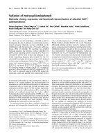
3.1. E2 upregulates PELP1 expression
E2 treatment of the MCF-7 cells induced PELP1 mRNA
to a level threefold greater than that in the untreated cells
(Fig. 1A). In contrast, cells pretreated with antiestrogen ICI182780 showed no increase in PELP1 levels suggesting that
E2-mediated upregulation of PELP1 levels occurs via the ER
(Fig. 1B). On the other hand, the treatment of the MCF-7
cells with cycloheximide, a translation inhibitor, did not
affect E2-mediated upregulation of PELP1 (Fig. 1C). However, treatment of these cells with actinomycin D, an inhibitor of transcription, completely prevented the E2-mediated
induction of PELP1, suggesting that E2 regulates PELP1
expression at the transcriptional level. To confirm that the
increase in PELP1 mRNA levels correlated with PELP1
protein levels, we treated the MCF-7 cells with E2 for
Fig. 1. E2 upregulated PELP1 expression. (A) Northern analysis of PELP1 expression in MCF-7 cells treated with E2 for 6 h. (B) Blockage of E2-mediated
PELP1 expression by ICI-182780 pretreatment. (C) Northern analysis of PELP1 expression in MCF-7 cells treated with E2 in the presence or absence of
cycloheximide (CHX) and or actinomycin-D (ACD). (D) MCF-7 cells were treated with E2 for various lengths of time, and PELP1 expression was analyzed by
Western blotting using a PELP1-specific antiserum.
118
S.K. Mishra et al. / Gene 330 (2004) 115–122
various times and analyzed PELP1 expression by Western
blotting (Fig. 1D). Our results showed that E2 increased the
expression of PELP1 protein by three- to fourfold and that
the PELP1 protein level increased after 6 h of E2 treatment.
3.2. E2 increases PELP1 promoter activity in breast cancer,
endometrial and osteosarcoma cells
To elucidate the mechanism by which E2 increases
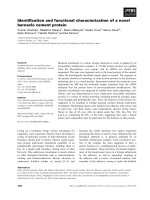
PELP1 expression at the transcriptional level, we analyzed
the sequence of the PELP1 promoter region (Fig. 2) and
found that it lacked consensus TATA and CCAAT motifs.
This 2-kb region also lacked consensus palindromic ERE
sites, although it did contain two ERE half sites, seven AP1
sites and five SP1 sites (Fig. 2). Other putative transcription
factor elements present in the PELP1 promoter included
thyroid receptor binding elements, GATA and E2F binding
sites. Sequence was submitted to the GenBank (AY427960).
To examine whether the cloned region ( À 2000 to + 1)
of the PELP1 promoter did indeed confer E2 inducibility,
we constructed and measured the activity of PELP1 promoter luc reporter. We found that E2 upregulated PELP1
promoter activity to threefold more in MCF-7 cells than in
the untreated cells. The E2-mediated increase in PELP1
promoter activity was also sensitive to antiestrogen ICI182780 treatment. To examine the generality of PELP1
promoter upregulation, we examined whether E2 also upregulated PELP1 promoter activity in the Ishikawa and
SAOS2 cells. The results showed that the PELP1 basal
activity varied among the three cell lines and that E2 was
able to induce the PELP1 promoter in the three cells lines
two to three times above their basal activity (Fig. 3).
Fig. 2. Sequence of the PELP1 promoter. (A) Nucleotide sequence of the 5Vflanking region, including part of the first exon of the PELP1 gene. The first exon
region is shown in bold, and the translation start site is shown by a box. The numbers shown to the left are relative to the putative start sites of PELP1 mRNA.
Consensus sequences for the AP1, SP1 and ERE half transcription factor sites are underlined. (B) Schematic representation of AP1, SP1 and ERE half sites in
the PELP1 promoter.
S.K. Mishra et al. / Gene 330 (2004) 115–122
119
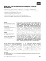
Fig. 3. Activity of the PELP1 promoter in different cell lines. PELP1 promoter ( À 2000 to + 1) fused to the luciferase reporter was transiently transfected into
(A) breast model cell line MCF-7, (B) endometrial model cell line Ishikawa, (C) and osteosarcoma cell line SAOS2. The cells were treated with E2 or E2 + ICI182,780, and after 24 h, the luciferase reporter activity was measured. Significant ( p < 0.05) induction by E2 is indicated with an asterisk.
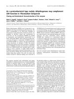
Fig. 4. Localization of the E2-responsive region in the PELP1 promoter. (A) MCF-7 cells were transfected with various PELP1 promoter deletion constructs,
treated with E2 for 24 h, and the PELP1-luciferase reporter activity was measured. (B) Chromatin immunoprecipitation analysis. ERs bound to the chromatin
were immunoprecipitated using an ERa-specific antibody, and its recruitment to the PELP1 promoter was analyzed using primers spanning the proximal ERE
half site of the PELP1 endogenous promoter. (C) Gel mobility shift assays using proximal ERE half site ( À 610/ À 550) containing oligonucleotides. Estrogentreated nuclear extract was incubated with 32P-labeled oligonucleotide. First lane shows free probe. Second, third and fourth lanes show complex formation with
5, 10 and 20 Ag of nuclear extract. A 100-fold excess of unlabeled oligonucleotide retarded the complex formation with the radiolabeled probe (lane 5). (D)
ERa-specific antibody was used to supershift the complex. Lane 1 shows free probe. Lane 2 shows complex formation with the nuclear extract. Lane 3, a 10fold excess of unlabeled oligonucleotide retarded the complex formation with the radiolabeled probe, and lane 4, anti-ERa antibody supershifted most of the
complex . (E) Activity of the PELP1 promoter À 600/ À 1 containing wild-type ERE half site or mutated ERE half site were transfected into MCF-7 cells,
treated with or without E2 and luciferase activity was measured. Significant ( p < 0.05) induction by E2 is indicated with an asterisk.
120
S.K. Mishra et al. / Gene 330 (2004) 115–122
3.3. Proximal ERE half site of PELP1 promoter confers E2
inducibility
To identify the E2-responsive region in the PELP1
promoter, we generated serial deletions of PELP1 promoter.
Deletion of the 1300-bp distal region containing one ERE
half site did not noticeably affect the E2 inducibility of the
PELP1 promoter containing the À 700 to + 1 region.
However, an additional deletion of 200 bp prevented E2
induction of the PELP1 promoter (Fig. 4A). Since the
PELP1 À 700 to À 500 promoter region contained one
ERE half site, these results suggested that this proximal
ERE half site conferred E2 inducibility (Fig. 4A). To
examine in vivo the possibility that ER is recruited to the
PELP1 promoter, we performed a chromatin immunoprecipitation assay using the primers spanning PELP1 promoter
region ( À 690 to À 294). Results showed that ER is
recruited to the PELP1 promoter region À 690 to À 294,
which contains proximal ERE half site, after E2 treatment in
a time-dependent manner (Fig. 4B). Gradual recruitment of
ER to the PELP1 promoter with peak at 60 min and lack of
its recruitment at 120 min suggests cyclical recruitment of
ER at the PELP1 promoter as predicted with other promoters including ER-responsive gene pS2 (Me´tivier et al.,
2003). We did not observe any recruitment of ER in
chromatin immunoprecipitation assay to the PELP1 promoter containing the distal ERE half site (data not shown).
To determine whether ER is directly recruited to the
region containing proximal ERE half site, 32P-labeled oligos
containing À 610/ À 550 region of the PELP1/MNAR promoter was incubated with increasing amounts of estrogentreated MCF-7 nuclear extract. Results showed formation of
higher order protein – DNA complexes, and the specific
band formed as a result of complex formation could be
competed out with 100-fold excess cold probe (Fig. 4C).
The same band could be supershifted with anti-ER-a
antibody (Fig. 4D). Mutation of the proximal ERE half site
(GGTCA to GTGCA) in the PELP1 À 600/ À 1 construct
substantially reduced the ability of estrogen to induce
reporter gene activity (Fig. 4E). Collectively, these results
suggest that proximal ERE half sites play an important role
in the estrogen-mediated upregulation of PELP1/MNAR
promoter activity.
3.4. PELP1 promoter is upregulated by both ERa and ERb
Since many E2-responsive tissues express distinct forms
of ER (ERa and ERh),we next examined whether both ERa
and ERh regulate PELP1 promoter activity. For this experiment, we have used HeLa cells, which have no detectable
levels of ERa or ERh. Cotransfection of ERa and ERh
along with the PELP1 ( À 2000 to + 1) luc reporter resulted
in a 2.6- and 2.2-fold induction, respectively, upon E2
stimulation (Fig. 5A). To confirm these results, we transfected PELP1 promoter construct into breast cancer cells
that selectively expressed either ERa (MCF-7 cells) or ERh
(MDA-MB-231). The cells were then treated with either
ERa-selective agonist PPT or ERh-selective agonist DPN.
The results showed that in the MCF-7 cells, which only
express ERa, PPT but not DPN upregulated PELP1 promoter activity (Fig. 5B). Similarly, in the MDA-MB-231
cells, which only express ERh, DPN but not PPT upregulated PELP1 promoter activity (Fig. 5C). These results
suggest that both ER isoforms have the potential to modulate PELP1 expression. Fig. 5D shows the expression of
ERa and ERh in MCF-7 and MDA-MB-231 cell lines.
3.5. Differential regulation of the PELP1 promoter by
selective estrogen receptor modulators
We next examined whether selective estrogen receptor
modulators regulate the expression of the PELP1 promoter.
In the MCF-7 cells, the selective estrogen receptor modulators did not upregulate PELP1 promoter activity; how-
Fig. 5. Regulation of PELP1 promoter activity by ERa and ERh. (A) HeLa cells were cotransfected with the PELP1-luciferase reporter gene alone with ERa or
ERh, and treated with E2 or E2 + ICI-182,780 for 24 h. The reporter activity was then measured. (B) ERa-positive MCF-7 cells and (C) ERh-positive MDAMD-231 cells were transfected with the PELP1-luciferase reporter ( À 2000 to + 1) and treated with PPT (ERa-specific ligand) or DPN (ERh-specific ligand).
After 24 h, the luciferase activity was measured. Significant ( p < 0.05) induction by E2/PPT/DPN is indicated with an asterisk.
S.K. Mishra et al. / Gene 330 (2004) 115–122
121
Fig. 6. Regulation of PELP1 promoter activity by selective estrogen receptor modulators. MCF-7 and Ishikawa cells were transfected with the PELP1luciferase reporter gene ( À 2000 to + 1), and the cells were treated with the indicated selective estrogen receptor modulators for 24 h. The luciferase activity
was measured. Significant ( p < 0.05) induction by E2, tamoxifen and raloxifene is indicated with an asterisk.
ever, in the endometrial Ishikawa cells, tamoxifen and
raloxifene upregulated PELP1 promoter activity (Fig. 6).
These results suggest that selective estrogen receptor modulators may modulate PELP1 expression in a tissue-dependent manner.
4. Discussion
The following results of this study strongly suggest that
PELP1 is an E2-inducible gene. (1) E2 upregulated PELP1
mRNA and PELP1 protein levels; (2) E2-mediated PELP1
expression was sensitive to antiestrogen ICI-182780; (3)
ERE half sites were present in PELP1 promoter, and ER was
recruited to the À 690/ À 294 region of PELP1 promoter
which contains proximal ERE half site; (4) EMSA and
supershift assays showing the ability of ER to recruit to
À 610/ À 550 oligonucleotide which contains proximal ERE
half site; (5) E2 upregulated the PELP1 promoter in breast
cancer, endometrial and osteosarcoma cell lines; and (6)
both ERa and ERh regulated the PELP1 promoter activity.
Ligand-bound ERs are thought to bind to the 13-bp ERE
element. Emerging evidence from several studies also suggested that ER regulates promoters containing ERE half sites
(Klinge et al., 1997; Lee and Mouradian 1999; Klinge 2001;
Martini and Katzenellenbogen, 2001, Ediger et al., 2002).
PELP1 promoter did not have a classical palindromic 13-bp
ERE site, although it did contained two ERE half sites, seven
AP1 and five SP1 sites. In this study, we have only focused
on the characterization of two ERE half sites present in the
promoter region. Our ChIP analysis indicated ER recruitment
in the promoter region containing À 690 to À 294. Mobility
shift assays support recruitment of ER to the proximal ERE
half site. Further, mutation of ERE half site in PELP1 À 600/
À 1 construct abolished the E2-mediated induction. These
results suggest that proximal ERE site contributes at least
50% of the E2-mediated induction. However, deletion of the
PELP1 promoter sequence upstream of PELP1 À 600/ À 1
also results in approximately 50% reduction in the fold of
activation of PELP1-luciferase activity. These data denote
the importance of the AP1 or SP1 sites located upstream the
proximal half ERE, in the E2-induced regulation of PELP1
promoter. Our ongoing studies are currently focused on the
characterizing the role of AP1 and SP1 in E2-mediated
induction of PELP1/MNAR promoter.
E2 regulates cell proliferation in a wide variety of tissues,
including breast tissue (Prall et al., 1998). The highest rate
of mitosis in ductal epithelial cells (the origin of most breast
cancer cells) is during the luteal phase, when the level of E2
is generally high (Foster et al., 2001). PELP1 expression is
high in mammary glands during pregnancy, when cell
proliferation is high (Vadlamudi et al., 2001). In a previous
study, we found that overexpression of PELP1 was accompanied by a persistent hyperphosphorylation of pRb in an
E2-dependent manner, and overexpression of PELP1 sensitized cells to G1/S progression (Balasenthil and Vadlamudi,
2003). Our finding in this study that E2 upregulates PELP1
expression suggests that upregulated PELP1 may in turn
help in E2-mediated cell cycle progression in physiological
settings. However, in pathological conditions such as breast
cancer, ER-positive tumors may upregulate PELP1 expression via activation of the E2-ER pathway. Since PELP1 is a
coactivator of ERs which modulates both the genomic and
nongenomic functions of ERs, deregulation of PELP1 may
contribute to excessive proliferation or hormonal independence in ER-positive tumors or to both.
In summary, we have shown that PELP1 expression is
modulated by the E2-ER pathway and that selective estrogens differentially regulate PELP1 reporter activity. The
ERE half site localized to the proximal region of PELP1
plays an important role in the E2-mediated regulation of
PELP1 expression.
122
S.K. Mishra et al. / Gene 330 (2004) 115–122
Acknowledgements
This study was supported in part by NIH grants
CA095681, CA90970 and 98823. We are grateful to Rakesh
Kumar, UTMDACC for thoughtful discussions and critical
reading of this manuscript. We thank Bruce A. Lessey, UNC
for Ishikawa cell line.
References
Anzick, S.L., Kononen, J., Walker, R.L., Azorsa, D.O., Tanner, M.M.,
Guan, X.Y., Sauter, G., Kallioniemi, O.P., Trent, J.M., Meltzer, P.S.,
1997. AIB1, a steroid receptor coactivator amplified in breast and ovarian cancer. Science 277, 965 – 968.
Apparao, K.B., Murray, M.J., Fritz, M.A., Meyer, W.R., Chambers, A.F.,
Truong, P.R., Lessey, B.A., 2001. Osteopontin and its receptor alphavbeta(3) integrin are coexpressed in the human endometrium during the
menstrual cycle but regulated differentially. J. Clin. Endocrinol. Metab.
86, 4991 – 5000.
Balasenthil, S., Vadlamudi, R.K., 2003. Functional interactions between the
estrogen receptor coactivator PELP1/MNAR and retinoblastoma protein. J. Biol. Chem. 278, 22119 – 22127.
Berry, M., Metzger, D., Chambon, P., 1990. Role of the two activating
domains of the oestrogen receptor in the cell-type and promoter-context
dependent agonistic activity of the anti-oestrogen 4-hydroxytamoxifen.
EMBO J. 9, 2811 – 2818.
Couse, J.F., Korach, K.S., 1999. Estrogen receptor null mice: what have we
learned and where will they lead us? Endocr. Rev. 20, 358 – 417.
Ediger, T.R., Park, S.E., Katzenellenbogen, B.S., 2002. Estrogen receptor
inducibility of the human Na+/H+ exchanger regulatory factor/ezrinradixin-moesin binding protein 50 (NHE-RF/EBP50) gene involving
multiple half-estrogen response elements. Mol. Endocrinol. 16,
1828 – 1839.
Foster, J.S., Henley, D.C., Ahamed, S., Wimalasena, J., 2001. Estrogens
and cell-cycle regulation in breast cancer. Trends Endocrinol. Metab.
12, 320 – 327.
Gaub, M.P., Bellard, M., Scheuer, I., Chambon, P., Sassone-Corsi, P., 1990.
Activation of the ovalbumin gene by the estrogen receptor involves the
fos-jun complex. Cell 63, 1267 – 1276.
Hermanson, O., Glass, C.K., Rosenfeld, M.G., 2002. Nuclear receptor
coregulators: multiple modes of modification. Trends Endocrinol.
Metab. 13, 55 – 60.
Katzenellenbogen, B.S., Sun, J., Harrington, W.R., Kraichely, D.M.,
Ganessunker, D., Katzenellenbogen, J.A., 2001. Structure-function relationships in estrogen receptors and the characterization of novel selective estrogen receptor modulators with unique pharmacological profiles.
Ann. N. Y. Acad. Sci. 949, 6 – 15.
Klinge, C.M., 2001. Estrogen receptor interaction with estrogen response
elements. Nucleic Acids Res. 29, 2905 – 2919.
Klinge, C.M., Bodenner, D.L., Desai, D., Niles, R.M., Traish, A.M., 1997.
Binding of type II nuclear receptors and estrogen receptor to full and
half-site estrogen response elements in vitro. Nucleic Acids Res. 25,
1903 – 1912.
Kumar, V., Chambon, P., 1988. The estrogen receptor binds tightly to
its responsive element as a ligand-induced homodimer. Cell 55,
145 – 156.
Kumar, V., Green, S., Stack, G., Berry, M., Jin, J.R., Chambon, P., 1987.
Functional domains of the human estrogen receptor. Cell 51, 941 – 951.
Lee, S.H., Mouradian, M.M., 1999. Up-regulation of D1A dopamine receptor gene transcription by estrogen. Mol. Cell. Endocrinol. 156, 151 – 157.
Mandal, M., Olson, D.J., Sharma, T., Vadlamudi, R.K., Kumar, R., 2001.
Butyric acid induces apoptosis by up-regulating Bax expression via
stimulation of the c-Jun N-terminal kinase/activation protein-1 pathway
in human colon cancer cells. Gastroenterology 120, 71 – 78.
Martini, P.G., Katzenellenbogen, B.S., 2001. Regulation of prothymosin
alpha gene expression by estrogen in estrogen receptor-containing
breast cancer cells via upstream half-palindromic estrogen response
element motifs. Endocrinology 142, 3493 – 3501.
Mazumdar, A., Wang, R.A., Mishra, S.K., Adam, L., Bagheri-Yarmand, R.,
Mandal, M., Vadlamudi, R.K., Kumar, R., 2001. Transcriptional repression of oestrogen receptor by metastasis-associated protein 1 corepressor. Nat. Cell Biol. 3, 30 – 37.
McDonnell, D.P., Norris, J.D., 2002. Connections and regulation of the
human estrogen receptor. Science 296, 1642 – 1644.
McKenna, N.J., Malley, B.W., 2002. Combinatorial control of gene expression by nuclear receptors and coregulators. Cell 108, 465 – 474.
Me´tivier, R., Penot, G., Hu¨bner, M.R., Reid, G., Brand, H., Kosˇ, M.,
Gannon, F., 2003. Estrogen receptor-a directs ordered, cyclical, and
combinatorial recruitment of cofactors on a natural target promoter. Cell
115, 751 – 763.
Osborne, C.K., Schiff, R., Fuqua, S.A., Shou, J., 2001. Estrogen receptor:
current understanding of its activation and modulation. Clin. Cancer
Res. 7, 4338s – 4342s.
Petz, L.N., Ziegler, Y.S., Loven, M.A., Nardulli, A.M., 2002. Estrogen
receptor alpha and activating protein-1 mediate estrogen responsiveness
of the progesterone receptor gene in MCF-7 breast cancer cells. Endocrinology 143, 4583 – 4591.
Prall, O.W., Rogan, E.M., Sutherland, R.L., 1998. Estrogen regulation of
cell cycle progression in breast cancer cells. J. Steroid Biochem. Mol.
Biol. 65, 169 – 174.
Safe, S., 2001. Transcriptional activation of genes by 17 beta-estradiol
through estrogen receptor-Sp1 interactions. Vitam. Horm. 62, 231 – 252.
Vadlamudi, R.K., Wang, R.A., Mazumdar, A., Kim, Y., Shin, J., Sahin, A.,
Kumar, R., 2001. Molecular cloning and characterization of PELP1, a
novel human coregulator of estrogen receptor alpha. J. Biol. Chem. 276,
38272 – 38279.
Warner, M., Nilsson, S., Gustafsson, J.A., 1999. The estrogen receptor
family. Curr. Opin. Obstet. Gynecol. 11, 249 – 254.
Webb, P., Lopez, G.N., Greene, G.L., Baxter, J.D., Kushner, P.J., 1992. The
limits of the cellular capacity to mediate an estrogen response. Mol.
Endocrinol. 6, 157 – 167.
Wong, C.W., McNally, C., Nickbarg, E., Komm, B.S., Cheskis, B.J., 2002.
Estrogen receptor-interacting protein that modulates its nongenomic
activity-crosstalk with Src/Erk phosphorylation cascade. Proc. Natl.
Acad. Sci. U. S. A. 99, 14783 – 14788.