Dynein light chain 1, a p21-activated kinase 1-interactingsubstrate, promotes cancerous phenotypes
Bạn đang xem bản rút gọn của tài liệu. Xem và tải ngay bản đầy đủ của tài liệu tại đây (753.46 KB, 11 trang )
A R T I C L E
Dynein light chain 1, a p21-activated kinase 1-interacting
substrate, promotes cancerous phenotypes
Ratna K. Vadlamudi,1 Rozita Bagheri-Yarmand,1 Zhibo Yang,1 Seetharaman Balasenthil,1
Diep Nguyen,1 Aysegul A. Sahin,2 Petra den Hollander,1 and Rakesh Kumar1,3,*
1
Department of Molecular and Cellular Oncology
Department of Pathology
3
Department of Biochemistry and Molecular Biology
The University of Texas M.D. Anderson Cancer Center, 1515 Holcombe Boulevard, Houston, Texas 77030
*Correspondence:
2
Summary
We identified dynein light chain 1 (DLC1) as a physiologic substrate of p21-activated kinase 1 (Pak1). Pak1-DLC1 interaction
plays an essential role in cell survival, which depends on Pak1’s phosphorylation of DLC1 on Ser88. Pak1 associates with
the complex of DLC1 and BimL, a proapoptotic BH3-only protein, and phosphorylates both proteins. Phosphorylation of
BimL by Pak1 prevents it from interacting with and inactivation of Bcl-2, an antiapoptotic protein. Overexpression of DLC1
but not DLC1-Ser88Ala mutant promotes cancerous properties of breast cancer cells. DLC1 protein level is elevated in
more than 90% of human breast tumors. The regulation of cell survival functions by Pak1-DLC1 interaction represents a
novel mechanism by which a signaling kinase might regulate the cancerous phenotypes.
Introduction
The p21-activated kinases (Paks), an evolutionarily conserved
family of serine/threonine kinases, are essential for a variety of
cellular functions, including cell morphogenesis, cell motility,
cell survival, angiogenesis, and mitosis (Bokoch, 2003; Kumar
and Vadlamudi, 2002). At present, the Pak family consists of
six members, Pak1 through Pak6. Pak1 has been identified as
one of the targets of the activated Rho GTPases Cdc42 and
Rac1, which stimulate Pak autophosphorylation and activity
(Manser et al., 1994). Stimulation of Pak1 activity results in
several phenotypic changes reminiscent of those produced by
Cdc42 and Rac1 (Sells et al., 1997; Vadlamudi et al., 2000; Li
et al., 2002; Thiel et al., 2002). Paks are widely expressed in
numerous tissues and are activated by a number of polypeptide
factors and extracellular signals in both a GTPase-dependent
manner via Rac1 or Cdc42 and a GTPase-independent manner
via its localization to membrane/focal adhesion (Bokoch et al.,
1996; Bagrodia and Cerione, 1999; Zhao et al., 2000). Paks are
also activated by lipids (Bokoch et al., 1998), tyrosine kinases
(McManus et al., 2000; Bagheri-Yarmand et al., 2001), novel
substrates such as filamin (Vadlamudi et al., 2002), and G proteins (Lian et al., 2001).
The activation of Pak1 by diverse signals leads to its autophosphorylation at multiple sites, including threonine 423
(T423), within the activation loop of the kinase (Daniels and
Bokoch, 1999). Expression of an activated Pak1 mutant (T423E)
triggers the dissolution of stress fibers and focal adhesion complexes, the formation of lamellipodia (Manser et al., 1997), and
reorganization of actin cytoskeleton. Some of the effects of
Pak1 on the actin cytoskeleton appear to be independent of
Pak1 kinase activity but dependent on protein-protein interactions (Turner et al., 1999). However, kinase activity is important
for directional motility (Sells et al., 2000). Nonetheless, Pak1
mutants with defective GTPase binding sites or kinase activity
retain the ability to induce the formation of membrane ruffles
and filopodia (Sells et al., 1997; Frost et al., 1998). Pak1 also
stimulates LIM kinase (LIMK) activity and, in turn, increases
phosphorylation and inactivation of cofilin, leading to reduction
in the depolymerization of actin filaments (Edwards and Gill,
1999; Edwards et al., 1999). Pak1 has been shown to influence
cell survival by phosphorylating Bad (Schurmann et al., 2000),
and overexpression of Paks in cancer cells increases cell migration potential and anchorage-independent growth and causes
abnormalities in mitosis (Banerjee et al., 2002; Li et al., 2002).
However, the molecular mechanisms by which Pak1 regulates
cell growth and cell survival functions remain elusive.
Dynein light chain 1 (DLC1), an 8 kDa component of the
cytoplasmic dynein complex (Hirokawa, 1998), is highly conserved among species, is widely expressed in a number of
S I G N I F I C A N C E
There is increasing evidence that p21-activated kinase 1 (Pak1) and dynein light chain 1 (DLC1), both components of the cytoskeleton
system, target different pathways to support cell survival. However, no DLC1 function has been associated with its phosphorylation,
and no signaling kinase has been shown to phosphorylate DLC1. We found that DLC1 is a physiologic interacting substrate of Pak1
and that the Pak1-DLC1 interaction is essential to its cell survival functions. We have discovered that deregulation of DLC1, but not
its Pak1 phosphorylation mutant, confers cell survival, anchorage independence, and tumorigenesis in nude mice and that DLC1
is widely overexpressed in human breast tumors. Thus, DLC1 can function as a tumor promoter and requires active Pak1 signaling.
CANCER CELL : JUNE 2004 · VOL. 5 · COPYRIGHT 2004 CELL PRESS
575
A R T I C L E
tissues, and is localized predominantly in the cytoplasm. In
addition to playing an essential role in dynein motor function,
DLC1 interacts with a number of proteins with diverse functions.
For example, DLC1 associates with neuronal nitric oxide synthase (nNOS) and inhibits its activity and proapoptotic function
in neuronal cells; hence, DLC1 has also been referred to as
protein inhibitor of NOS (PIN) (Jaffrey and Snyder, 1996). DLC1
also interacts and interferes with the proapoptotic Bcl-2 family
protein BimL (Puthalakath et al., 1999; Puthalakath and Strasser,
2002). Although the functional role of DLC1 in cell survival has
been observed, no function of DLC1 has been assigned to its
phosphorylation, and no upstream signaling kinase has been
shown to phosphorylate DLC1 and influence these functional
outcomes.
To identify novel Pak1 binding proteins, we performed a
yeast two-hybrid screen of the mammary gland cDNA expression library using Pak1 N-terminal amino acids 1–270 as a bait.
We identified DLC1 as a Pak1-interacting protein. We showed
that DLC1 is a physiologic, interacting substrate of Pak1. The
Pak1-DLC1 interactions play an essential role in the cell-survival
functions of both Pak1 and DLC1, and the underlying mechanism involves Pak1’s phosphorylation of DLC1 on serine 88
(Ser88). Unexpectedly, we discovered that DLC1 expression is
commonly elevated in human breast tumors and that deregulation of DLC1, but not DLC1 mutant lacking Ser88, promotes
the tumorigenic potential of breast cancer cells. The regulation
of cell survival and tumorigenic functions by Pak1-DLC1 interaction represents a novel mechanism by which a signaling kinase
might control these essential processes in cancer cells.
Results
Pak1 interacts with DLC1
A yeast two-hybrid screening of the mammary gland cDNA
expression library was performed to identify Pak1-interacting
proteins using the Pak1 N-terminal amino acids 1–270 as bait.
This bait contains several protein binding motifs that differ from
one Pak family member to another. This analysis resulted in the
identification of a substantial number of known Pak1 binding
proteins (i.e., Cdc42, Rac1, Pix, and Nck) and several previously
uncharacterized Pak1-interacting proteins. Sequence analysis
of several isolates from one of the novel interacting clones was
identical to that of DLC1 (GenBank accession number
NM_003746). The specificity of the Pak1-DLC1 interactions was
verified by cotransfection of Pak1 and DLC1 plasmids into the
yeast cells (Figure 1A). Cotransfection of DLC1 with the Pak1
N-terminal amino acids 1–270 but not control vector enabled
the transformed colonies to grow in medium lacking adenosine,
histidine, leucine, and tryptophan (AHLT) and reacted positively
in -galactosidase assay (Figure 1A).
To verify the specificity of the Pak1-DLC1 interaction, we
next evaluated the ability of in vitro translated Pak1 protein to
bind with the DLC1-GST fusion protein. The DLC1-GST fusion
protein, but not GST, efficiently interacted with 35S-labeled fulllength Pak1 protein (Figure 1B, left). Conversely, in vitro translated 35S-labeled DLC1 protein specifically interacted with the
GST-Pak1 (Figure 1B, right). The in vivo interaction of the endogenous DLC1 with Pak1 was confirmed by immunoprecipitation
of Pak1 and DLC1 using lysates from MCF-7 breast cancer
cells treated with or without epidermal growth factor (EGF),
and results show that Pak1 interacted with DLC1 upon ligand
576
stimulation (Figure 1C). Similarly, immunoprecipitation of the
endogenous DLC1 along with a catalytically active Pak1 (HAtagged T423E Pak1) further confirmed the existence of Pak1DLC1 interaction in vivo (Figure 1D). In brief, these findings
strongly suggested that Pak1 interacts with DLC1 in a physiologically relevant setting.
DLC1 interacts with Pak1 via its C-terminal region
We next mapped the DLC1 binding site in Pak1 using the GST
fusion domains of Pak1 (Figure 2A). Results showed that the
Pak1 amino acids (aa) 132–270, but not aa 1–132, efficiently
interacted with DLC1. Furthermore, the Cdc42/Rac interactive
binding (CRIB) domain (aa 53–132), the Pak1 autoinhibitory domain (aa 75–149), and the kinase domain of Pak1 (aa 270–545)
did not interact with DLC1, suggesting that the binding sites
for DLC1 are localized within the aa 150–270 of Pak1. Earlier
studies showed that the Pak1 aa 150–270 region contains the
PIX binding site (a noncanonical proline-rich region, aa 192–203)
(Daniels et al., 1999). Therefore, to further examine whether the
DLC1 binding region in Pak1 overlaps with the PIX binding site,
we created additional fine deletions in this region (Figure 2B).
GST protein containing the PIX binding domain (aa 182–203)
showed no interaction with DLC1, suggesting that the Pak1
binding site does not overlap with the PIX binding site. However,
GST fusion containing Pak1 aa 203–270 showed distinct binding
to DLC1, implying that the DLC1 binding site in Pak1 is localized
to the 67 amino acid region spanning aa 203–270 of Pak1.
Next, using a series of DLC1 deletion constructs, we identified the minimal region of DLC1 required for its interaction with
Pak1. As illustrated in Figure 2C, deletion of N-terminal regions
had no effect on Pak1 binding, whereas deletion of the
C-terminal 22 amino acids (aa 67–89) completely abolished
Pak1 binding (Figure 2C, lanes 6 and 7). GST fusion of DLC1
C-terminal 19 amino acids alone was sufficient for Pak1 binding
(Figure 2C, lane 5), suggesting that the Pak1 binding site in
DLC1 is in the C terminus.
DLC1 is a physiological substrate of Pak1
To determine whether DLC1 is a substrate of Pak1, we explored
the possibility that DLC1 could be phosphorylated in vivo by
physiologic signals, such as EGF, that activate Pak1 (Kumar
and Vadlamudi, 2002). MCF-7 cells were metabolically labeled
with [32P]orthophosphoric acid and stimulated with EGF, and
DLC1 was immunoprecipitated with an anti-DLC1 antibody. The
autoradiogram showed a substantial increase in the level of
DLC1 phosphorylation in EGF-treated cells (Figure 3A). To examine the specific role of Pak1 in growth factor-mediated phosphorylation of DLC1, we used an siRNA specific to Pak1 (Wang
et al., 2002) to selectively knock down its expression. Introduction of Pak1 siRNA in MCF-7 cells resulted in 70% reduction
in the level of Pak1 protein and was accompanied by a 60%
reduction in the ability of EGF to stimulate DLC1 phosphorylation compared with its activity in control cells (Figure 3A, lane
3). To independently validate these findings, we repeated the
experiment using a Pak1 autoinhibitory fragment, Pak1 aa 83–
149, a well-established inhibitor of Pak1 (Vadlamudi et al., 2002).
MCF-7 cells were transiently transfected with T7-tagged DLC1
with or without the Pak1 inhibitory fragment. As shown in Figure
3B, EGF treatment rapidly stimulated DLC1 phosphorylation in
a Pak1-sensitive manner, and it was substantially blocked by
coexpression of Pak1 autoinhibitory fragment aa 83–149. Next,
CANCER CELL : JUNE 2004
A R T I C L E
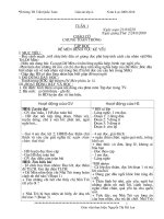
Figure 1. Identification of DLC1 as a Pak1 binding
protein
A: Yeast cells were cotransfected with control
GAD vector or GAD-DLC1, GAD-Rac1 along with
GBD vector, or GBD-Pak1 (aa 1–270) or GBDPak1 (aa 271–545). Cotransformants were plated
on selection plates lacking leucine and tryptophan (LT) or adenosine, histidine, leucine, and
tryptophan (AHLT). Growth was recorded after
72 hr. For -galactosidase assay, filter lift assays
were performed (middle). Blue color indicates
specific interaction of two proteins. Rac1 was
used as a positive control.
B: Pak1 and DLC1 interaction in the GST pulldown assays. DLC1 or Pak1 cDNAs were translated in vitro, and 35S-labeled proteins were incubated with either GST-Pak1 or GST-DLC1 and
analyzed by SDS-PAGE and autoradiography.
C: In vivo interaction of Pak1 with endogenous DLC1. MCF-7 cells were serum starved for 48 hr and treated with epidermal growth factor (EGF) for 30 min,
and cell lysates were immunoprecipitated with either Pak1 or DLC1 antibody. DLC1 association was analyzed by Western blotting with Pak1 or DLC1
antibody.
D: Activated Pak1 interacts with DLC1. Tet on-vector or Tet on-HA-Pak1-T423E expressing MCF-7 cells grown in 10% serum were lysed, and lysates containing
equal amounts of protein were immunoprecipitated with HA antibody and immunoblotted with antibodies against DLC1 and Pak1.
we examined whether expression of catalytically active Pak1
(Pak1T423E) would mimic EGF-mediated DLC1 phosphorylation. MCF-7 cells were cotransfected with T7-tagged DLC1 and
myc-tagged constitutively active T423E Pak1 or kinase dead
Pak1 K299R and labeled with [32P]orthophosphoric acid. Immunoprecipitation of DLC1 with an anti-T7 monoclonal antibody
showed increased phosphorylation of DLC1 in cells expressing
myc-T423E Pak1 but not in cells expressing myc-K299R Pak1
Figure 2. Mapping of interaction domains of Pak1 and DLC1
A: Identification of Pak1 domains that interact with DLC1. GST-Pak1 fusion
proteins containing wild-type (aa 1–545), kinase domain (aa 271–545),
N-terminal domain (aa 1–132), PIX binding domain (aa 182–203), Nck binding domain (aa 1–75), Cdc42/Rac-interactive binding (CRIB) domain (aa
52–132), and autoinhibitory domain (aa 75–149) were incubated with in vitro
translated DLC1 (aa 1–89), and binding was analyzed by GST pull-down
assay.
B: GST pull-down assay in lanes 9–13 shows various deletions of Pak1 in the
region of 132–270.
C: Identification of the DLC1 region involved in the binding of Pak1. GST
fusions of various lengths of DLC1 were used in the GST pull-down assay
using 35S-labeled Pak1 (amino acids 1–545).
CANCER CELL : JUNE 2004
(Figure 3C), suggesting a role of Pak1 in the phosphorylation
of DLC1 in vivo.
To map the Pak1 phosphorylation site in DLC1, we performed in vitro kinase assays using GST-DLC1 as a substrate.
The BAD, a well-known Pak1 substrate, was used as a positive
control. The Pak1 enzyme efficiently phosphorylated GST-DLC1
as well as BAD (Figure 3D). We next mapped the Pak1 phosphorylation site in DLC1 using various deletion constructs of DLC1.
GST fusion containing the C-terminal 19 amino acids of DLC1
alone was efficiently phosphorylated by Pak1 (Figure 3E, lane
5), suggesting the presence of a putative phosphorylation site
in the C-terminal region. Analysis of the C-terminal 19 amino
acids sequence revealed one serine at amino acid 88. Deletion
of two C-terminal amino acids of DLC1 (aa 88 and aa 89) completely abolished the ability of Pak1 to phosphorylate DLC1
(Figure 3E, lane 7). To confirm these findings, we substituted
Ser88 to alanine (DLC1 Ser88A). As expected from the deletion
studies, Pak1 failed to phosphorylate DLC1 Ser88A, confirming
that Pak1 phosphorylates DLC1 on Ser88 (Figure 3F). To verify
that Ser88 is the only DLC1 site that is phosphorylated by growth
factors in vivo, we performed a transient transfection experiment
with wild-type and Ser88A mutants of DLC1 (Figure 3G). Mutation of Ser88 on DLC1 to alanine completely abolished growth
factor-mediated phosphorylation of DLC1. Collectively, these
observations confirmed that DLC1 is a physiologic substrate of
Pak1 and that Ser88 on DLC1 is the Pak1 phosphorylation site.
DLC1 regulation of cell growth
To further delineate the potential effects of DLC1 on the biology
of breast cancer cells, we next examined the status of DLC1
expression in a number of breast cancer cell lines. Breast cancer
cells contained a higher level of DLC1 than nontumorigenic
breast cancer cell lines, such as MCF10A and HC11 (Figure 4A).
Because the ZR75 cells showed lower levels of DLC1 expression
than other breast cancer cell lines used here, we next established stable ZR75 pooled clones expressing T7-tagged wildtype DLC1 or mutant DLC1del88–89 or control vector (Figure 4B).
In general, these clones contained DLC1 levels 2- to 4-fold
higher than the endogenous level of DLC1. Because both Pak1
577
A R T I C L E
Figure 3. Pak1 phosphorylation of DLC1
A: MCF-7 cells were transfected with Pak1-specific or control siRNA, metabolically labeled with [32P]orthophosphoric acid, and treated with or without EGF.
Endogenous DLC1 was immunoprecipitated, and the phosphorylation status of DLC1 was analyzed by autoradiography. Total lysates were analyzed for
Pak1, Pak2, and vinculin by Western blotting.
B: MCF-7 cells were transfected with T7-DLC1 with or without Pak1 autoinhibitory domain (Pak1 aa 83–149). Cells were metabolically labeled with [32P]orthophosphoric acid and treated with or without EGF (1 nM) for 1 hr; then the phosphorylation of T7-DLC1 was analyzed by autoradiography.
C: MCF-7 cells were cotransfected with wild-type T7-DLC1 and T423E-Pak1 or K299R-Pak1. Cells were metabolically labeled with [32P]orthophosphoric acid.
T7-DLC1 was immunoprecipitated, and the phosphorylation status was visualized by autoradiography.
D: In vitro kinase assay using GST-DLC1 as a substrate and purified bacterially expressed Pak1 as an enzyme. GST was used as negative control, and HisBAD was used as a positive control.
E: In vitro Pak1 kinase assay using GST fusion of DLC1 fragments containing the indicated serial deletions of DLC1.
F: In vitro Pak1 kinase assay using DLC1 wild-type or mutant DLC1S88A as substrates.
G: MCF-7 cells transfected with wild-type DLC1 or mutant DLC1S88A were labeled in vivo with [32P]orthophosphoric acid, serum starved, treated with EGF
for 60 min, and immunoprecipitated; then phosphorylation status of DLC1 or its mutant was visualized by autoradiography.
and DLC1 are both shown to have a role in cell growth and
survival (Puthalakath et al., 1999; Chang et al., 2000; Schurmann
et al., 2000), we next examined whether Pak1 and DLC1 interaction have a role in cell growth and cell survival pathways. Interestingly, deregulated DLC1 expression increased the proliferation rate of the ZR75 cells (Figure 4C). The increased proliferation
of the ZR75 cells could have reflected either an increased proliferation rate or protection of the cells from apoptosis. Flow cytometry analysis of serum-starved ZR75 clones revealed that
deregulation of wild-type DLC1, but not mutant DLC1del88-89, was
accompanied by increased G1→S progression of breast cancer
cells. Interestingly, ZR75 cells expressing DLC1del88-89 exhibited
a significant increase in the number of cells in the pre-G0 phase
of the cell cycle (Figure 4D). Consistent with a potential role of
DLC1 in the G1/S transition, we found an increased expression
of cyclin E in the clones with deregulated DLC1 but not in
control pcDNA clones (Figure 4E). Further analysis using CDK2
phospho-specific antibody revealed a substantial enhancement
of CDK2 phosphorylation in DLC1 expressing clones. In addition, biochemical analysis of the cyclin E immunocomplex confirmed the presence of elevated CDK2 kinase activity in the
DLC1 clones, as well as increased phosphorylation of pRb at
Ser795, a known CDK2/cyclin E phosphorylation site (Figure
4E). These findings provided clues about a potential role of
Pak1-mediated DLC1 phosphorylation in optimal cell survival.
578
Interference with this Pak1-DLC1 pathway might trigger spontaneous apoptosis.
An essential role of DLC1 in Pak1-mediated cell survival
Earlier studies have shown that Pak family kinases protect murine NIH3T3 cells from ultraviolet (UV)-induced apoptosis (Jakobi et al., 2001). Using this well-established model system, we
next investigated the potential role of Pak1-DLC1 interaction in
protecting NIH3T3 cells against UV-induced apoptosis. NIH3T3
cells were cotransfected with RFP-tagged DLC1 or the mutant
Ser88A DLC1 and catalytically active Pak1 (myc-T423E Pak1)
or kinase-dead Pak1 (myc-K299LL Pak1), and cells were exposed to UV irradiation. After 10 hr, apoptosis was quantitated
by scoring the integrity of the nuclear membrane in cells expressing both RFP (red) and myc-epitope tags to detect DLC1
and Pak1, respectively (Figure 5A). Results indicated that UVinduced apoptosis was substantially reduced in cells expressing
DLC1 and catalytically active Pak1 but not in Ser88A DLC1
or kinase-dead Pak1 or the combination of these (Figure 5A).
Interestingly, overexpression of active Pak1 could not protect
the cells transfected with the mutant Ser88A DLC1. Similarly,
overexpression of DLC1 failed to protect the NIH3T3 cells overexpressing the kinase-dead Pak1 from UV-induced apoptosis
(Figure 5A).
As an additional evidence of a role of DLC1 in the cell survival
and a broader significance of our findings in human cells in
CANCER CELL : JUNE 2004
A R T I C L E
Figure 4. DLC1 expression and cell growth
A: Expression of DLC1 in a number of normal and
breast cancer cells analyzed by Western blot
analysis.
B: Expression of T7-DLC1 wild-type or T7DLC1del88-89 in stable clones analyzed by Western
blot analysis.
C: Equal number of ZR75 stable cells expressing
DLC1 wild-type or DLC1del88-89 mutant were cultured. At indicated intervals, cells were trypsinized and counted using a Coulter Counter to
determine cell proliferation. Each clone was
plated in triplicate, and the experiment was repeated twice with similar results.
D: Cell cycle status of DLC1 wild-type, mutant,
and pcDNA clones as analyzed by flow cytometry.
E: pcDNA and DLC1 stable clones were analyzed
by Western blotting for the levels of cyclin E (left),
for the levels of phospho CDK2 (middle) and the
activity of CDK2 by immunoprecipitation followed by in vitro kinase assay using histone H1
as substrate, and also by Western blotting of total
lysate using phospho pRb Ser795-specific antibody (right).
general, we next downregulated Pak1 in IMR-90 human fibroblasts by transiently expressing GFP vector or GFP vector expressing Pak1-siRNA. In some cultures, cells were cotransfected with GFP-tagged dominant active Pak1-T423E along
with control or DLC-specific siRNA (Figure 5B). After 48 hr, cells
were exposed to UV (100 J/m2). The onset of apoptosis was
monitored by scoring the nuclear integrity using lamin A/C antibody. GFP staining was used to localize the positive transfected
cells. We found that as expected, expression of activated Pak1
increased the cell survival after UV exposure, while knockdown
of the expression of DLC1 resulted in a substantial increase in
the ratio of apoptotic cells after UV damage. Interestingly, the
noticed apoptotic response due to lack of DLC1 could not be
rescued by activated Pak1 (Figure 5C). These findings suggest
that Pak1-DLC1 interactions play an important role in their survival functions.
Pak1-DLC1 interaction promotes cell survival
by blocking BimL’s apoptotic functions
Because DLC1 is known to interact and sequester proapoptotic
BimL in the cytoplasm (Puthalakath et al., 1999) and because
Pak1 phosphorylates DLC1 in the region that interacts with
BimL (Puthalakath et al., 1999), we next examined whether Pak1
phosphorylation of BimL interferes with BimL apoptotic functions. Initially, we examined the effect of Pak1 phosphorylation
on the ability of DLC1 to interact with BimL. GST-DLC1 was
CANCER CELL : JUNE 2004
phosphorylated with Pak1 enzyme and subjected to the GST
pull-down assay with 35S-labeled BimL. Our results suggest
that unphosphorylated but not phosphorylated DLC1 efficiently
interacts with BimL, raising the possibility that Pak1 phosphorylation might interfere with the ability of DLC1 to interact with
BimL (Figure 6A). To confirm these observations, we next mutated the Pak1 phosphorylation site Ser88 either to alanine or
glutamic acid. GST pull-down assays showed that a Ser88 to
alanine mutation has no effect on BimL binding to DLC1 (Figure
6B). However, a Ser88 to glutamic acid mutation, which mimics
the phosphorylated form of DLC1 due to the addition of negative
charge, abolished DLC1 interaction with BimL. Similarly, deletion of the C-terminal amino acids 88 and 89, which contain
Ser88, also abolished BimL binding (Figure 6C). These results
support the earlier view that DLC1 interacts with BimL via the
C-terminal region (Puthalakath et al., 1999) and now offers an
additional regulatory role of the Pak1 phosphorylation of DLC1.
In normal physiologic conditions, BimL is sequestered by
DLC1, and DLC1-BimL dimers are released upon stimulation
of cells with apoptotic stimuli. The DLC1-BimL dimers then
interact with Bcl-2 and thus inhibit cell survival functions of
Bcl-2 (Puthalakath et al., 1999; Puthalakath and Strasser, 2002).
Therefore, we next explored whether Pak1 recognizes DLC1BimL dimers. Results from the GST pull-down studies demonstrated that Pak1 recognizes DLC1 as well as DLC1 bound to
BimL (Figure 6D). We then examined whether apoptotic signals
579
A R T I C L E
Figure 5. Requirement of DLC1 and Pak1 interaction for cell survival
A: Immunofluorescence staining of NIH3T3 cells
cotransfected with various combinations of
DLC1 wild-type or DLC1S88A mutant and various
Pak1 constructs, exposed to UV radiation 100
mJ/s and after 10 hr, costained for myc-tag (for
Pak1 constructs, blue), RFP-DLC1 wild-type, or
mutants (DLC1 constructs, red) and for lamin A/C
(green). Cells expressing the transfected plasmids were identified using appropriate filter settings and then characterized for their nuclear
integrity using morphologic features identified by
lamin A/C staining. For example, a smooth nuclear membrane was scored as nonapoptotic
as opposed to membrane blebbing or rupture,
which was scored as apoptotic. Nuclei committed to enter mitosis were not scored in either of
these groups because they were easily identified
for their specific “pulverized” pattern of lamin
A/C staining. Ten randomly selected fields were
analyzed for each condition. The experiment
was repeated twice with similar results.
B: Functionality of DLC siRNA was analyzed by
Western analysis.
C: Immunofluorescence staining of IMR90 human fibroblast cells cotransfected with GFPtagged T423E Pak1 or GFP-tagged Pak1 siRNA
or DLC1 siRNA or in combination. After 48 hr, cells
were exposed to UV radiation 100 mJ/s and after
10 hr, costained for GFP-Pak1T423E (green), GFPPak1 siRNA (green), or endogenous DLC1 (blue)
and for lamin A/C (red). Apoptotic cells were
scored as described in (A). Yellow arrow points
to an apoptotic cell and white arrow points to
a cell that was protected by expression of active
Pak1.
such as UV and survival signals such as EGF modulate the
status of phosphorylation of DLC1 and BimL. UV treatment
alone did not induce phosphorylation of DLC1 or BimL; rather,
a slight decrease in the phosphorylation was observed compared to control (Figure 6E). Interestingly, the addition of survival
factors such as EGF followed by UV treatment resulted in a
substantial increase in the phosphorylation of both DLC1 and
BimL. Transfection of cells with Pak1 inhibitory domain (Pak1
aa 83–149) effectively inhibited the noticed upregulation of BimL
phosphorylation by EGF under condition of UV treatment (data
not shown), suggesting an additional regulatory role of Pak1 in
BimL phosphorylation. In support of this notion, we next found
that Pak1 could indeed directly phosphorylate BimL (Figure 6F).
We then examined whether Pak1 phosphorylates BimL when
present as a dimer with DLC1 and when attached to the dynein
complex via dynein intermediate chain (DIC) as a trimeric complex (DIC-DLC1-BimL) using in vitro Pak1 kinase assay. Results
show that Pak1 preferentially phosphorylates BimL present in
the dimeric form (DLC1-BimL) compared to BimL present in
trimeric form (DIC-DLC1-BimL) (Figure 6G).
Physiologic cell survival signals are known to downregulate
580
the levels of BimL expression via its phosphorylation and thus
counteract proapoptotic functions of BimL (Ley et al., 2003;
Luciano et al., 2003; Reginato et al., 2003; Seward et al., 2003).
Because growth factors activate Pak1 (Kumar and Vadlamudi,
2002) and because Pak1 phosphorylated BimL in this study,
we next examined the effect of Pak1 activation on the status of
BimL expression using a well-established Tet-inducible MCF-7
model system (Vadlamudi et al., 2000). Treatment of cells with
doxycycline induced the levels of constitutively active Pak1 in
a time-dependent manner. Analysis of BimL expression under
this condition showed a decrease in the expression of BimL
levels with an increase in the expression of active Pak1 (Figure
6H). As a positive control, we analyzed the status of cyclin D1
expression, which is known to be upregulated by Pak1 (Balasenthil et al., 2004). As expected, cyclin D1 expression was upregulated with an increase in active Pak1 levels. To examine whether
Pak1 kinase activity indeed is involved in the downregulation
of BimL, we transfected activated Pak1 (T423E Pak1) or GTPase
inactive kinase-dead Pak1 (LLK299R) into MCF-7 cells, and the
expression of BimL was analyzed after 48 hr. Our results showed
a decrease in the level of BimL expression in cells with activated
CANCER CELL : JUNE 2004
A R T I C L E
Figure 6. Pak1 phosphorylation regulates BimL and DLC1 interactions
A: Effect of Pak1 phosphorylation on the binding of DLC1 with BimL. GST-DLC1 was phosphorylated with Pak1 enzyme, and the GST pull-down assay was
performed using phosphorylated GST-DLC1 or wild-type GST-DLC1. Autoradiogram showing the phosphorylation of DLC1 is shown in the right panel.
B: Ability of DLC1-Ser88A or DLC1-Ser88E mutants to interact with BimL in GST pull-down assay.
C: Ability of GST-DLC1 (aa 1–87) to interact with BimL was analyzed by GST pull-down assay.
D: Ability of Pak1 to interact with DLC1-BimL dimers. 35S-labeled DLC1 and BimL were incubated with GST-Pak1, and GST pull-down assay was performed.
E: MCF-7 cells were labeled with 32P-orthophosphoric acid, and cells were treated with UV or pretreated with EGF (100 ng/ml) followed by UV treatment.
Cell lysates were immunoprecipitated with antibodies against DLC1 or BimL, and the phosphorylation status of proteins was analyzed by autoradiography.
F: Phosphorylation of GST-BimL by Pak1 in in vitro kinase assay.
G: Purified GST-BimL was incubated with purified beads bound GST-DLC1 along with or without purified GST-dynein intermediate chain proteins. After 60
min of incubation, dimeric and trimeric complexes were purified by GST-DLC1 pull-down assay and used as a substrate in in vitro Pak1 kinase assay.
H: Downregulation of BimL levels by constitutively active Pak1. MCF-7 cells expressing T423E-Pak1 under the control of Tet-regulated promoter were treated
with doxycycline for 12 or 24 hr. Expression of BimL, cyclin D1, and HA-tagged T423E Pak1 was analyzed by Western blotting.
I: MCF-7 cells were transiently transfected with CMV vector or constitutively active Pak1 (CMV-myc-T423EPak1) or kinase-dead Pak1 (CMV-myc-LLK299Rpak1).
After 48 hr of transfection, cell lysates were analyzed for BimL expression. Actin was used as a loading control. Myc-epitope antibody was used to monitor
the expression of myc-tagged Pak1 constructs.
J: Model for Pak1 regulation of DLC1 and BimL functions. Under physiologic conditions, DLC1 sequesters BimL to the microtubules. Under apoptotic conditions,
DLC1-BimL dimers are released and interact with Bcl-2. Survival factors activate Pak1, which in turn phosphorylates DLC1-BimL dimers and, thus, prevents
BimL interaction with Bcl-2.
Pak1 but not in kinase-dead Pak1, suggesting Pak1 kinase
activity is involved in the downregulation of BimL expression
(Figure 6I). These findings suggested a mechanistic role of DLC1
Ser88 phosphorylation in conferring a survival advantage by
modulating the levels of DLC1-BimL dimers, which are dissociated upon apoptotic signaling and, thus, may have a role in
enhanced cell survival and, consequently, increased growth rate
(Figure 6J).
Deregulation of DLC1 promotes tumorigenesis
Because DLC1 expression promoted G1→S transition as well
as cell survival (Figures 4C and 4D) and because Pak1 overexpression is known to support the anchorage-independent
CANCER CELL : JUNE 2004
growth of breast cancer cells (Vadlamudi et al., 2000), we examined the ability of the ZR75/DLC1 clones to grow in an anchorage-independent manner. DLC1 wild-type but not the DLC1
mutant expression substantially enhanced the number of colonies in soft agar (Figure 7A), suggesting a role of DLC1 in anchorage-independent growth.
To examine the potential correlation of DLC1 deregulation
with the progression of breast cancer to more aggressive phenotypes, we next analyzed DLC1 expression in lysates from exponentially growing cell lines derived from the MCF10AT model
system. This model contains a spectrum of cell lines that allows
us to examine the expression of genes during the progression
of breast malignancy (Heppner et al., 2000; Santner et al., 2001).
581
A R T I C L E
Figure 7. DLC1 deregulation potentiates anchorage-independent growth and promotes tumorigenesis
A: Effect of DLC1 wild-type or mutant overexpression on the ability of ZR75 cells to grow in an
anchorage-independent manner. The anchorage-independent growth potential of DLC1expressing clones was measured by the ability
of the cells to form colonies on soft agar. Similar
results were obtained in three independent experiments. Error bars represent standard error of
mean.
B: Western blot analysis of DLC1 and Pak1 levels
in lysates from cells derived from MCF10AT model
system. MCF10A, nonmalignant human breast
cancer cells; MCF10AT, weakly tumorigenic;
MCF10DCIS, Comedo-type DCIS, highly proliferative, aggressive, invasive; and MCF10CA, undifferentiated carcinomas, metastatic.
C: Nude mice were injected with 5 ϫ 106 cells
stably expressing pcDNA or wild-type DLC1
(pooled clone #2) or mutant pooled clone
DLC1del88-89, and tumor growth was measured at
weekly intervals.
D: A representative picture of DLC1-induced tumor in nude mice. Tumor growth was observed
only in cells expressing wild-type DLC1 but not
detected in mutant-expressing cells.
E: Morphology of tumor and expression of T7DLC1 in tumors as evaluated by hematoxylin and
eosin and anti-T7 staining, respectively.
Figure 7B shows a clearly progressive upregulation of DLC1
expression as a function of tumorigenesis. Consistent with the
aggressive growth characteristics of the DLC1-expressing
breast cancer cells, the ZR/DLC1 cells (pooled clone 2) exhibited
increased tumorigenic potential in nude mice in the absence of
any exogenous estradiol treatment (Figures 7C and 7D), suggesting that deregulation of DLC1 might be sufficient to promote
tumorigenic phenotypes. Interestingly, DLC1 mutant lacking the
Pak1 phosphorylation site did not exhibit any tumorigenic potential in nude mice (Figure 7C). The clonal origin of T7-DLC1deregulated cells from tumors was confirmed by staining the
cells with an anti-T7 monoclonal antibody (Figure 7E). These
findings suggested the existence of a close relationship between
DLC1 deregulation and its phosphorylation by Pak1 and tumorigenesis.
DLC1 upregulation in human breast cancer
We next examined the level of DLC1 in 15 paired samples of
human breast tumors and adjacent normal tissues. The status
of DLC1 expression was considerably higher in 14 out of 15
breast tumor specimens than in adjacent normal tissues (Figures
8A and 8B). The anti-DLC1 antibody also works well in formalinfixed, paraffin-embedded human breast tissues, as shown in a
representative DLC1 immunohistochemical (IHC) staining (Figure 8C). Further IHC examination of a tumor array containing
60 breast tumor specimens revealed widespread upregulation
of DLC1 expression in 54 out of 60 breast tumors (Figure 8D).
Because breast tumor specimens used here have recently been
characterized for the levels of Pak1 expression (Balasenthil et
582
al., 2004), we found that 34 out of 60 specimens had overexpressed both Pak1 and DLC1. To examine whether DLC1
protein in tumors is phosphorylated, we have used a recently
available phospho-serine-specific antibody, which specifically
recognizes serine-phosphorylated proteins (Gronborg et al.,
2002). We have used an affinity column of this antibody to purify
serine-phosphorylated proteins from the human breast tumors
and adjacent normal tissues. Elutes from columns were precipitated and then Western blotted with DLC1-specific antibody.
As a proof of principle, we have analyzed six normal and six
tumor samples. Results showed that five out of six tumors have
more serine-phosphorylated form of DLC1 compared to control
(Figure 8E). These results further support our hypothesis that
increased DLC1 protein expression in tumors also translates into
its more phosphorylated form. Together, results from Figures 7
and 8 suggested that deregulation of DLC1 and Pak1 might be
a common event in cancer progression and that these molecules
may be involved in the development and/or maintenance of the
malignant phenotypes in tumors.
Discussion
In this study, we discovered that DLC1 is a Pak1-interacting
protein. This conclusion is supported by the ability of Pak1 to
interact with DLC1 in yeast two-hybrid, in GST pull-down, and in
reciprocal immunoprecipitation assays. Furthermore, our results
show that DLC1 is a physiologic substrate of Pak1. We have
identified Ser88 of DLC1 as the Pak1 phosphorylation site and
demonstrated an essential role of this pathway in growth factormediated cell survival.
CANCER CELL : JUNE 2004
A R T I C L E
Figure 8. DLC1 deregulation in human breast tumors
A: Breast tumor lysates were analyzed by Western blot analysis for DLC1
expression (top) and subsequently reprobed with vinculin antibody as a
loading control.
B: Quantitation of DLC1 expression in normal and tumor samples.
C: Immunohistochemical analysis of DLC1 in breast tissue samples. The left
panel shows normal mammary gland tissue, and the right panel shows
tumor from the same patient. Tumor shows very strong nuclear and cytoplasmic DLC1 immunoreactivity.
D: Quantitation of DLC1 expression in breast tumor array.
E: Phosphorylation of DLC1 in normal and tumor samples. Serine phosphorylated proteins in normal and tumor lysates (2 mg) were purified using phosphoserine purification kit (Qiagen) as described in Experimental Procedures,
and purified proteins were precipitated using trichloroacetic acid. Levels
of DLC1 in the precipitate were then measured by Western blotting using
DLC1-specific antibody.
DLC1 is ubiquitously expressed in different cell types and
highly conserved throughout evolution. DLC1 is not only associated with myosin V (Naisbitt et al., 2000) and dynein (Hirokawa,
1998) motors, but it also binds to nNOS (Jaffrey and Snyder,
1996; Fan et al., 1998), the proapoptotic Bcl-2 family protein
BimL (Puthalakath et al., 1999), the Drosophila mRNA localization protein Swallow (Schnorrer et al., 2000), transcriptional regulator IkB (Crepieux et al., 1997), and the postsynaptic scaffold
protein GKAP (Naisbitt et al., 2000). These observations suggest
that DLC1 is a multifunctional regulatory protein. Previously
structural studies have shown that DLC1 exists as a dimer and
contains two identical target binding grooves on the opposite
faces of the protein dimer interface (Fan et al., 2001). In this
context, DLC1 has been suggested to function as a linker protein
by binding two different proteins (Liang et al., 1999). The ability
of DLC1 to interact with proteins with diverse cellular functions
suggests that DLC1, in principle, might recruit Pak1 to other
signaling proteins.
Pak1 has been shown to promote cell survival by phosphorylating and inactivating the proapoptotic functions of Bad (Schurmann et al., 2000). Overexpression of a constitutively active
T423E Pak1 mutant promotes the survival of NIH3T3 murine
fibroblasts. Similar to Pak1, Drosophila DLC1 plays a role in the
CANCER CELL : JUNE 2004
inhibition of apoptosis (Dick et al., 1996). In human cells, DLC1
interacts with BimL and promotes cell survival by inhibiting its
proapoptotic activity (Puthalakath et al., 1999). In addition,
cyclooxygenase 2 promotes cell survival by stimulating DLC1
expression and inhibiting the neuronal NO pathway (Chang et
al., 2000). Under physiologic conditions, BimL is sequestered
to dynein motor via the DIC-DLC1-BimL complex (Puthalakath
et al., 1999; Puthalakath and Strasser, 2002). Apoptotic stimuli
release dimer-containing DLC1-BimL, which then translocates
and interacts with Bcl-2, leading to the inactivation of the antiapoptotic function of Bcl-2 and promotion of the apoptotic function of BimL. Growth factors have been shown to increase phosphorylation and degradation of BimL and thus modulate the
apoptotic functions of BimL. Although the mechanism by which
growth factors regulate BimL functions remains elusive, emerging evidence suggests a role of phosphorylation in the degradation of BimL by growth factors (Ley et al., 2003; Luciano et al.,
2003; Seward et al., 2003). In this context, our findings that
Pak1 phosphorylates BimL and that activated Pak1 reduces
BimL levels are important because they suggest that Pak1 might
act as a downstream effector of growth factor-mediated modulation of BimL levels. The ability of Pak1 to recognize and phosphorylate DLC1-BimL dimer also strongly supports the possibility that Pak1 acts as a growth factor sensor to detect the
DLC1-BimL dimers generated by apoptotic signals and that
Pak1 might dissociate these complexes via phosphorylation.
Emerging evidence implies that Pak1 might play an important role in tumorigenesis (Kumar and Vadlamudi, 2002). Deregulation of Pak1 expression is reported in breast and ovarian
cancers (Vadlamudi et al., 2000; Balasenthil et al., 2004; Schraml
et al., 2003). We showed that Pak1 interacted with DLC1 and
that such interactions are essential for cell cycle progression
and survival functions. Accordingly, overexpression of DLC1
substantially increased the growth potential of the breast cancer
cells, allowing the cells to survive in an anchorage-independent
manner and exhibit increased tumorigenic potential of breast
cancer cells. Interestingly, DLC1 and Pak1 expression levels
were more elevated in breast tumors than in normal control
specimens, raising the possibility that DLC1 has a role in tumorigenesis.
In summary, our results showed that DLC1 is an interacting
substrate of Pak1, that DLC1 is overexpressed in human breast
cancers, and that Pak1 phosphorylation of DLC1 on Ser88 plays
a critical role in tumorigenic phenotypes of DLC1 in breast cancer cells.
Experimental procedures
Cell cultures, reagents, transfection, and cell extracts
MCF-7 breast cancer cells (Vadlamudi et al., 2000) were maintained in Dulbecco’s modified Eagle’s medium (DMEM)-F12 (1:1) supplemented with
10% fetal calf serum. Antibodies DLC1 were purchased from Santa Cruz
Biotechnology (Santa Cruz, CA) and BD Biosciences (San Jose, CA). Antibody against T7 was purchased from Novagen (Milwaukee, WI). BimL antibody was purchased from Chemicon (Melbourne, Australia). Antibodies for
Pak1, phos Ser795 pRb, Phos CDK2, and siRNA against Pak1 (#6361) and
control FITC siRNA (#6201) were purchased from Cell Signaling (Boston,
MA). SMART POOL siRNA for DLC1 and DLC2 was synthesized by Dharmacon (Lafayette, CO). Transient transfection studies were performed using
a FuGENE-6 kit (Roche Molecular Biochemicals, Indianapolis, IN) in accordance with the manufacturer’s instructions. Cell lysates were resolved on
an 18% tricine gels. ZR75 cells were transfected with pcDNA3.1, pcDNAT7-DLC1, or dsREDGFP-DLC1 via the calcium phosphate method. Forty-
583
A R T I C L E
eight hours after transfection, cells were selected in medium containing
G418 (500 g/ml).
Two-hybrid library screening and plasmid construction
Pak1 baits were constructed by amplifying amino acids 1–270 and amino
acids 271–545 by polymerase chain reaction (PCR) and then subcloned
into Gal4 binding domain vector pGBD vector (Clontech, Palo Alto, CA). A
mammary gland cDNA library fused to Gal4 activation domain was purchased from Clontech and screened using N-terminal Pak1 (aa 1–270) as
bait in accordance with the manufacturer’s instructions. A total of 2 ϫ
106 clones were screened. Positive interactors were verified by one-onone transformations followed by selection of agar plates lacking adenosine,
histidine, leucine, and tryptophan (AHLT) and also by -galactosidase assay.
To generate epitope-tagged constructs of DLC1, the open reading frame of
DLC1 was amplified by PCR and subcloned into pcDNA 3.1 (Invitrogen,
Carlsbad, CA) or dsRED (Clontech) vectors. Mutation of Ser88A was created
by site-directed mutagenesis (Stratagene, Cedar Creek, TX). GST fusions
of various DLC1 mutants were generated by PCR cloning into PGEX vector
(Amersham Pharmacia Biotech, Piscataway, NJ). Pak1 GST constructs were
described earlier (Vadlamudi et al., 2002).
Pak kinase assay
In vitro kinase assays using myelin basic protein (MBP) or glutathione-Stransferase (GST)-DLC1 protein (4 g each) were performed in HEPES buffer
(50 mM HEPES, 10 mM MgCl2, 2 mM MnCl2, 0.2 mM DTT) containing 1 g
of purified GST-Pak1 enzyme, 10 Ci of [␥-32P]ATP, and 25 M cold ATP.
Purified human Pak1 enzyme (#GLO110-100) was purchased from Alexis
Biochemicals (San Diego, CA). His-BAD (#14-357) was purchased from Upstate Biotechnology (Waltham, MA) and used as a positive control.
Immunohistochemistry
For immunohistochemical detection of DLC1 and T7, sections were deparaffinized with xylene and rehydrated using graded ethanol. Sections were
incubated in 0.3% H2O2 and methanol for 30 min to inactivate endogenous
peroxidase. The sections were then boiled for 10 min in 0.01 M citrate buffer
and cooled for 30 min at room temperature to expose antigenic epitopes.
The sections were incubated with 2% normal goat serum in 1% bovine
serum albumin and PBS for 30 min and then with an anti-DLC1 antibody
(1:50 dilution) or anti-T7 antibody (1:800 dilution) and incubated overnight
at room temperature. The sections were washed 3 times with 0.05% Tween
in PBS for 10 min and incubated with secondary antibody, developed with
DAB-H2O2, and counter stained with Mayer’s hematoxylin.
Acknowledgments
We thank Drs. D. Wu and A. Strasser for generously providing pSuperEGFP-Pak1-siRNA and BimL cDNAs, respectively, and Kapil Mehta for
MCF10A model cell lysates. We also thank Mahitosh Mandal for cell cycle
analysis, Liana Adam for the initial microscopic experiments, and Feng Li
for GST pull-down assays. This study was supported by NIH grants CA80066
and CA90970 (R.K.).
Received: December 2, 2003
Revised: March 16, 2004
Accepted: April 20, 2004
Published: June 14, 2004
References
GST pull-down assay
In vitro transcription and translation of the Pak1 and DLC1 proteins were
performed using the TNT T7 Quick Coupled Transcription/Translation system
(Promega, Madison, WI) as previously described (Vadlamudi et al., 2002).
Cell proliferation, soft agar, and tumorigenicity assays
Soft agar colony-growth assays were performed as previously described
(Vadlamudi et al., 2000). Briefly, 1 ml of 0.6% DIFCO agar in DMEM supplemented with 10% fetal bovine serum and insulin was layered onto tissue
culture plates. Test cells (104) mixed with 1 ml of 0.36% bactoagar solution
in DMEM were layered on top of the 0.6% bactoagar layer. The plates were
incubated at 37ЊC in 5% CO2 for 21 days. For xenograft studies, 5 ϫ 106
cells were implanted subcutaneously into the mammary fat pads of 10 nude
mice as previously described (Bagheri-Yarmand et al., 2001) and allowed
to grow for 20 weeks. Tumor size was then measured.
Immunofluorescence and confocal studies
The cellular location of proteins was determined using indirect immunofluorescence, as described previously (Kumar et al., 2002). Cells were incubated
with primary antibodies for 2 hr, washed 3 times in PBS, and then incubated
with 546-Alexa- (red), 633-Alexa- (blue), or 488-Alexa- (green)-labeled secondary antibodies (Molecular Probes, Eugene, OR). The procedure involved
processing the same section for each detector (the two excitations corresponding to 546 and 488 or 633 nm) and making a pixel-by-pixel comparison.
Human samples
Tissue samples from patients who had undergone routine surgery for breast
cancer were snap frozen in liquid nitrogen and stored at Ϫ80ЊC in the
M.D. Anderson Breast Core Pathology Laboratory and have been described
previously (Balasenthil et al., 2004). Purification of phosphoserine proteins
in normal and tumor tissues was done using phospho protein purification
kit (#37101) and phosphoserine antibody (#37430) purchased from Qiagen
(Valencia, CA) as per manufacturer instructions. In brief, tissue samples
were lysed in zwitterionic detergent CHAPS-, phosphatase inhibitor-, and
nuclease-containing lysis buffer to disrupt protein-protein and protein-DNA
complexes. Cleared cell lysates were then passed through a column containing immobilized phosphoserine antibody provided in the kit. Elutes from
columns were precipitated with TCA and washed four times with acetone,
and the precipitated proteins were separated on 18% tricine gels. Blots
were then Western blotted with DLC1-specific antibody.
584
Bagheri-Yarmand, R., Mandal, M., Taludker, A.H., Wang, R.A., Vadlamudi,
R.K., Kung, H.J., and Kumar, R. (2001). Etk/Bmx tyrosine kinase activates
Pak1 and regulates tumorigenicity of breast cancer cells. J. Biol. Chem. 276,
29403–29409.
Bagrodia, S., and Cerione, R.A. (1999). Pak to the future. Trends Cell Biol.
9, 350–355.
Balasenthil, S., Sahin, A.A., Barnes, C.J., Wang, R.A., Pestell, R.G., Vadlamudi, R.K., and Kumar, R. (2004). p21-activated kinase-1 signaling mediates
cyclin D1 expression in mammary epithelial and cancer cells. J. Biol. Chem.
279, 1422–1428.
Banerjee, M., Worth, D., Prowse, D., and Nikolic, M. (2002). Pak1 phosphorylation on t212 affects microtubules in cells undergoing mitosis. Curr. Biol.
12, 1233–1239.
Bokoch, G.M. (2003). Biology of the p21-activated kinases. Annu. Rev. Biochem. 72, 743–781.
Bokoch, G.M., Wang, Y., Bohl, B.P., Sells, M.A., Quilliam, L.A., and Knaus,
U.G. (1996). Interaction of the Nck adapter protein with p21-activated kinase
(PAK1). J. Biol. Chem. 271, 25746–25749.
Bokoch, G.M., Reilly, A.M., Daniels, R.H., King, C.C., Olivera, A., Spiegel,
S., and Knaus, U.G. (1998). A GTPase-independent mechanism of p21activated kinase activation. Regulation by sphingosine and other biologically
active lipids. J. Biol. Chem. 273, 8137–8144.
Chang, Y.W., Jakobi, R., McGinty, A., Foschi, M., Dunn, M.J., and Sorokin,
A. (2000). Cyclooxygenase 2 promotes cell survival by stimulation of dynein
light chain expression and inhibition of neuronal nitric oxide synthase activity.
Mol. Cell. Biol. 20, 8571–8579.
Crepieux, P., Kwon, H., Leclerc, N., Spencer, W., Richard, S., Lin, R., and
Hiscott, J. (1997). I kappaB alpha physically interacts with a cytoskeletonassociated protein through its signal response domain. Mol. Cell. Biol. 17,
7375–7385.
Daniels, R.H., and Bokoch, G.M. (1999). p21-activated protein kinase: a
crucial component of morphological signaling? Trends Biochem. Sci. 24,
350–355.
Daniels, R.H., Zenke, F.T., and Bokoch, G.M. (1999). alphaPix stimulates
p21-activated kinase activity through exchange factor-dependent and -independent mechanisms. J. Biol. Chem. 274, 6047–6050.
CANCER CELL : JUNE 2004
A R T I C L E
Dick, T., Ray, K., Salz, H.K., and Chia, W. (1996). Cytoplasmic dynein (ddlc1)
mutations cause morphogenetic defects and apoptotic cell death in Drosophila melanogaster. Mol. Cell. Biol. 16, 1966–1977.
Edwards, D.C., and Gill, G.N. (1999). Structural features of LIM kinase that
control effects on the actin cytoskeleton. J. Biol. Chem. 274, 11352–11361.
Edwards, D.C., Sanders, L.C., Bokoch, G.M., and Gill, G.N. (1999). Activation
of LIM-kinase by Pak1 couples Rac/Cdc42 GTPase signalling to actin cytoskeletal dynamics. Nat. Cell Biol. 1, 253–259.
Fan, J.S., Zhang, Q., Li, M., Tochio, H., Yamazaki, T., Shimizu, M., and
Zhang, M. (1998). Protein inhibitor of neuronal nitric-oxide synthase, PIN,
binds to a 17-amino acid residue fragment of the enzyme. J. Biol. Chem.
273, 33472–33481.
Fan, J., Zhang, Q., Tochio, H., Li, M., and Zhang, M. (2001). Structural basis
of diverse sequence-dependent target recognition by the 8 kDa dynein light
chain. J. Mol. Biol. 306, 97–108.
Frost, J.A., Khokhlatchev, A., Stippec, S., White, M.A., and Cobb, M.H.
(1998). Differential effects of PAK1-activating mutations reveal activitydependent and -independent effects on cytoskeletal regulation. J. Biol.
Chem. 273, 28191–28198.
Gronborg, M., Kristiansen, T.K., Stensballe, A., Andersen, J.S., Ohara, O.,
Mann, M., Jensen, O.M., and Pandey, A. (2002). A mass spectrometry-based
proteomic approach for identification of serine/threonine-phosphorylated
proteins by enrichment with phosphospecifc antibodies. Mol. Cell. Proteomics 1, 517–527.
Heppner, G.H., Miller, F.R., and Shekhar, P.M. (2000). Nontransgenic models
of breast cancer. Breast Cancer Res. 2, 331–334.
Hirokawa, N. (1998). Kinesin and dynein superfamily proteins and the mechanism of organelle transport. Science 279, 519–526.
Jaffrey, S.R., and Snyder, S.H. (1996). PIN: an associated protein inhibitor
of neuronal nitric oxide synthase. Science 274, 774–777.
Jakobi, R., Moertl, E., and Koeppel, M.A. (2001). p21-activated protein kinase
gamma-PAK suppresses programmed cell death of BALB3T3 fibroblasts.
J. Biol. Chem. 276, 16624–16634.
Kumar, R., and Vadlamudi, R.K. (2002). Emerging functions of p21-activated
kinases in human cancer cells. J. Cell. Physiol. 193, 133–144.
Kumar, R., Wang, R.A., Mazumdar, A., Talukder, A.H., Mandal, M., Yang,
Z., Bagheri-Yarmand, R., Sahin, A., Hortobagyi, G., Adam, L., et al. (2002).
A naturally occurring MTA1 variant sequesters oestrogen receptor-alpha in
the cytoplasm. Nature 418, 654–657.
Ley, R., Balmanno, K., Hadfield, K., Weston, C., and Cook, S.J. (2003).
Activation of the ERK1/2 signaling pathway promotes phosphorylation and
proteasome-dependent degradation of the BH3-only protein, Bim. J. Biol.
Chem. 278, 18811–18816.
Li, F., Adam, L., Vadlamudi, R.K., Zhou, H., Sen, S., Chernoff, J., Mandal,
M., and Kumar, R. (2002). p21-activated kinase 1 interacts with and phosphorylates histone H3 in breast cancer cells. EMBO Rep. 3, 767–773.
Lian, J.P., Crossley, L., Zhan, Q., Huang, R., Coffer, P., Toker, A., Robinson,
D., and Badwey, J.A. (2001). Antagonists of calcium fluxes and calmodulin
block activation of the p21-activated protein kinases in neutrophils. J. Immunol. 166, 2643–2650.
Liang, J., Jaffrey, S.R., Guo, W., Snyder, S.H., and Clardy, J. (1999). Structure
of the PIN/LC8 dimer with a bound peptide. Nat. Struct. Biol. 6, 735–740.
Luciano, F., Jacquel, A., Colosetti, P., Herrant, M., Cagnol, S., Pages, G.,
and Auberger, P. (2003). Phosphorylation of Bim-EL by Erk1/2 on serine 69
promotes its degradation via the proteasome pathway and regulates its
proapoptotic function. Oncogene 22, 6785–6793.
McManus, M.J., Boerner, J.L., Danielsen, A.J., Wang, Z., Matsumura, F.,
and Maihle, N.J. (2000). An oncogenic epidermal growth factor receptor
signals via a p21-activated kinase-caldesmon-myosin phosphotyrosine
complex. J. Biol. Chem. 275, 35328–35334.
Naisbitt, S., Valtschanoff, J., Allison, D.W., Sala, C., Kim, E., Craig, A.M.,
Weinberg, R.J., and Sheng, M. (2000). Interaction of the postsynaptic density-95/guanylate kinase domain-associated protein complex with a light
chain of myosin-V and dynein. J. Neurosci. 20, 4524–4534.
Puthalakath, H., and Strasser, A. (2002). Keeping killers on a tight leash:
transcriptional and post-translational control of the pro-apoptotic activity of
BH3-only proteins. Cell Death Differ. 9, 505–512.
Puthalakath, H., Huang, D.C., Reilly, L.A., King, S.M., and Strasser, A. (1999).
The proapoptotic activity of the Bcl-2 family member Bim is regulated by
interaction with the dynein motor complex. Mol. Cell 3, 287–296.
Reginato, M.J., Mills, K.R., Paulus, J.K., Lynch, D.K., Sgroi, D.C., Debnath,
J., Muthuswamy, S.K., and Brugge, J.S. (2003). Integrins and EGFR coordinately regulate the pro-apoptotic protein Bim to prevent anoikis. Nat. Cell
Biol. 5, 733–740.
Santner, S.J., Dawson, P.J., Tait, L., Soule, H.D., Eliason, J., Mohamed,
A.N., Wolman, S.R., Heppner, G.H., and Miller, F.R. (2001). Malignant
MCF10CA1 cell lines derived from premalignant human breast epithelial
MCF10AT cells. Breast Cancer Res. Treat. 65, 101–110.
Schnorrer, F., Bohmann, K., and Nusslein, V. (2000). The molecular motor
dynein is involved in targeting swallow and bicoid RNA to the anterior pole
of Drosophila oocytes. Nat. Cell Biol. 2, 185–190.
Schraml, P., Schwerdtfeger, G., Burkhalter, F., Raggi, A., Schmidt, D., Ruffalo, T., King, W., Wilber, K., Mihatsch, M.J., and Moch, H. (2003). Combined
array comparative genomic hybridization and tissue microarray analysis suggest Pak1 at 11q13.5-q14 as a critical oncogene target in ovarian carcinoma.
Am. J. Pathol. 163, 985–992.
Schurmann, A., Mooney, A.F., Sanders, L.C., Sells, M.A., Wang, H.G., Reed,
J.C., and Bokoch, G.M. (2000). p21-activated kinase 1 phosphorylates the
death agonist bad and protects cells from apoptosis. Mol. Cell. Biol. 20,
453–461.
Sells, M.A., Knaus, U.G., Bagrodia, S., Ambrose, D.M., Bokoch, G.M., and
Chernoff, J. (1997). Human p21-activated kinase (Pak1) regulates actin organization in mammalian cells. Curr. Biol. 7, 202–210.
Sells, M.A., Pfaff, A., and Chernoff, J. (2000). Temporal and spatial distribution of activated Pak1 in fibroblasts. J. Cell Biol. 151, 1449–1458.
Seward, R.J., von Haller, P.D., Aebersold, R., and Huber, B.T. (2003). Phosphorylation of the pro-apoptotic protein Bim in lymphocytes is associated
with protection from apoptosis. Mol. Immunol. 39, 983–993.
Thiel, D., Reeder, M., Pfaff, A., Coleman, T., Sells, M., and Chernoff, J.
(2002). Cell cycle-regulated phosphorylation of p21-activated kinase 1. Curr.
Biol. 12, 1227–1232.
Turner, C.E., Brown, M.C., Perrotta, J.A., Riedy, M.C., Nikolopoulos, S.N.,
McDonald, A.R., Bagrodia, S., Thomas, S., and Leventhal, P.S. (1999). Paxillin LD4 motif binds PAK and PIX through a novel 95-kD ankyrin repeat, ARFGAP protein: a role in cytoskeletal remodeling. J. Cell Biol. 145, 851–863.
Vadlamudi, R.K., Adam, L., Wang, R.A., Mandal, M., Nguyen, D., Sahin, A.,
Chernoff, J., Hung, M.C., and Kumar, R. (2000). Regulatable expression
of p21-activated kinase-1 promotes anchorage-independent growth and
abnormal organization of mitotic spindles in human epithelial breast cancer
cells. J. Biol. Chem. 275, 36238–36244.
Vadlamudi, R.K., Li, F., Adam, L., Nguyen, D., Ohta, Y., Stossel, T.P., and
Kumar, R. (2002). Filamin is essential in actin cytoskeletal assembly mediated
by p21-activated kinase 1. Nat. Cell Biol. 4, 681–690.
Manser, E., Leung, T., Salihuddin, H., Zhao, Z.S., and Lim, L. (1994). A brain
serine/threonine protein kinase activated by Cdc42 and Rac1. Nature 367,
40–46.
Wang, R.A., Mazumdar, A., Vadlamudi, R.K., and Kumar, R. (2002). P21-activated kinase-1 phosphorylates and transactivates estrogen receptor-alpha and
promotes hyperplasia in mammary epithelium. EMBO J. 21, 5437–5447.
Manser, E., Huang, H.Y., Loo, T.H., Chen, X.Q., Dong, J.M., Leung, T., and
Lim, L. (1997). Expression of constitutively active alpha-PAK reveals effects
of the kinase on actin and focal complexes. Mol. Cell. Biol. 17, 1129–1143.
Zhao, Z.S., Manser, E., and Lim, L. (2000). Interaction between PAK and
nck: a template for Nck targets and role of PAK autophosphorylation. Mol.
Cell. Biol. 20, 3906–3917.
CANCER CELL : JUNE 2004
585