Primitive duplicate hox clusters in the european eels genome
Bạn đang xem bản rút gọn của tài liệu. Xem và tải ngay bản đầy đủ của tài liệu tại đây (736.47 KB, 12 trang )
5/2/2018
Primitive Duplicate Hox Clusters in the European Eel's Genome
Primitive Duplicate Hox Clusters in the European Eel's
Genome
Christiaan V. Henkel
, Erik Burgerhout, Daniëlle L. de Wijze, Ron P. Dirks, Yuki Minegishi, Hans J. Jansen,
Herman P. Spaink, Sylvie Dufour, FinnArne Weltzien, Katsumi Tsukamoto, Guido E. E. J. M. van den Thillart
Published: February 24, 2012
/>
Abstract
The enigmatic life cycle and elongated body of the European eel (Anguilla anguilla L., 1758) have long motivated scientific enquiry.
Recently, eel research has gained in urgency, as the population has dwindled to the point of critical endangerment. We have
assembled a draft genome in order to facilitate advances in all provinces of eel biology. Here, we use the genome to investigate the
eel's complement of the Hox developmental transcription factors. We show that unlike any other teleost fish, the eel retains fully
populated, duplicate Hox clusters, which originated at the teleostspecific genome duplication. Using mRNAsequencing and in situ
hybridizations, we demonstrate that all copies are expressed in early embryos. Theories of vertebrate evolution predict that the
retention of functional, duplicate Hox genes can give rise to additional developmental complexity, which is not immediately apparent
in the adult. However, the key morphological innovation elsewhere in the eel's life history coincides with the evolutionary origin of its
Hox repertoire.
Citation: Henkel CV, Burgerhout E, de Wijze DL, Dirks RP, Minegishi Y, Jansen HJ, et al. (2012) Primitive Duplicate Hox
Clusters in the European Eel's Genome. PLoS ONE 7(2): e32231. />Editor: Michael Schubert, Ecole Normale Supérieure de Lyon, France
Received: August 11, 2011; Accepted: January 25, 2012; Published: February 24, 2012
Copyright: © 2012 Henkel et al. This is an openaccess article distributed under the terms of the Creative Commons
Attribution License, which permits unrestricted use, distribution, and reproduction in any medium, provided the original author
and source are credited.
Funding: This work was supported by the Norwegian School of Veterinary Science and the Research Council of Norway
(184851), by Centre National de la Recherche Scientifique and L'Agence Nationale de la Recherche (08BLAN0173), and by
private resources from ZFscreens B.V., Leiden University and The University of Tokyo. The funders had no role in study
design, data collection and analysis, decision to publish, or preparation of the manuscript.
Competing interests: The authors have read the journal's policy and have the following conflicts: HPS and GEEJMvdT are
founders and shareholders of ZFscreens B.V. CVH, EB, RPD and HJJ are employees of ZFscreens B.V. This does not alter
the authors' adherence to all the PLoS ONE policies on sharing data and materials.
Introduction
The life history of the European eel (Anguilla anguilla L., 1758) involves two distinct oceandwelling larval stages, a protracted
juvenile phase in European continental freshwater, and finally sexual maturation coincident with migration to spawning grounds in
the Atlantic Ocean, presumably the Sargasso Sea (Figure 1) [1]. The complexity and geographical range of this life cycle have long
inspired evolutionary and physiological studies, especially on the structure of the eel's single, randomly mating (panmictic)
population [2], interspecific hybridization with the American eel (A. rostrata, which shares the same oceanic spawning grounds [3]),
its hidden migrations [4]–[6], and the development of fertility [6].
/>
1/12
5/2/2018
Primitive Duplicate Hox Clusters in the European Eel's Genome
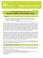
Figure 1. The life cycle of the European eel.
After hatching, presumably in the Sargasso Sea, cylindrical larvae develop into leafshaped leptocephalus larvae, which after
drifting on the Gulf Stream for approximately one year metamorphose into glass eels close to the European coast. The glass
eels may stay at the coast or migrate upriver, where they stay as juveniles (elvers and yellow eel) for many years (depending
on the region: males 4–6 years, females 8–12 years). Finally, they develop into migrating silver eels; the cause and timing of
silvering is not well understood. They mature during or after migration to the spawning grounds.
/>Its catadromous migratory behaviour, long life, serious habitat reduction, pollution, and overfishing may be amongst the causes of
the catastrophic collapse of the European eel population observed over the past decades [7]. So far, Anguilla species have resisted
efforts directed at efficient and sustainable artificial breeding [8]. As knowledge on the eel's genetic makeup is sparse, physiological
studies aimed at understanding maturation, reproduction and the sustenance of successive larval stages have not been able to
take full advantage of gene expression profiling. In order to alleviate this shortcoming, we have sequenced and assembled its
genome.
While the draft genome will be an important tool in reproduction research, it also offers new perspectives for fundamental studies in
eel biology, as well as a resource for the comparative interpretation of model fish genomes (e.g. zebrafish and medaka). Here, we
investigate its repertoire of Hox genes in a comparative genomics context.
The Hox genes encode transcription factors, which throughout the animal kingdom are involved in the developmental patterning of
the body plan. In vertebrates, Hox genes are tightly organized into clusters which exhibit colinearity between gene position and
temporal and spatial expression along the primary body axis: genes at the 3′ ends of clusters are expressed earlier in development,
and more anterior, than genes at the 5′ ends of clusters [9]. The organization of Hox clusters has been extensively documented for
many groups of vertebrates [10].
A. anguilla is a member of the superorder Elopomorpha [11], [12], a major teleost group of 856 species [13]. As such, elopomorphs
presumably share the inferred wholegenome duplication at the base of the teleost lineage [14], [15]. This teleostspecific genome
duplication (TSGD) event is most apparent when considering the Hox genes in extant species [10], [16], [17]. In tetrapods and
coelacanths, approximately forty genes are organized in four ancestral vertebrate clusters. In theory, teleosts could have retained
eight duplicate clusters. However, whereas tetrapod Hox loci are relatively stable, teleost genomes show dramatic gene loss, such
that all species examined in detail retain at most seven of these clusters, each with on average about half their original gene
content [9], [10]. A PCRbased survey of the Hox clusters of the Japanese eel A. japonica found evidence for the conservation of
eight clusters and 34 genes [18].
As the Elopomorpha represent an early branch on the teleost tree [12], the eel Hox gene complement may expose constraints on
the evolution of morphological complexity in teleost fish, and in vertebrates in general. Furthermore, analysis of the eel's Hox
clusters may shed light on the developmental mechanisms and evolutionary history of its life cycle and body plan. In particular, they
may provide evidence regarding the evolutionary novelty of the eel's indirect development.
Results
Genome assembly of the European eel
We have sequenced and assembled the genome of a female juvenile A. anguilla specimen caught in the brackish Lake Veere, the
Netherlands in December 2009. Its haploid genome size was determined to be 1.1 Gbp. Because of the impossibility of breeding A.
anguilla, no genetic linkage information is available. We therefore employed Illumina Genome Analyzer sequencing technology only
in the assembly of a draft genome. Based on a de novo genome assembly, we constructed 923 Mbp of scaffolds with a length
weighed median fragment length (N50) of 78 Kbp (Figure S1 and Table S1). An additional 179 Mbp of initial contigs, which are
either very small or highly repetitive, were excluded from scaffolding, but included in all further analyses.
Identification of Hox transcripts and genes
To identify A. anguilla Hox genes, we used a de novo assembled transcriptome of a 27 hours postfertilization (hpf) embryo of the
shortfinned eel, A. australis. This species is closely related to A. anguilla [19], yet produces viable embryos more easily [20]. We
compared Hoxlike sequences from the transcriptome to the genome assembly using Blast [21], which yielded ten genomic
scaffolds (Table S2) that were further examined for the presence of additional genes. This resulted in the identification of 73 Hox
genes (twice as many as found in A. japonica in a previous study using PCR fragments [18]), including three pseudogenes,
organized in eight clusters (Figure 2 and Table S3). The flanking regions of these eight clusters contain an additional 24 predicted
genes (Figure 2). No further proteincoding genes were found within the Hox clusters.
/>
2/12
5/2/2018
Primitive Duplicate Hox Clusters in the European Eel's Genome
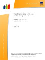
Figure 2. Genomic organization of the Hox gene clusters of the European eel.
Scaffolds are indicated by black lines and asterisks represent two gaps between scaffolds. Hox genes are indicated by
colored arrows that are numbered according to their paralogous groups. Three pseudogenes are indicated by the symbol ψ.
Neighboring genes are indicated by grey arrows. Conserved microRNA genes are indicated by triangles: miR196 (closed
triangles) and miR10 (open triangles).
/>Conserved microRNAs were discovered using Blast searches with human and zebrafish homologues (Figure 2). miR10 is present
posterior of Hox4 in six clusters (all except Aa and Ab). miR196 is found between Hox9 and Hox10 in five clusters (all except Bb,
Da and Db). This arrangement is consistent with that found in other vertebrates [22], [23].
Hox cluster identity
We based preliminary identification of clusters on homology between A. anguilla and Danio rerio protein sequences and
comparisons with all sequences in the NCBI nonredundant protein database (Table S3). Whereas the two A. anguilla HoxA
clusters can easily be matched to their corresponding HoxAa and HoxAb orthologues in D. rerio, each of the two HoxB and HoxC
clusters of A. anguilla most closely resembles D. rerio HoxBa and HoxCa, respectively. Both A. anguilla HoxD clusters predictably
match D. rerio HoxDa only, since the zebrafish HoxDb cluster has lost all proteincoding genes [24].
To more precisely assign the Hox genes to proper cluster orthologues, we generated unrooted maximum likelihood phylogenetic
trees for paralogous group 9 (Figures 3 and S2), of which A. anguilla possesses all eight copies. These confirmed the preliminary
classification in A, B, C and D paralogous groups, but failed to validate the identity of teleost a and b cluster duplicates (with the
exception of HoxAa and HoxAb). Likewise, phylogenetic trees based on multigene alignments do not conclusively indicate either a
or b cluster membership for HoxB, HoxC and HoxD (Figure 4). In general, there appears to be a lack of sequence divergence
between eel Hox gene duplicates, which makes classification based on coding sequence alone inaccurate.
Figure 3. Classification of the European eel Hox clusters.
/>
3/12
5/2/2018
Primitive Duplicate Hox Clusters in the European Eel's Genome
An unrooted phylogenetic tree showing the relationships between A. anguilla and fish Hox9 paralogues. Numbers indicate
bootstrap support.
/>
Figure 4. Phylogeny of Hox clusters of the European eel.
Unrooted phylogenetic trees based on alignments combining multiple Hox genes per cluster. A) Cluster A relationships, based
on HoxA9, HoxA11 and HoxA13 genes. B) Cluster B relationships, based on HoxB1, HoxB5 and HoxB6 genes. C) Cluster C
relationships, based on HoxC6, HoxC11, HoxC12 and HoxC13 genes. D) Cluster D relationships, based on HoxD4 and
HoxD9 genes. Species included: A. anguilla, Salmo salar (Atlantic salmon), Danio rerio (zebrafish), Oryzias latipes (medaka),
and Tetraodon nigroviridis (green spotted puffer). Asterisks indicate bootstrap support >90%.
/>Final orthologous relationships could only be established on the basis of conserved local synteny between Hox clusters and
flanking genes (Figure 5). In addition to both HoxA clusters, eel HoxBa and HoxBb appear orthologous with their respective teleost
equivalents. This identification is further supported by the absence of miR196 from both D. rerio and A. anguilla HoxBb clusters.
The affinities of HoxC and HoxD duplicates remain difficult to resolve because of conserved synteny around a and b cluster
duplicates, and extensive cluster reduction and deletion in other teleosts (Figure 5c, d).
Figure 5. Synteny around Hox clusters.
Conservation of flanking genes supports the classification of A. anguilla clusters into different orthologous subgroups. The eel
clusters and up to seven flanking genes are compared with the genomic organization in zebrafish (Danio rerio) and medaka
(Oryzias latipes). Coloured box outlines indicate preserved synteny between eel and the two other species, dotted outlines
denote flanking genes found on extended eel scaffolds (see Methods). Interpretation should take into account residual
synteny between a and b paralogous clusters. Limited data is available on HoxCb (lost in O. latipes, possibly misassembled in
D. rerio) and HoxDb (lost in D. rerio) clusters.
/>Hox gene expression
In order to confirm the transcriptional activity of the Hox genes, we determined relative expression levels by aligning transcriptomic
reads of the 27 hpf embryo against the Hox proteincoding regions (Figure 6a). Transcriptome reads mapped unambiguously to 71
out of 73 Hox genes, including one pseudogene (ψHoxD3b), suggesting that all A. anguilla Hox proteincoding genes are
functional. The relative expression levels vary over five orders of magnitude with the lowest expression observed for the posterior
paralogous groups 12 and 13, and the highest expression for paralogous groups 7–9, but with particularly high expression levels for
HoxB1a, HoxB1b, HoxB4b and HoxD1a.
/>
4/12
5/2/2018
Primitive Duplicate Hox Clusters in the European Eel's Genome
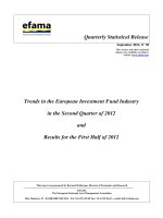
Figure 6. Hox gene expression in A. australis embryos.
A) mRNAseqbased gene expression in a 27 hpf embryo. B) Whole mount in situ hybridizations showing the expression of
HoxB9a, HoxD12b and HoxC13a. HoxB9a expression can be detected in 26 hpf (dorsal view) and 48 hpf (lateral tail region
view) embryos. HoxD12b and HoxC13a display expression in the tail region (lateral views) at 96 hpf, but not at 48 hpf. White
arrowheads indicate anterior expression boundaries. Scale bars correspond to 100 µm.
/>Full mRNAseq read alignment to the entire Hox clusters indicated that transcriptional activity is not restricted to protein coding
regions (Figure S3). In fact, intergenic expression sometimes exceeds intragenic levels, supporting the observation that complete
Hox clusters function as metagenes [9], [25].
At 27 hpf, expression of posterior Hox genes is very low (Figure 6a). We therefore confirmed transcriptional activity of posterior Hox
paralogues by whole mount in situ hybridizations (Figure 4b). HoxB9a is expressed at 26 and 48 hpf, with an anterior expression
boundary coinciding with somite number 5/6. Expression of HoxD12b and HoxC13a is not yet detectable at 48 hpf, but clearly
visible at 96 hpf with anterior expression boundaries located at somite numbers 65/70 and 90/95 for HoxD12b and HoxC13a,
respectively. For these Hox genes, expression in the eel embryo appears to conform to the expected spatiotemporal pattern
(colinearity between cluster organization and developmental timing and localization), with expression of Hox12 and Hox13
paralogues appearing later in development, and more posterior than Hox9.
The evolution of Hox cluster organization
The early branching of the Elopomorpha from the main teleost trunk allows a new reconstruction of ancestral Hox cluster
architectures (Figure 7), which are strongly constrained by the limited organizational divergence between eel HoxB, C and D
duplicates.
Figure 7. Model for the evolution of teleost Hox gene organization.
/>
5/12
5/2/2018
Primitive Duplicate Hox Clusters in the European Eel's Genome
Schematic Hox clusters [10], [26], [52] are superimposed on a species phylogeny with estimates of divergence times [53], [54]
– which vary considerably between studies [33]. Ancestral architectures are inferred on the basis of maximum parsimony, i.e.
the number of cluster duplications and gene loss events is minimized. Salmo salar (Atlantic salmon) has presumably lost
several duplicate clusters [26] (not shown). Deduced gene loss in a lineage is illustrated by a cross, question marks denote
uncertainty about cluster gene content in the preTSGD actinopterygian Polypterus senegalus (bichir). Arrows indicate the
possible origins of the leptocephalus body plan.
/>Since teleost fish are believed to have experienced the TSGD event early in their evolutionary history [14], [15], their genomes
should in theory possess up to eight cluster duplicates. However, all teleosts examined in detail retain at most seven clusters of
proteincoding genes [9]: a HoxC duplicate was lost in the lineage leading to medaka and pufferfish, a HoxD duplicate in the lineage
represented by zebrafish. The high number of clusters in salmon is the result of relatively recent further duplications [26].
The main teleost lineages diverged briefly after the TSGD [16]. The reconstruction in Figure 7 demonstrates that nearly all post
duplication gene loss events in the eel's ancestry occurred within this interval, followed by millions of years of stasis. Only the
HoxAb cluster appears to have accumulated major changes in prebranching, postgenome duplication teleosts. Alternative
hypotheses, in which a wholegenome duplication is not shared between elopomorphs and advanced teleosts, or in which the
genome duplication is followed by successive deletion and duplication of specific clusters in the eel, are less parsimonious and not
consistent with local conservation of synteny (Figure 5).
Discussion
Two rounds of Hox cluster duplications in chordates are believed to be responsible for important vertebrate novelties (e.g. brains,
heads, jaws) and increases in complexity [27]. A plausible mechanism invokes a temporary relaxation of metagenic cluster
constraints after duplication, paving the way for innovation [28], [29]. In contrast, the TSGDassociated third duplication of
vertebrate Hox clusters theoretically endowed teleost fish not with additional complexity within individuals, but with increased
prospects for morphological diversification between individuals and species [9], [10]. In support of this hypothesis, advanced
teleosts have extensively pruned their Hox surplus, leading to significant diversity in cluster structure (Figure 7). In all examined
representatives (with the exception of salmon [26]), the residual number of Hox genes is not much higher than the nonduplicated
count in tetrapods. The resulting teleost Hox cluster architectures have been interpreted as an evolutionary choice for
developmental flexibility in a tradeoff with robustness [9]. By proving that it is possible for a vertebrate to stably preserve eight
densely populated (Figure 2) and functional (Figure 6) Hox clusters, the eel genome presents an exception to these models, and a
third alternative in the evolution of vertebrate complexity.
For hundreds of millions of years, A. anguilla and its ancestors have maintained the highest ontogenic potential of any vertebrate,
indicative of continuous selective pressure. However, as adults, they do not display markedly more complex bodies than other fish
or tetrapods. The eel's distinctive life cycle and body plan suggest three (not mutually exclusive) explanations for this cryptic
complexity.
Hox genes are involved in the primary patterning of the body axis, which implies a functional role for A. anguilla's Hox surplus in
axial elongation. Alterations in Hox genes have been associated with elongated body plans [30], [31], however the changes
observed are of a regulatory nature, and do not involve extra genes. For example, elongation of the body axis in snakes has been
linked to a spatial relaxation in the posterior end of Hox clusters facilitated by the insertion of transposable elements between genes
[31]. In addition, even the elongate members of the Elopomorpha (which also includes nonelongated tarpons, bonefish and others)
display considerable diversity in the developmental mechanisms resulting in axial lengthening [32]. Hence, the eel's adult body plan
cannot explain the preservation of primitive Hox clusters between the TSGD (226–316 million years ago [33]) and the origin of the
genus Anguilla, estimated at 20–50 million years ago [19]. Similarly, if the European eel may at present experience singular
evolutionary forces because of its panmictic population [2], any explanation these offer does not extend beyond the genus Anguilla
of freshwater eels [34].
Even if for most of their lives eels are eelshaped, as oceandwelling larvae [35] their body plan is radically different (Figure 1). In
fact, until the late nineteenth century, these large, longlived, laterally compressed leptocephali were considered to be autonomous
pelagic species [36]. Fully transparent and slowly metabolizing, a leptocephalus provides considerable survival benefits [37], [38].
After approximately one year, they undergo a dramatic metamorphosis [39], including extensive tissue remodelling and shortening
of the body, resulting in cylindrical juveniles. In the early embryos investigated here (Figure 6), nearly all Hox genes are expressed
and presumably functionally involved in determining cell fate. Logically, a high gene and cluster count can be explained by the
assumption that the eel's two body plans are simultaneously outlined at this stage.
Leptocephali are the fundamental developmental innovation shared by all Elopomorpha [11]–[13], and therefore arose either before
or soon after the TSGD, or at the base of the lineage (arrows in Figure 7). The last alternative is the most parsimonious (no loss of
developmental complexity in advanced teleosts), especially since no member of the Elopomorpha is known to have ever discarded
the leptocephalous larval stage [11], [13]. Regardless, either of the postTSGD origins is compatible with an intercalation model of
indirect development [40], in which a temporary excess of developmental potential was permanently recruited for the conception of
an additional body plan. Although speculative, an explanation invoking the morphological challenges associated with a complex life
history is consistent with the stable high Hox gene and cluster count found in the anadromous Atlantic salmon [26].
Further functional studies on eel development will become possible once A. anguilla's life cycle can be completed in captivity. In
particular, there exists considerable variation in development (timing, number of somites) between leptocephali of related and
interbreeding Anguilla species [1], [35], which can only be studied when these larvae can be raised under controlled conditions [8],
[41].
Methods
/>
6/12
5/2/2018
Primitive Duplicate Hox Clusters in the European Eel's Genome
Eel embryos
Wild female and male silver shortfinned silver eels (A. australis) from Lake Ellesmere, New Zealand, were held together in a 2,300
L recirculation system with seawater (30 ppt salinity) at 21°C. Sexual maturation was induced as described [20]. Briefly, males
received nine weekly injections with 250 IU human chorionic gonadotropin and females were injected once a week with 20 mg
salmon pituitary extract. Eggs and milt were stripped and the eggs were dry fertilized. Embryos were reared in glass beakers with
UVsterilized seawater (35 ppt) at 21°C. At 26, 48 and 96 hpf embryos were fixed in 4% paraformaldehyde and stored in 100%
methanol.
Total RNA was isolated from 27 hpf embryos using the Qiagen miRNeasy kit according to the manufacturer's instructions (Qiagen
GmbH, Hilden, Germany), and analyzed with an Agilent Bioanalyzer 2100 total RNA Nano series II chip (Agilent, Santa Clara). A
transcriptome library was prepared from 10 µg total RNA, using the Illumina mRNASeq Sample Preparation Kit according to the
manufacturer's instructions (Illumina Inc., San Diego, USA).
Genome size determination
Blood samples taken from two eels (A. anguilla and A. australis) were washed with physiological salt and fixed in cold ethanol. Prior
to analysis the cells were collected, resuspended in physiological salt and stained with propidium iodide. After 30 minutes of
incubation the cells were analyzed by FACS, using human blood cells as a size reference (3.05 Gbp haploid). The eel genome size
was calculated by (human size)/(mean fluorescence human)×(mean fluorescence eel). Both Anguilla genomes were determined to
be 1.1 Gbp in size (haploid).
Genomic DNA libraries
Genomic DNA was isolated from blood of a female yellow European eel (A. anguilla, caught in Lake Veere, The Netherlands) using
the Qiagen Blood and Tissue DNeasy kit according to the manufacturer's description. Pairedend libraries were prepared from 5 µg
of isolated gDNA using the PairedEnd Sequencing Sample Prep kit according to the manufacturer's description. Either a 200 bp
band or a 600 bp band was cut from the gel (libraries PE200 and PE600, Table S1). After amplification for 10 cycles the resulting
libraries were analyzed with a Bioanalyzer 2100 DNA 1000 series II chip.
Mate pair libraries were prepared from 10 µg of isolated gDNA using the Mate Pair Library Prep Kit v2 (Illumina Inc.). Either a 3,000
bp band or a 10,000 bp band was cut from gel (libraries MP3K and MP10K, Table S1). After the first gel purification the fragment
length was analyzed using a Agilent Bioanalyzer 2100 DNA 12000 chip. After circularization, shearing, isolation of biotinylated
fragments, and amplification, the 400 to 600 bp fraction of the resulting fragments was isolated from gel. Finally, the libraries were
examined with an Agilent Bioanalyzer 2100 DNA 1000 series II chip.
Illumina sequencing
All libraries were sequenced using an Illumina GAIIx instrument according to the manufacturer's description. Genomic pairedend
libraries were sequenced with a read length of 2×76 nucleotides (to ∼20fold genome coverage), genomic matepair libraries with a
read length of 2×51 nucleotides (to ∼33fold genome span), and the mRNASeq library with a read length of 2×76 nucleotides
(Table S1). Image analysis and base calling was done by the Illumina pipeline.
Genome assembly
Sequencing reads from both pairedend libraries were used in building the initial contigs (Figure S1). Both sets were preprocessed
to eliminate low quality and adapter contamination. Whenever possible, PE200 pairs were merged into longer single reads. For
initial contig assembly, we employed the De Bruijn graphbased de novo assembler implemented in the CLC bio Genomics
Workbench version 3.6.5 (CLC bio, Aarhus, Denmark). A run with a kmer length of 25 nt resulted in an assembly a total length of
969 Mbp and a contig N50 of 1672 bp.
Initial contigs were oriented in larger supercontigs (scaffolds) using SSPACE [42]. In scaffolding the contigs, we decided to exclude
lowquality and highly repetitive contigs as much as possible. SSPACE was used in a hierarchical fashion, employing first links
obtained from the PE600 library to generate intermediate supercontigs, which were used as input for subsequent runs with links
from the MP3K and MP10K libraries, respectively. At each stage, a minimum of three nonredundant links was required to join two
contigs. This procedure resulted in a final scaffold set with a total length of 923 Mbp and an N50 of 77.8 Kbp (Table S1).
AUGUSTUS (version 2.4) was used to predict genes [43], which were provisionally annotated using Blast2GO (version 2.4.8) [44].
The draft assembly is available at www.eelgenome.org.
In order to obtain more information on flanking genes for the analysis of conserved synteny (Figure 5), scaffolds were subjected to
a further round of linking by SSPACE using reduced stringency (two instead of three nonredundant links required to join scaffolds).
This resulted in extended scaffolds with an N50 of 169 Kbp.
Hox genes
Hox contigs in the shortfinned eel embryonic transcriptome (generated using CLC bio's de novo assembler) were identified via
Blast [21] searches at the NCBI website (www.ncbi.nlm.nih.gov). European eel genomic scaffolds were annotated using CLC bio's
DNA Workbench. Remaining Hox genes and genes flanking the Hox clusters were identified using Blast, based on
AUGUSTUS/Blast2GO predictions. Annotated Hox scaffolds have been submitted to GenBank (accession numbers JF891391–
JF891400).
MicroRNAs were identified by Blast using H. sapiens and D. rerio miR10 and miR196 sequences (precursors and mature)
retrieved from miRBase release 18 (www.mirbase.org, [45]).
Phylogenetic methods
/>
7/12
5/2/2018
Primitive Duplicate Hox Clusters in the European Eel's Genome
Species and Hox gene accession numbers used are listed in Table S4. Amino acid sequences of Hox genes were aligned using
Clustal X [46] and checked manually. After excluding ambiguous alignments, ProtTest 2.4 [47] was used to choose an optimum
substitution model, based on the Akaike information criterion. The aligned sequences were subjected to maximum likelihood
analysis using RAxML version 7.2.6 [48] with 1000 rapid bootstrap replicates (f a option).
For the analysis of Hox9 genes (Figure 3), 70 aligned residues were used and analyzed using a JTT+I+Γ model [49]. All other
alignments were fitted using a JTT+Γ model. The multigene analyses of HoxA, HoxB, HoxC and HoxD (Figure 4) were based on
alignments of 427, 493, 935 and 308 amino acid residues, respectively. The phylogenetic trees of sarcopterygian and
actinopterygian Hox9 paralogues (Figure S2) were based on 151 (HoxA9), 210 (HoxB9), 248 (HoxC9), and 136 (HoxD9) residues.
Synteny was analyzed using D. rerio and O. latipes genomic contexts extracted from Ensembl release 65 (www.ensembl.org),
based on the Zv9 and MEDAKA1 genome assemblies, respectively (Table S5). Pairwise alignments were generated by NCBI
tblastx and analyzed using genoPlotR [50].
Whole mount in situ hybridization
Chromosomal DNA was isolated from A. australis blood using a DNeasy Blood & Tissue Kit (Qiagen). Riboprobe template
fragments, including a T7 RNA polymerase promoter, were PCR amplified from chromosomal DNA using the following primer sets:
HoxB9a forward (5′TGAAACCGAAGACCCGAC3′), HoxB9a reverse (5′GAAATTAATACGACTCACTATAGGGCTGAGGAAGACTC
CAA), HoxD12b forward (5′TAATCTTCTCAGTCCTGGCTATG3′), HoxD12b reverse (5′GAAATTAATACGACTCACTATAGATCCAA
GTTTGAAAATTCATATTTGC3′), HoxC13a forward (5′CACCTTGATGTACGTGTATGAAAA3′), HoxC13a reverse (5′GAAATTAAT
ACGACTCACTATAGGCTCCGTGTATTTCTCTGACG3′). Digoxygeninlabelled riboprobes were made according to standard
protocols using T7 RNA polymerase. Whole mount in situ hybridization with labelled riboprobes was performed at 70°C, according
to a slightly modified version of a standard protocol [51]. Hybridizing riboprobes were made visible using antiDigoxigenin AP and
BM Purple AP substrate (Roche). Stained embryos were bleached using hydrogen peroxide (SigmaAldrich) and photographed
using a Leica M205 FA stereo microscope.
Supporting Information
Figure S1.
Genome assembly pipeline. See Methods section for details.
/>(TIF)
Figure S2.
Unrooted maximum likelihood phylogenetic trees of actinopterygian and sarcopterygian Hox9 genes. See Methods section
for details. Sequences used are listed in Table S4.
/>(TIF)
Figure S3.
Metagenic expression of Hox clusters. mRNAseq reads of the A. australis embryo were aligned to entire Hoxcontaining
scaffolds, demonstrating large amounts of mRNA production from intronic and intergenic regions.
/>(TIF)
Table S1.
Statistics of European eel genome and shortfinned embryonic eel transcriptome.
/>(DOC)
Table S2.
Hox transcriptome contigs. All de novo assembled Hox contigs of a 27hour A. australis embryo map to ten A. anguilla genome
scaffolds.
/>(DOC)
Table S3.
A. anguilla Hox genes. Complete list of A. anguilla Hox genes, predicted protein sizes, matching A. australis embryo contigs and
best blastp hits.
/>(DOC)
Table S4.
Hox genes used in phylogeny reconstruction. List of the Hox gene sequences used in this study.
/>
/>
8/12
5/2/2018
Primitive Duplicate Hox Clusters in the European Eel's Genome
(DOC)
Table S5.
Hox clusters used in synteny analysis. Genomic locations of D. rerio and O. latipes Hox clusters. HoxCb is absent from O.
latipes, and HoxDb from D. rerio. However, the genomic loci can still be identified based on the presence of flanking gene
duplicates or conserved microRNA (D. rerio HoxDb).
/>(DOC)
Acknowledgments
We thank the following colleagues for generously contributing to our work: Bas Brittijn for production of shortfinned eel embryos,
Nabila Bardine and Tony Durston for help with in situ hybridizations, Tony Durston and Joost Woltering for critical reading of the
manuscript, Pieter Slijkerman for project financial management, Marten Boetzer and Walter Pirovano for help with genome
assembly.
Author Contributions
Conceived and designed the experiments: CVH EB RPD HPS SD FAW KT GEEJMvdT. Performed the experiments: EB DLdW
HJJ. Analyzed the data: CVH RPD YM. Wrote the paper: CVH RPD.
References
1. Tesch FW (2003) The eel. Oxford: Blackwell Publishing. FW Tesch2003The eelOxfordBlackwell Publishing
2. Als TD, Hansen MM, Maes GE, Castonguay M, Riemann L, et al. (2011) All roads lead to home: panmixia of the European eel in the Sargasso Sea. Mol
Ecol 20: 1333–1346.TD AlsMM HansenGE MaesM. CastonguayL. Riemann2011All roads lead to home: panmixia of the European eel in the Sargasso
Sea.Mol Ecol2013331346
View Article
PubMed/NCBI
Google Scholar
3. Avise JC, Nelson WS, Arnold J, Koehn RK, Williams GC, et al. (1990) The evolutionary genetic status of Icelandic eels. Evolution 44: 1254–1262.JC
AviseWS NelsonJ. ArnoldRK KoehnGC Williams1990The evolutionary genetic status of Icelandic eels.Evolution4412541262
View Article
PubMed/NCBI
Google Scholar
4. Schmidt J (1923) Breeding places and migration of the eel. Nature 111: 51–54.J. Schmidt1923Breeding places and migration of the eel.Nature1115154
View Article
PubMed/NCBI
Google Scholar
5. Aarestrup K, Okland F, Hansen MM, Righton D, Gargan P, et al. (2009) Oceanic spawning migration of the European eel (Anguilla anguilla). Science 325:
1660.K. AarestrupF. OklandMM HansenD. RightonP. Gargan2009Oceanic spawning migration of the European eel (Anguilla anguilla).Science3251660
View Article
PubMed/NCBI
Google Scholar
6. Van den Thillart G, Dufour S, Rankin JC, editors. (2009) Spawning migration of the European eel. Dordrecht: SpringerVerlag. G. Van den ThillartS.
DufourJC Rankin2009Spawning migration of the European eelDordrechtSpringerVerlag
7. Freyhof J, Kottelat M (2010) Anguilla anguilla. J. FreyhofM. Kottelat2010Anguilla anguilla.IUCN red list of threatened species, version 2010.4. Available:
. IUCN red list of threatened species, version 2010.4. Available: .
8. Tanaka H, Kagawa H, Ohta H, Unuma T, Nomura K (2003) The first production of glasseel in captivity: fish reproductive physiology facilitates great
progress in aquaculture. Fish Physiol Biochem 28: 493–497.H. TanakaH. KagawaH. OhtaT. UnumaK. Nomura2003The first production of glasseel in
captivity: fish reproductive physiology facilitates great progress in aquaculture.Fish Physiol Biochem28493497
View Article
PubMed/NCBI
Google Scholar
9. Duboule D (2007) The rise and fall of Hox gene clusters. Development 134: 2549–2560.D. Duboule2007The rise and fall of Hox gene
clusters.Development13425492560
View Article
PubMed/NCBI
Google Scholar
10. Kuraku S, Meyer A (2009) The evolution and maintenance of Hox gene clusters in vertebrates and the teleostspecific genome duplication. Int J Dev Biol
53: 765–773.S. KurakuA. Meyer2009The evolution and maintenance of Hox gene clusters in vertebrates and the teleostspecific genome duplication.Int
J Dev Biol53765773
View Article
PubMed/NCBI
Google Scholar
11. Greenwood PH, Rosen DE, Weitsman SH, Myers GS (1966) Phyletic studies of teleostean fishes, with a provisional classification of living forms. Bull
Am Mus Nat Hist 131: 339–456.PH GreenwoodDE RosenSH WeitsmanGS Myers1966Phyletic studies of teleostean fishes, with a provisional
classification of living forms.Bull Am Mus Nat Hist131339456
View Article
PubMed/NCBI
Google Scholar
/>
9/12
5/2/2018
Primitive Duplicate Hox Clusters in the European Eel's Genome
Inoue JG, Miya M, Tsukamoto K, Nishida M (2004) Mitogenomic evidence for the monophyly of elopomorph fishes (Teleostei) and the evolutionary origin
12. of the leptocephalus larva. Mol Phylogenet Evol 32: 274–286.JG InoueM. MiyaK. TsukamotoM. Nishida2004Mitogenomic evidence for the monophyly of
elopomorph fishes (Teleostei) and the evolutionary origin of the leptocephalus larva.Mol Phylogenet Evol32274286
View Article
PubMed/NCBI
Google Scholar
13. Nelson JS (2006) Fishes of the world, 4th edition. Hoboken: Wiley. JS Nelson2006Fishes of the world, 4th editionHobokenWiley
14. Jaillon O, Aury JM, Brunet F, Petit JL, StangeThomann N, et al. (2004) Genome duplicaton in the teleost fish Tetraodon nigroviridis reveals the early
vertebrate protokaryotype. Nature 431: 946–957.O. JaillonJM AuryF. BrunetJL PetitN. StangeThomann2004Genome duplicaton in the teleost fish
Tetraodon nigroviridis reveals the early vertebrate protokaryotype.Nature431946957
View Article
PubMed/NCBI
Google Scholar
15. Meyer A, Van de Peer Y (2005) From 2R to 3R: evidence for a fishspecific genome duplication (FSGD). BioEssays 27: 937–945.A. MeyerY. Van de
Peer2005From 2R to 3R: evidence for a fishspecific genome duplication (FSGD).BioEssays27937945
View Article
PubMed/NCBI
Google Scholar
16. Crow KD, Stadler PF, Lynch VJ, Amemiya C, Wagner GP (2006) The “fishspecific” Hox cluster duplication is coincident with the origin of teleosts. Mol
Biol Evol 23: 121–136.KD CrowPF StadlerVJ LynchC. AmemiyaGP Wagner2006The “fishspecific” Hox cluster duplication is coincident with the origin of
teleosts.Mol Biol Evol23121136
View Article
PubMed/NCBI
Google Scholar
17. Amores A, Force A, Yan YL, Joly L, Amemiya C, et al. (1999) Zebrafish hox clusters and vertebrate genome evolution. Science 282: 1711–1714.A.
AmoresA. ForceYL YanL. JolyC. Amemiya1999Zebrafish hox clusters and vertebrate genome evolution.Science28217111714
View Article
PubMed/NCBI
Google Scholar
18. Guo B, Gan X, He S (2010) Hox genes of the Japanese eel Anguilla japonica and Hox cluster evolution in teleosts. J Exp Zool (Mol Dev Evol) 314B:
135–147.B. GuoX. GanS. He2010Hox genes of the Japanese eel Anguilla japonica and Hox cluster evolution in teleosts.J Exp Zool (Mol Dev
Evol)314B135147
View Article
PubMed/NCBI
Google Scholar
19. Minegishi Y, Aoyama J, Inoue JG, Miya M, Nishida M, et al. (2005) Molecular phylogeny and evolution of the freshwater eels genus Anguilla based on
the whole mitochondrial genome sequences. Mol Phylogenet Evol 24: 134–146.Y. MinegishiJ. AoyamaJG InoueM. MiyaM. Nishida2005Molecular
phylogeny and evolution of the freshwater eels genus Anguilla based on the whole mitochondrial genome sequences.Mol Phylogenet Evol24134146
View Article
PubMed/NCBI
Google Scholar
20. Burgerhout E, Brittijn SA, Kurwie T, Decker P, Dirks RP, et al. (2011) First artificial hybrid of the eel species Anguilla australis and Anguilla Anguilla. BMC
Dev Biol 11: 16.E. BurgerhoutSA BrittijnT. KurwieP. DeckerRP Dirks2011First artificial hybrid of the eel species Anguilla australis and Anguilla
Anguilla.BMC Dev Biol1116
View Article
PubMed/NCBI
Google Scholar
21. Altschul SF, Gish W, Miller W, Myers EW, Lipman DJ (1990) Basic local alignment search tool. J Mol Biol 215: 403–410.SF AltschulW. GishW. MillerEW
MyersDJ Lipman1990Basic local alignment search tool.J Mol Biol215403410
View Article
PubMed/NCBI
Google Scholar
22. Yekta S, Tabin CJ, Bartel DP (2008) MicroRNAs in the Hox network: an apparent link to posterior prevalence. Nat Rev Genet 9: 789–796.S. YektaCJ
TabinDP Bartel2008MicroRNAs in the Hox network: an apparent link to posterior prevalence.Nat Rev Genet9789796
View Article
PubMed/NCBI
Google Scholar
23. He X, Yan YL, Eberhart JK, Herpin A, Wagner TU, et al. (2011) miR196 regulates axial patterning and pectoral appendage initiation. Dev Biol 357: 463–
477.X. HeYL YanJK EberhartA. HerpinTU Wagner2011miR196 regulates axial patterning and pectoral appendage initiation.Dev Biol357463477
View Article
PubMed/NCBI
Google Scholar
24. Woltering JM, Durston AJ (2006) The zebrafish hoxDb cluster has been reduced to a single microRNA. Nat Genet 38: 601–602.JM WolteringAJ
Durston2006The zebrafish hoxDb cluster has been reduced to a single microRNA.Nat Genet38601602
View Article
PubMed/NCBI
Google Scholar
25. Mainguy G, Koster J, Woltering J, Jansen H, Durston A (2007) Extensive polycistronism and antisense transcription in the mammalian Hox clusters.
PLoS ONE 2: e356.G. MainguyJ. KosterJ. WolteringH. JansenA. Durston2007Extensive polycistronism and antisense transcription in the mammalian
Hox clusters.PLoS ONE2e356
View Article
PubMed/NCBI
Google Scholar
26. Mungpakdee S, Seo HS, Angotzi AR, Dong X, et al. (2008) Differential evolution of the 13 Atlantic salmon Hox clusters. Mol Biol Evol 25: 1333–1343.S.
MungpakdeeHS SeoAR AngotziX. Dong2008Differential evolution of the 13 Atlantic salmon Hox clusters.Mol Biol Evol2513331343
View Article
PubMed/NCBI
Google Scholar
/>
10/12
5/2/2018
Primitive Duplicate Hox Clusters in the European Eel's Genome
27. Holland PW, GarciaFernàndez J, Williams SA, Sidow A (1994) Gene duplications and the origins of vertebrate development. Development Supplement
123–133.PW HollandJ. GarciaFernàndezSA WilliamsA. Sidow1994Gene duplications and the origins of vertebrate development.Development
Supplement123133
View Article
PubMed/NCBI
Google Scholar
28. Wagner GP, Amemiya C, Ruddle F (2003) Hox cluster duplications and the opportunity for evolutionary novelties. Proc Natl Acad Sci USA 100: 144603–
14606.GP WagnerC. AmemiyaF. Ruddle2003Hox cluster duplications and the opportunity for evolutionary novelties.Proc Natl Acad Sci
USA10014460314606
View Article
PubMed/NCBI
Google Scholar
29. Lynch VJ, Wagner GP (2008) Resurrecting the role of transcription factor change in developmental evolution. Evolution 62: 2131–2154.VJ LynchGP
Wagner2008Resurrecting the role of transcription factor change in developmental evolution.Evolution6221312154
View Article
PubMed/NCBI
Google Scholar
30. Woltering JM, Vonk FJ, Müller H, Bardine N, Tuduce IL, et al. (2009) Axial patterning in snakes and caecilians: evidence for an alternative interpretation
of the Hox code. Dev Biol 332: 82–89.JM WolteringFJ VonkH. MüllerN. BardineIL Tuduce2009Axial patterning in snakes and caecilians: evidence for an
alternative interpretation of the Hox code.Dev Biol3328289
View Article
PubMed/NCBI
Google Scholar
31. DiPoï N, MontoyaBurgos JI, Miller H, Pourquié O, Milinkovitch MC, et al. (2010) Changes in Hox genes' structure and function during the evolution of
the squamate body plan. Nature 464: 99–103.N. DiPoïJI MontoyaBurgosH. MillerO. PourquiéMC Milinkovitch2010Changes in Hox genes' structure and
function during the evolution of the squamate body plan.Nature46499103
View Article
PubMed/NCBI
Google Scholar
32. Mehta RS, Ward AB, Alfaro ME, Wainwright PC (2010) Elongation of the body in eels. Integr Comp Biol 50: 1091–1105.RS MehtaAB WardME AlfaroPC
Wainwright2010Elongation of the body in eels.Integr Comp Biol5010911105
View Article
PubMed/NCBI
Google Scholar
33. Hurley IA, Lockridge Mueller R, Dunn KA, Schmidt EJ, Friedman M, et al. (2007) A new timescale for rayfinned fish evolution. Proc R Soc B 274: 489–
498.IA HurleyR. Lockridge MuellerKA DunnEJ SchmidtM. Friedman2007A new timescale for rayfinned fish evolution.Proc R Soc B274489498
View Article
PubMed/NCBI
Google Scholar
34. Inoue JG, Miya M, Miller MJ, Sado T, Hanel R, et al. (2010) Deepocean origin of the freshwater eels. Biol Lett 6: 363–366.JG InoueM. MiyaMJ MillerT.
SadoR. Hanel2010Deepocean origin of the freshwater eels.Biol Lett6363366
View Article
PubMed/NCBI
Google Scholar
35. Miller MJ (2009) Ecology of anguilliform leptocephali: remarkable transparent fish larvae of the ocean surface layer. AquaBioSci Monogr (ABSM) 2, No
4: 1–94.MJ Miller2009Ecology of anguilliform leptocephali: remarkable transparent fish larvae of the ocean surface layer.AquaBioSci Monogr (ABSM)2,
No 4194
View Article
PubMed/NCBI
Google Scholar
36. Cunningham JT (1897) The discovery of the larva of the common eel. Nature 55: 467–468.JT Cunningham1897The discovery of the larva of the common
eel.Nature55467468
View Article
PubMed/NCBI
Google Scholar
37. Pfeiler E (1999) Developmental physiology of elopomorph leptocephali. Comp Biochem Physiol A 123: 113–128.E. Pfeiler1999Developmental physiology
of elopomorph leptocephali.Comp Biochem Physiol A123113128
View Article
PubMed/NCBI
Google Scholar
38. Bishop RE, Torres JJ (1999) Leptocephalus energetics: metabolism and excretion. J Exp Biol 202: 2485–2493.RE BishopJJ Torres1999Leptocephalus
energetics: metabolism and excretion.J Exp Biol20224852493
View Article
PubMed/NCBI
Google Scholar
39. Otake T (2003) Metamorphosis. In: Aida K, Tsukamoto K, Yamauchi K, editors. Eel biology. Tokyo: SpringerVerlag. pp. 61–74.T.
Otake2003Metamorphosis.K. AidaK. TsukamotoK. YamauchiEel biologyTokyoSpringerVerlag6174
40. Raff RA (2008) Origins of the other metazoan body plans: the evolution of larval forms. Phil Trans R Soc B 363: 1473–1479.RA Raff2008Origins of the
other metazoan body plans: the evolution of larval forms.Phil Trans R Soc B36314731479
View Article
PubMed/NCBI
Google Scholar
41. Oliveira K, Hable WE (2010) Artificial maturation, fertilization, and early development of the American eel (Anguilla rostrata). Can J Zool 88: 1121–
1128.K. OliveiraWE Hable2010Artificial maturation, fertilization, and early development of the American eel (Anguilla rostrata).Can J Zool8811211128
View Article
PubMed/NCBI
Google Scholar
42. Boetzer M, Henkel CV, Jansen HJ, Butler D, Pirovano W (2011) Scaffolding preassembled contigs using SSPACE. Bioinformatics 27: 578–579.M.
BoetzerCV HenkelHJ JansenD. ButlerW. Pirovano2011Scaffolding preassembled contigs using SSPACE.Bioinformatics27578579
/>
11/12
5/2/2018
Primitive Duplicate Hox Clusters in the European Eel's Genome
View Article
PubMed/NCBI
Google Scholar
43. Stanke M, Diekhans M, Baertsch R, Haussler D (2008) Using native and syntenically mapped cDNA alignments to improve de novo gene finding.
Bioinformatics 24: 637–644.M. StankeM. DiekhansR. BaertschD. Haussler2008Using native and syntenically mapped cDNA alignments to improve de
novo gene finding.Bioinformatics24637644
View Article
PubMed/NCBI
Google Scholar
44. Götz S, GarcíaGómez JM, Terol J, Williams TD, Nagaraj SH, et al. (2008) Highthroughput functional annotation and data mining with the Blast2GO
suite. Nucleic Acids Res 36: 3420–3435.S. GötzJM GarcíaGómezJ. TerolTD WilliamsSH Nagaraj2008Highthroughput functional annotation and data
mining with the Blast2GO suite.Nucleic Acids Res3634203435
View Article
PubMed/NCBI
Google Scholar
45. Kozomara A, GriffithsJones S (2011) miRBase: integrating microRNA annotation and deepsequencing data. Nucl Acids Res 39: D152–D157.A.
KozomaraS. GriffithsJones2011miRBase: integrating microRNA annotation and deepsequencing data.Nucl Acids Res39D152D157
View Article
PubMed/NCBI
Google Scholar
46. Larkin MA, Blackshields G, Brown NP, Chenna R, McGettigan PA, et al. (2007) Clustal W and Clustal X version 2.0. Bioinformatics 23: 2947–2948.MA
LarkinG. BlackshieldsNP BrownR. ChennaPA McGettigan2007Clustal W and Clustal X version 2.0.Bioinformatics2329472948
View Article
PubMed/NCBI
Google Scholar
47. Abascal F, Zardoya R, Posada D (2005) ProtTest: selection of bestfit models of protein evolution. Bioinformatics 21: 2104–2105.F. AbascalR. ZardoyaD.
Posada2005ProtTest: selection of bestfit models of protein evolution.Bioinformatics2121042105
View Article
PubMed/NCBI
Google Scholar
48. Stamatakis A, Hoover P, Rougemont J (2008) A rapid bootstrap algorithm for the RAxML Web servers. Syst Biol 57: 758–771.A. StamatakisP. HooverJ.
Rougemont2008A rapid bootstrap algorithm for the RAxML Web servers.Syst Biol57758771
View Article
PubMed/NCBI
Google Scholar
49. Jones DT, Taylor WR, Thornton JM (1992) The rapid generation of mutation data matrices from protein sequences. Comput Appl Biosci 8: 275–282.DT
JonesWR TaylorJM Thornton1992The rapid generation of mutation data matrices from protein sequences.Comput Appl Biosci8275282
View Article
PubMed/NCBI
Google Scholar
50. Guy L, Kultima JR, Andersson SG (2010) genoPlotR: comparative gene and genome visualization in R. Bioinformatics 26: 2334–2335.L. GuyJR
KultimaSG Andersson2010genoPlotR: comparative gene and genome visualization in R.Bioinformatics2623342335
View Article
PubMed/NCBI
Google Scholar
51. Hiroi J, Maruyama K, Kawazu K, Kaneko T, OhtaniKaneko R, et al. (2004) Structure and developmental expression of hatching enzyme genes of the
Japanese eel Anguilla japonica: an aspect of the evolution of fish hatching enzyme gene. Dev Genes Evol 214: 176–184.J. HiroiK. MaruyamaK.
KawazuT. KanekoR. OhtaniKaneko2004Structure and developmental expression of hatching enzyme genes of the Japanese eel Anguilla japonica: an
aspect of the evolution of fish hatching enzyme gene.Dev Genes Evol214176184
View Article
PubMed/NCBI
Google Scholar
52. Raincrow JD, Dewar K, Stocsits C, Prokasha SJ, Amemiya CT, et al. (2011) Hox clusters of the bichir (Actinopterygii, Polypterus senegalus) highlight
unique patterns in sequence evolution in gnathostome phylogeny. J Exp Zool (Mol Dev Evol) 316B: 451–464.JD RaincrowK. DewarC. StocsitsSJ
ProkashaCT Amemiya2011Hox clusters of the bichir (Actinopterygii, Polypterus senegalus) highlight unique patterns in sequence evolution in
gnathostome phylogeny.J Exp Zool (Mol Dev Evol)316B451464
View Article
PubMed/NCBI
Google Scholar
53. Inoue JG, Miya M, Venkatesh B, Nishida M (2005) The mitochondrial genome of Indonesian coelacanth Latimera menadoensis (Sarcopterygii:
Coelacanthiformes) and divergence time estimation between the two coelacanths. Gene 349: 227–235.JG InoueM. MiyaB. VenkateshM. Nishida2005The
mitochondrial genome of Indonesian coelacanth Latimera menadoensis (Sarcopterygii: Coelacanthiformes) and divergence time estimation between the
two coelacanths.Gene349227235
View Article
PubMed/NCBI
Google Scholar
54. Matschiner M, Hanel R, Salzburger W (2011) On the origin and trigger of the notothenioid adaptive radiation. PLoS ONE 6: e18911.M. MatschinerR.
HanelW. Salzburger2011On the origin and trigger of the notothenioid adaptive radiation.PLoS ONE6e18911
View Article
PubMed/NCBI
Google Scholar
/>
12/12