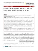
The aetiology, histopathology, and ultrastructural features of perianal erythema (red anus syndrome) in the european eel (anguilla anguilla)
Bạn đang xem bản rút gọn của tài liệu. Xem và tải ngay bản đầy đủ của tài liệu tại đây (849.54 KB, 7 trang )
5/2/2018
The Aetiology, Histopathology, and Ultrastructural Features of Perianal Erythema (Red Anus Syndrome) in the European Eel (Anguilla anguilla)
The Aetiology, Histopathology, and Ultrastructural Features of
Perianal Erythema (Red Anus Syndrome) in the European Eel
(Anguilla anguilla)
Omar A. S. Tamam
Published: March 7, 2014
/>
Abstract
The European eel (Anguilla anguilla) is a critically endangered species. Red anus syndrome (RAS) is known to be associated with
parasitic infections of the eel, particularly with Anguillicola crassus, but the full range of causative pathogenic organisms has not
been systematically investigated. Here we examined the infective organisms and histopathological and ultrastructural features of
seventy eels with RAS. In total, nine different pathogens were detected in association with RAS: Pseudomonas aeruginosa were
present in twelve specimens (17%), the metacercaria of Euclinostromum heterostomum in three cases (4%), Gastrostome
(Bucephalidae family) in seven cases (10%), A. crassus in fortyfive cases (64%), Bothriocephalus in seventeen cases (24%), and
Proteocephalus in twentythree cases (32%). Yeast, amoeba, and myxoboluslike pathogens were seen in the anal skin in all cases
when examined in combination with electron microscopy. Histopathologically, the lesions appeared as anoproctitis of varying
severity from mild anusitis to severe haemorrhagic anoproctitis, with severe perianal oedema, haemorrhage, and proctoptosis. Gut
inflammation ranged from mild catarrhal enteritis to severe haemorrhagic enteritis with mucosal sloughing. RAS is associated with a
range of parasitic infections, not only A. crassus, some of which we describe here for the first time. Since RAS is not associated
with direct invasion by parasites, it is likely that RAS is a secondary phenomenon caused by superadded infection on a background
of generalised immunosuppression, or indirect local toxic effects. RAS may be used as a noninvasive indicator of underlying
parasitic infection, but further investigations are required to establish the causative organisms for effective fishery management.
Citation: Tamam OAS (2014) The Aetiology, Histopathology, and Ultrastructural Features of Perianal Erythema (Red Anus
Syndrome) in the European Eel (Anguilla anguilla). PLoS ONE 9(3): e90790. />Editor: Pikul Jiravanichpaisal, National Science and Technology Development Agency, Thailand
Received: May 21, 2013; Accepted: February 4, 2014; Published: March 7, 2014
Copyright: © 2014 Omar A. S. Tamam. This is an openaccess article distributed under the terms of the Creative Commons
Attribution License, which permits unrestricted use, distribution, and reproduction in any medium, provided the original author
and source are credited.
Funding: The author has no support or funding to report.
Competing interests: The author has declared that no competing interests exist.
Introduction
The European eel, Anguilla anguilla, is common to the waters of Europe, Asia, and the Mediterranean basin. The European eel has
undergone a dramatic decline in population since the 1970s due to overfishing, pollution, habitat loss, and susceptibility to infection
[1]; consequently, the European eel is a critically endangered species. The eels remain an important source of economic value to
many communities and they are an important source of food. Therefore, understanding the factors that might influence or be useful
to sustainable fishing practices remains an important area of study.
Perianal erythema, also termed the ‘red anus syndrome’ (RAS), is a recognised external morphological manifestation of eel
pathology, in particular infection with parasites [2]. Perianal erythema is best described in association with infection with the
swimbladder nematode Anguillicolla crassus, which causes retarded growth, increased susceptibility to other disease, and, in
severe cases, death of the fish [3]. In one study, A. crassus was present in as many as 80% of eels with perianal discoloration, and
therefore this feature alone may be a useful nondiagnostic tool for use in fisheries management [2].
However, perianal erythema has also been described in association with other eel infections, such as the nematode Camallanus
lacustris, and therefore the feature is unlikely to be specific for one particular infection [4]. We therefore sought to undertake a
systematic analysis of the organisms associated with RAS in a large number of European eels with anal discoloration from the
Domiat Coast region of Egypt. In addition, we comprehensively examined the histopathological and ultrastructural features of the
gastrointestinal tracts of eels with RAS, in order to better understand the pathogenesis of this collection of diseases. Together,
these analyses provide a valuable contribution to understanding the causes and effects of eel pathologies, so that fisheries can
better formulate management strategies for sustainable aquaculture.
/>
1/7
5/2/2018
The Aetiology, Histopathology, and Ultrastructural Features of Perianal Erythema (Red Anus Syndrome) in the European Eel (Anguilla anguilla)
Methods
Ethics statement
Although A. anguilla is a critically endangered species, there remains widespread legal fishing activity for human food consumption
in Egypt and worldwide; hence the need to conduct this study to guide effective fishery management and help restore stocks. The
eels were obtained from fish market on their way for human consumption as part of the legal eel fishing industry in Egypt. The eels
were euthanized by decapitation by fishermen working under the regulated authority of the General Authority for Fish Resources
Development, Egypt. Since the study did not use laboratory animals, was performed as part of the investigation of natural disease,
and used animals already in the food chain, no ethical approval from an animal ethics committee was required.
Fish samples and histopathology
Seventy live European eels with RAS were obtained from fish market between March 2010 and December 2011; 0–3 fish were
examined during any one period. Eels originated from the Domiat region of the Nile Delta in Egypt. Only fish with visible perianal
erythema were selected for examination (see Figure 1). The selected fish were characterized by the presence of either red, reddish
brown, or grey zones around the anal opening; in addition, anal prolapse was apparent in some eels.
Figure 1. The appearances of the eel anal opening.
Normal (A), different degrees of perianal erythema (B–G) with the presence of a blood worm (Chironomid midge larva) on the
skin of the eel in sample (F). Perianal oedema and proctoptosis (H–J) and perianal black pigmentation (K, L).
/>Each specimen underwent a full postmortem examination, as described previously [5]. In particular, the intestine was opened
longitudinally for detailed examination. Samples from the anal opening, rectum, large intestine, small intestine, and swim bladder
were immediately fixed in 10% phosphatebuffered formalin. Fixed tissues were processed using standard fish histology protocols,
sectioned at 5 µm, and stained with haematoxylin and eosin and periodic acid Schiff (PAS) for subsequent microscopic analysis
and photomicrography using a digital camera [6].
Scanning electron microscopy (SEM)
Three 0.5 cm thick fresh tissue samples were taken from the anal opening to include the rectal mucosa and placed in 5% cold
buffered glutaraldehyde for two days. Samples were then washed in cacodylated buffer for thirteen minutes three times, postfixed
in 1% osmium tetroxide for two hours, and then dehydrated in a graded ethanol series (30%, 50%, 70%, and 90%) over a period of
two hours. Samples were left in 100% ethanol for two days, and then amyl acetate for a further two days. Finally, liquid carbon
dioxide was used to achieve criticalpoint drying, samples were stuck to metallic blocks using silver paint in a sputtering apparatus,
and samples were evenly coated with gold (15 nm). SEM was performed using the JEOL JSM 5400 LV scanning electron
microscope at 15 kV.
Microbiological examination
Swabs were obtained from the anal opening using aseptic technique and were cultured in nutrient broth for one day then culture on
blood agar in order to culture and detect pathogenic bacteria. Colonies were picked and restreaked onto new plates of
Pseudomonas isolation agar as described in [7]. The bacterial isolates were identified according to the biochemical reaction
scheme provided by Finegold and Martin [8]. Fungal organisms were assessed using light microscopy.
Parasitological examination
The intestine was opened longitudinally and its contents examined for parasites using a digital stereomicroscope. Live parasites
were collected from the intestines of infected eels and fixed and processed using standard methods [9], [10]. Identification was
made on wholemount sections according to the criteria shown in Table 1.
/>
2/7
5/2/2018
The Aetiology, Histopathology, and Ultrastructural Features of Perianal Erythema (Red Anus Syndrome) in the European Eel (Anguilla anguilla)
Table 1. Criteria used for the identification of the main parasites seen in the eels.
/>
Results
Gross external examination and diagnosis of red anus syndrome
The normal eel anus is identified as a small, punctate depression on the underside of the fish. A healthy specimen will show no
signs of erythema or swelling (Figure 1A), with anal colour the same as the lateral surface skin colour. In fish with red anus
syndrome, a range of appearances is apparent, depending on severity. In mild lesions there were telangiectatic or punctate
erythematous changes confined to the edge of the anal orifice (Figure 1 B–G). In more advanced cases, there was marked perianal
oedema, haemorrhage, and proctoptosis (Figure 1 H–J). Chronic lesions were identified by brownblack discoloration and the anal
tissue acquired a hard texture with irregular, but well defined, borders (Figure 1 (K, L). Although no ectoparasites were observed,
occasional blood worms (chironomid midge larvae) were present and adherent to the eel skin as nonpathogenic water
contaminants (Figure 1F).
Parasites and bacteria associated with red anus syndrome
Pseudomonas aeruginosa organisms were isolated from the anal smears of twelve specimens (17%). This pathogen was always
associated with severe anoproctitis. No other pathogenic bacteria were detected.
In total, five different metazoal parasite species were detected. The metacercariae of Euclinostomum heterostomum were detected
in the intestinal lumen in three cases (4%; Figure 2A); Gastrostome, a member of the Bucephalidae family, in seven cases (10%;
Figure 2B); A. crassus in 45 cases (64%; Figure 2 C, D); Bothriocephalus in 17 cases (24%; Figure 2 E, G, I); and Proteocephalus
in 23 cases (32%; Figure 2 F and H). Yeast, amoeba, and myxoboluslike pathogens were noted in all cases. Myxosporeanlike
parasite cysts were recorded in all cases, which mainly invaded the perianal subcutaneous tissues (Figure 2J).
Figure 2. The parasites found in the eels.
(A) Metacercaria of Euclinostromum heterostomum, (B) Gastrostome, a member of Bucephalidae, (C) Dissected swimbladder
and Anguillicola crassus, (D) Swimbladder fluid containing numerous Anguillicola crassus larvae, (E) Bothriocephalus scolex,
(F) Proteocephalus scolex, (G) Bothriocephalus gravid segment, (H) Proteocephalus mature segment, (I) Bothriocephalus
mature segment, (J) Myxoboluslike cysts invading the skin.
/>Mixed infections were common, as can be seen in Figure 3. In all cases, amoeba, myxoboluslike organisms, and yeast were
detected using the combination of techniques. A. crassus was associated with other infections in nearly 50% (22/45) of cases;
Bothriocephalus were found in seven cases, Pseudomonas aeruginosa in five cases, and Proteocephalus in ten cases.
Proteocephalus and Gastrostome infections were present together (as coinfections) in two eels, and Euclinostomum, in itself a
rare disease, always occurred in isolation (i.e., with no other concurrent infection).
/>
3/7
5/2/2018
The Aetiology, Histopathology, and Ultrastructural Features of Perianal Erythema (Red Anus Syndrome) in the European Eel (Anguilla anguilla)
Figure 3. Red anus syndrome is complex and multifactorial.
This diagram shows the distribution of the different infective organisms, and highlights that infection with 3 or more organisms
is not uncommon. The intensity of parasitic infection in the fish is indicated by + <3, ++ 3–10, +++ >10 parasites per host.
/>Histopathologic features of red anus syndrome
In the small intestine, mild catarrhal enteritis was observed in fish with trematode infection; this may have been due to the
trematodes themselves or one of the coinfections. Small intestinal villi were covered with mucus, inflammatory cells, and epithelial
cell debris from excessive sloughing. In fish with cestodal infection, there was severe haemorrhagic enteritis, with sloughing of the
mucosal epithelium, severe congestion of submucosal blood vessels, and a proteinrich exudate containing active chronic
inflammatory cells including macrophages, lymphocytes, and eosinophil granulocytes the surface (Figure 4B). Necrotic changes
involved the mucosa and extended into the submucosa.
Figure 4. Photomicrographs of the intestine of an eel.
(A) normal (H&E x10). (B) Cross section of eel intestine with specimens of Protocephalus cestode free in the lumen (P). An
apical erosion of a villus and complete sloughing of the intestinal mucosa can be seen (arrows), with severe degeneration and
necrotic changes in the mucosa and submucosa (H&E x40). (C, D) Cross section of eel anal opening showing sloughing of
the anal epithelium (arrow) with bleeding and congested vessels (H&E x20). (E) Submucosa shows dilated congested
capillaries (H&E x40). (F) Severe oedema separating the connective tissue fibres, inflammatory infiltrate, and red blood cells
in the anal mucosa (x40) (G) Longitudinal section through the anal opening, with small milletsized nodules containing a large
number of Myxobolus species (H&E x10), and arrows indicating focal necrosis. (H) Anal skin showing severe degenerative
and necrotic changes, mucus, and club cells (arrows) of the epidermis with oedema and an inflammatory cell infiltrate (H&E
x40). (I) Tissue rupture and degeneration of the swimbladder, containing numerous larvae. The larvae are surrounded by cell
debris from the necrotic submucosa (H&E x10).
/>In the large intestine in the majority of cases, but in particular Proteocephalus infestation with Pseudomonas infection,
demonstrated massive epithelial and mucosal necrosis, submucosal oedema, widespread haemorrhage, and an associated active
chronic inflammatory cell infiltrate.
At the anal opening there was clear evidence of anusitis. There was sloughing of the anal canal epithelium with rupture of
superficial capillaries (Figure 4C and D). Submucosal capillaries were ectatic and congested (Figure 4E), and the lamina propria
was oedematous with extravasation of red blood cells and separation of the connective tissue fibres (Figure 4F). Myxoboluslike
cysts were seen invading the perianal dermis to cause focal necrosis (Figure 4G), with the skin itself showing severe degenerative
and necrotic changes in the epithelium with ‘club cells’ and an associated inflammatory exudate (Figure 4H).
The changes in the swimbladder were only seen with anguillicolosis, as expected and previously described [11]. There was
widespread extravasation of red blood cells. In some cases, there was also evidence of old haemorrhage in the form of
hemosiderin deposition, and eosinophils were occasionally seen. Due to the presence of exudate, the muscle layer was loose with
separated the muscle fibres and dilated capillaries. The submucosa of the swim bladder contained numerous larvae, which were
surrounded by cell debris from the necrotic and degenerate submucosa (Figure 4I).
Electron microscopic features of red anus syndrome
/>
4/7
5/2/2018
The Aetiology, Histopathology, and Ultrastructural Features of Perianal Erythema (Red Anus Syndrome) in the European Eel (Anguilla anguilla)
A wide spectrum of anorectal mucosal abnormalities were observed using SEM, as shown in Figure 5A–F, and the technique was
particularly useful for revealing the 3dimensional view of the surface environment. In areas without observable lesions, the mucosa
was not clearly visible when viewed at low magnification due to an intact mucus layer (Figure 5A). However, at higher
magnification, spherical yeast cells and cocci were clearly visible in the mucus covering the intestinal epithelium. In areas with
visible abnormalities, the mucosal organization was altered due to the presence of mixed infection with yeast, bacteria, and
amoeba demonstrating characteristic pseudopodia and membrane blebbing (Figure 5B). Yeast cells were intimately associated with
the intestinal mucosa (Figure 5C). The glandular borders were oedematous, and the gland lumen was dilated due to epithelial loss
(Figure 5D). In addition, the enterocytes were covered with a proteinrich pseudomembrane containing material composed of a
network of filaments with a fibrinlike structure (Figure 5E). When condensed and dehydrated, this material was most apparent as
fibrous strands (Figure 5F).
Figure 5. Scanning electron micrographs of the lower intestine of an eel with anal redness.
Enterocytes are not clearly visible due to mucus coating (M). (A) Spherical yeast cells and cocci (arrow) captured in the
mucus covering the intestinal epithelium. (B) Typical view of mixed infection by yeast, bacteria, and amoebae with
characteristic pseudopodia and membrane blebbing (arrow) (C) Close association of yeast cells and intestinal mucosa. (D)
Derangement of the enterocytes, especially on the right as the cell borders were not clearly defined with epithelial loss. (E)
The surface is partially covered with mucus and a fibrinoid pseudomembrane. (F) The enterocytes are covered with mucus
fibrinoid pseudomembrane, which after dehydration appear as fibrous strands of material.
/>
Discussion
This study is the first comprehensive analysis of the aetiology, histopathology, and ultrastructural features of European eels with
RAS. As expected, A. crassus was frequently observed, followed by Proteocephalus (32%), Bothriocephalus (24%), Gastrostome
(10%), and Euclinostomum heterostomum (4%). All eels were infected with more than one organism, confirming that RAS is a
manifestation of a number of different infections that frequently coexist.
Although RAS has previously been described in association with A. crassus, other organisms have also been implicated in the
pathogenesis. The nematode Camallanus lacustris has also specifically been described in association with RAS [4], while others
have described the phenomenon in more general terms; Van Banning and Haenen attributed RAS to bacterial infection [12], while
Rodjunk and Shelenkova reported that RAS is secondary to parasite–induced enteritis, without naming specific organisms [13].
Here, for the first time, we describe RAS in association with Euclinostomum heterostomum, Gastrostome, and Bothriocephalus.
Other parasites, such as Myxdium spp., Trichodina spp, Dactylogyrus pseudodactylogyrus, Anguillicola cruses, and Pseudoergostla
have all been described in European eel in Egyptian waters, but these were not observed in association with RAS in this study [11].
Most strikingly, nearly 50% of eels with A. crassus infection also harboured another specific infection, demonstrating the complex
and multifactorial aetiology of RAS. RAS is certainly not specific for A. crassus, and fisheries need to perform more specific tests if
the causative organisms need to be established for management decisions.
In addition to the metazoal species identified, 17% of cases were shown to harbour P. aeruginosa by microbiologic culture; no other
bacterial species were identified on routine culture. Gramnegative infections, including Pseudomonas, are not uncommon in
warmwater fish and in order to successfully colonise and be pathogenic must overcome host defences, as appears to have been
the case here [14], [15]. Myxobolus species have been described in the European eel since the early 1900s and are a common
infection of the fish over a wide range of geographic locations from Europe to Japan, but poor morphologic and taxonomic
characterization makes subclassification difficult; hence the term ‘myxoboluslike’ is used in this paper [16], [17]. Although the
specific types of amoebae and yeast were not formally identified in this study, various types of amoebae are common secondary
infections in fish [4] and fungal infections, including with yeasts, have been reported to be pathogenic in eels in fish farms [18].
However, to my knowledge this is the first description of these organisms in association with RAS.
The prevalence of A. crassus (64%) was entirely consistent with the previously described association with RAS [2], and at the high
end of the prevalence reported for the general eel population (from 11% to 80%), as would be expected from a population enriched
in disease [19], [20]. A. crassus causes damage to the swimbladder by sucking blood from its wall, causing direct damage to the
mucosa as seen in this study [21], [22]. The mechanism of perianal erythema, which is distant from the primary site of infection, is
less well understood. It has been suggested that the red and swollen anus is due the release of eggs and larvae to the intestine via
the pneumatic duct, which then induce severe inflammation, or due to bacterial infections in the posterior region of the abdomen
[12]. Our SEM data suggest that the perianal erythema is not directly associated with the presence of eggs or larvae of A. crassus,
which were not present in the perianal skin, but is due to superadded infection with amoeba, fungi, and myxoboluslike organisms,
which were evident in every case. It therefore appears that the perianal erythema induced by A. crassus is a secondary
phenomenon, either due to generalised immune compromise, or local mucosal weakness cause by luminal toxins or organisms.
Although the parasitic infections affecting the European eel have been extensively studied, the histopathological features of the
damage they cause are less well described. Abdelnomen et al. recently presented a comprehensive overview of the distribution and
histological features of European eel infected with parasites, and described similar features of A. crassus infection of acute on
chronic inflammation of the swimbladder with erosion and ulceration in the most severe cases [11], [22]. Likewise, the other RAS
/>
5/7
5/2/2018
The Aetiology, Histopathology, and Ultrastructural Features of Perianal Erythema (Red Anus Syndrome) in the European Eel (Anguilla anguilla)
associated organism detected in their study, Proteocephalus spp., showed features of mild enteritis. Even when infected with the
same organism, European eels show a spectrum of pathological abnormalities; for instance, in one study, infections with A. crassus
resulted in pathological abnormalities in the swimbladder in only 28% of infected cases [19]. Since all eels with RAS and A. crassus
had visible pathological abnormalities in this study, it may be that RAS only becomes associated with underlying parasitic infection
in the most severe cases.
Conclusions
Red anus syndrome commonly affects European eels. Far from only being associated with A. crassus infection, RAS is associated
with a range of infections, some of which we describe here for the first time. Parasitic infections cause a range of severity of
enteritis, but most strikingly the ultrastructural studies reveal that perianal erythema is always associated with superadded fungal,
bacterial, and amoebic infections. It is therefore likely that RAS is a secondary phenomenon caused either by generalised
immunosuppression, or indirect local toxic effects caused by the presence of parasites that allow superadded infection to occur.
RAS may be used as a noninvasive indicator of underlying parasitic infection, but further investigations are required to establish
the causative organisms for effective fishery management.
Acknowledgments
The author is grateful to Professor Alam Nafadie, Professor Adel Peker, Professor H. M. Elsheikha, and Dr Hilda Tresz for their
technical assistance and scientific collaboration. The author would like to gratefully acknowledge editorial assistance from
Nextgenediting (www.nextgenediting.com).
Author Contributions
Conceived and designed the experiments: OAST. Performed the experiments: OAST. Analyzed the data: OAST. Contributed
reagents/materials/analysis tools: OAST. Wrote the paper: OAST.
References
1. Kennedy CR (2007) The pathogenic helminth parasites of eels. J Fish Dis 30: 319–334.
View Article
PubMed/NCBI
Google Scholar
2. Crean SR, Dick JTA, Evans DW, Elwood RW, Rosell RS (2003) Anal redness in European eels as an indicator of infection by the swimbladder nematode,
Anguillicola crassus. J Fish Biol 62: 482–485.
View Article
PubMed/NCBI
Google Scholar
3. Kennedy CR, Fitch DJ (1990) Colonization, larval survival and epidemiology of the nematode Anguillicola crassus, parasitic in the eel, Anguilla anguilla,
in Britain. J Fish Biol 36: 117–131.
View Article
PubMed/NCBI
Google Scholar
4. Woo PTK, Bruno DW, Leatherland JF (1995) Fish Diseases and Disorders: Volume 1: Protozoan and Metazoan Infections: CABI.
5. Lucký Z (1977) Methods for the Diagnosis of Fish Diseases: Amerind Pub.
6. Culling CFA (1974) Handbook of histopathological techniques, including museum techniques.
7. Buller NB (2004) Bacteria from fish and other aquatic animals [electronic resource]: a practical identification manual: CABI.
8. Fingold S, Martin W (1982) Bailey and Scott's diagnostic microbiology. Mosby, St Louis: 128–143.
9. Fernando CH (1972) Methods for the Study of Freshwater Fish Parasites: Department of Biology, University of Waterloo.
10. Roberts RJ (2012) Fish Pathology: Wiley.
11. Abdelmonem AA, Metwally MM, Hussein HS, Elsheikha HM (2010) Gross and microscopic pathological changes associated with parasitic infection in
European eel (Anguilla anguilla, Linnaeus 1758). Parasitol Res 106: 463–469.
View Article
PubMed/NCBI
Google Scholar
12. Banning PV, Haenen O, Perkins F, Cheng T (1990) Effects of the swimbladder nematode Anguillicola crassus in wild and farmed eel, Anguilla anguilla.
Academic Press Inc.: 317–330.
13. Rodjuk G, Shelenkova O (2006) Parasite fauna of the European eel (Anguilla anguilla L, 1758) from the Russian part of the Vistula Lagoon (Baltic Sea).
Wiad Parazytol 52: 121–125.
View Article
PubMed/NCBI
Google Scholar
14. Thune RL, Stanley LA, Cooper RK (1993) Pathogenesis of gramnegative bacterial infections in warmwater fish. Annual Review of Fish Diseases 3: 37–
68.
View Article
PubMed/NCBI
Google Scholar
/>
6/7
5/2/2018
The Aetiology, Histopathology, and Ultrastructural Features of Perianal Erythema (Red Anus Syndrome) in the European Eel (Anguilla anguilla)
15. Trust TJ (1986) Pathogenesis of Infectious Diseases of Fish. Annual Review of Microbiology 40: 479–502.
View Article
PubMed/NCBI
Google Scholar
16. Copland JW (1982) Myxobolus dermatobia (Ishii, 1915) infection of wild eels, Anguilla anguilla L., in England. Journal of Fish Diseases 5: 549–552.
View Article
PubMed/NCBI
Google Scholar
17. Molnár K, Lom J, Malik E (1986) A skin disease of the eels caused by Myxobolus kotlami n. sp. Journal of Applied Ichthyology 2: 42–48.
View Article
PubMed/NCBI
Google Scholar
18. Mellergaard S, Dalsgaard I (1987) Disease problems in Danish eel farms. Aquaculture 67: 139–146.
View Article
PubMed/NCBI
Google Scholar
19. Wurtz J, Knopf K, Taraschewski H (1998) Distribution and prevalence of Anguillicola crassus (Nematoda) in eels Anguilla anguilla of the rivers Rhine and
Naab, Germany. Dis Aquat Organ 32: 137–143.
View Article
PubMed/NCBI
Google Scholar
20. Pilcher MW, Moore JF (1993) Distribution and prevalence of Anguillicola crassus in eels from the tidal Thames catchmentb. J Fish Biol 43: 339–344.
View Article
PubMed/NCBI
Google Scholar
21. Höglund J, Andersson J, Härdig J (1992) Haematological responses in the European eel, Anguilla anguilla L., to sublethal infestation by Anguillicola
crassus in a thermal effluent of the Swedish Baltic. J Fish Dis 15: 507–514.
View Article
PubMed/NCBI
Google Scholar
22. Wurtz J, Taraschewski H (2000) Histopathological changes in the swimbladder wall of the European eel Anguilla anguilla due to infections with
Anguillicola crassus. Dis Aquat Organ 39: 121–134.
View Article
PubMed/NCBI
Google Scholar
23. Jhansilakshmibai K, Madhavi R (1997) Euclinostomum heterostomum (Rudolphi, 1809)(Trematoda): lifecycle, growth and development of the
metacercaria and adult. Systematic Parasitology 38: 51–64.
View Article
PubMed/NCBI
Google Scholar
24. Yamaguti S (1971) Synopsis of digenetic trematodes of vertebrates. Vols I and II. Synopsis of digenetic trematodes of vertebrates Vols I and II.
25. Wardle RA, McLeod JA (1952) The zoology of tapeworms. The zoology of tapeworms.
26. Scholz T, Drábek R, Hanzelová V (1998) Scolex morphology of Proteocephalus tapeworms (Cestoda: Proteocephalidae), parasites of freshwater fish in
the Palaearctic Region. Folia Parasitologica 45: 27–43.
View Article
PubMed/NCBI
Google Scholar
27. Hedrick RP, ElMatbouli M, Bartholomew J, Wilson J (2002) Review: recent advances with taxonomy, life cycle, and development of Myxobolus
cerebralis in the fish and oligochaete hosts. American Fisheries Society: 45–54.
/>
7/7