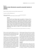
CHARACTERISTICS OF MENINGITIS CAUSED BY ESCHERICHIA COLI IN CHILDREN OLDER THAN ONE MONTH
Bạn đang xem bản rút gọn của tài liệu. Xem và tải ngay bản đầy đủ của tài liệu tại đây (1.82 MB, 31 trang )
CHARACTERISTICS OF MENINGITIS CAUSED BY
ESCHERICHIA COLI IN CHILDREN OLDER THAN ONE MONTH
IN THE INFECTIOUS DISEASE WARD OF CHILDREN’S HOSPITAL 1
FROM 2013 TO 2018
Nguyễn Hoàng Thiên Hương, Nguyễn An Nghĩa
Dư Tuấn Quy, Trương Hữu Khanh
1
OUTLINE
1. INTRODUCTION
2. MATERIALS AND METHODS
3. RESULTS AND DISCUSSION
4. CONCLUSION
5. SUGGESTION
2
1. INTRODUCTION
This
was conducted
to answer
the question:
▪ E.
colistudy
meningitis
neonates
(premature/low
birth weight) and
What were the features of E. coli meningitis in children >1 month of age in
infants (with/without risk factors).
Children’s Hospital 1 from 2013 to 2018?
▪ Basmaci et al. (2015) E. coli meningitis mortality 9.2%.
▪ E. coli meningitis: important cause of mortality, high incidence,
severe neurologiacl sequelae in children globally
▪ Vietnam: limited contemporary data on E. coli meningitis
3
OBJECTIVES
Secondary objective
Primary objective
▪ To
determine
the proportion
E. coli among
bacterial
pathogens
To
identify
the clinical
features, of
laboratory
findings,
treatment,
and
outcome ofinE.children
coli meningitis
in children >1 moth old
of meninigitis
in our setting
admitted to Children’s Hospital 1 from 2013 to 2018
▪ To describe the clinical features, laboratory findings, treatment,
and outcome of E. coli meningitis in children in our setting
▪ To identify the proportion of factors that were potentially
associated with mortality of children with E. coli meningitis
4
2. MATERIALS AND METHODS
▪ STUDY DESIGN:
Case series
▪ STUDY POPULATION:
✓ Target population: hospitalised children >1 month of age having
a diagnosis of E. coli meningitis
✓ Sampling population: hospitalised children >1 month of age
having a diagnosis of E. coli meningitis in Children’s Hospital 1 from
1st Jan 2013 to 30th Jun 2018
5
2. MATERIALS AND METHODS
Diagnostic criteria
for confirmed
E. coli
meningitis
▪ ➢PARTICIPANT
RECRUITMENT:
all hospitalised
children
fulfil the
Clinical
fever and meningitis syndrome, and
inclusionrelevance:
criteria
3, and
CSF
≥10
leucocytes/mm
❖Inclusion criteria: in-patient children >1 moth, admitted to
Children’s
withE.suspected/confirmed
Positive
CSF Hospital
culture 1,
with
coli identification.E. coli meningitis,
2013-2018. criteria for suspected E. coli meningitis
➢ Diagnostic
❖Exclusion
criteria:
informed
consent were
not provided.
Clinical
relevance:
fever
and meningitis
syndrome,
and
CSF ≥10 leucocytes/mm3, and
CSF Latex with detected E. coli and
Negative CSF culture.
6
2. MATERIALS AND METHODS
• DATA COLLECTION
An investigator recorded and collected information to case report forms
• DATA ANALYSIS
✓ Data from these records were subsequently entered into EpiData 3.1
✓ Data were analysed using Stata 13.0
✓ Continuous variables were presented in the forms of mean, SD, median, IQR
✓ Categorical variables were presented in percentage
7
3. RESULTS AND DISCUSSION
3.1. Proportion of E. coli among pathogens of menigitis
3.2. Clinical features of E. coli meningitis
➢ Administrative and demographic information
➢ Clinical manifestation
➢ Laboratory and imaging findings
➢ Treatment
➢ Comparisons of features between died and survival groups
3.3. Factors potentially associated with mortality in E. coli
meningitis
8
PROPORTION OF E. COLI MENIGITIS
▪ 144 confirmed bacterial meningitis in children
▪ 41 confirmed E. coli meningitis: 28.4%
Latex (+), CSF culture
(-), 34.1%
Latex and CSF
culture (+),
34.1%
Latex (-), CSF culture (+), 31.8%
9
DEMOGRAPHIC FEATURES
▪ Age: 3,4 ± 3,3 months old
▪ Male:female ratio = 2,7
24%
76%
1-3 month old
> 3 month - 5 year old
10
CLINICAL FEATURES
CHIEF COMPLAINTS(%)
9.7
2.4
2.4
2.4
19.5
63.4
Fever
Seizure
Diarrhea
Irritation
Anorexia
Others
• 100% patients continued to have fever after admission
• Time from fever onset to admission: 3 days (2-5 days); min 1 day, max 16 days
• Fever duration: 12 days (9-19 days), min 5 days, max: 27 days
• 80% had high fever (≥39◦C) with body temparature 39,5±0,5◦C
11
CLINICAL FEATURES
Features
(N=41)
Seizure
n (%)
28 (68.2%)
Localised seizure
21 (75.0%)
Generalised seizure
24 (85.7%)
Post-seizure impaired consciousness
26 (92.8%)
Impaired consciousness
Lethargy
14 (34.1%)
10 (71.4%)
Coma
3 (21.4%)
Semi-coma
1 (7.1%)
12
CLINICAL FEATURES
Meningitis signs
100%
80%
60%
40%
20%
0%
Opened-fontanelle group
Meningitis signs (+)
Closed-fontanelle group
Meningitis signs (-)
13
LABORATORY FINDINGS
FULL BLOOD COUNT
N
n(%)/Median (IQR)
Min
Max
28.31
Leucocytes (1000/mm3)
41
10.59 (6.21-12.32)
2.28
Neutrophil (1000/mm3)
41
3.50 (1.58-5.15)
0.49
19.66
Hemoglobin (g/dL)
41
9.10 (8.3-9.8)
7.2
25
Hematocrit (%)
41
26.9 (24.4-29.7)
22.4
83.1
Platelet (1000/mm3)
41
376 (206-519)
25
940
C-RP hs (mg/L)
40
178.25 (107.25-187.25)
0.7
197.6
14
LABORATORY FINDINGS
BIOCHEMISTRY
Arterial blood gases
n (%)
(ABG)
Blood electrolyte
n (%)
panel
Normal ABG
28 (68.3%)
Abnormal
27 (65.8%)
Abnormal ABG
13 (31.7%)
Normal
14 (34.2%)
Electrolyte disturbance
Abnormal ABG (n=13)
Metabolic acidosis
Respiratory acidosis
6 (46.1%)
4 (30.8%)
Respiratory alkalosis
2 (15.3%)
Elevated AaDO2
1 (7.8%)
Hyper K+
16 (59.2%)
Hypo Na+
6 (22.2%)
Hypo K+
2 (7.4%)
Hyper Na+ and hypo K+
2 (7.4%)
Hyper K+ and hypo Na+
1 (3.7%)
15
LABORATORY FINDINGS
CSF FEATURES
CSF features
n(%)/ Median
(N=41)
17%
WBC (cells)
7%
20%
Neutrophil (%)
Monocyte (%)
Min
Max
(IQR)
729 (240-1735)12%
15
71111
Glucose CSF/plasma ≤ 0,1
33 (80.4%)
83%
Protein (g/L)
CSF Glucose
(mmol/l)
CSF Protein
>1g/L
73%
8 (19.6%)
1.92 (1.14-3.44)
CSF Protein0.50
≤1g/L
Glucose CSF/plasma ≤0,5
88%
Glucose CSF/plasma >0,5
0.47
CSF Lactate ≥3 mmol/l
(0.08-1.56)
0
Glucose CSF/Plasma
Lactate (mmol/L)
7.88 (5.60-9.55)
CSF Protein
8.17
CSF Lactate 3.72
<3 mmol/l
1.12
CSF
Lactate
15.6
16
LABORATORY FINDINGS
CSF FEATURES
CSF Culture
Negative
E. coli K1
11 (26.8%)
11 (26.8%)
Gram(-) bacillus
2 (4.8%)
16 (39%)
Others bacteria
1 (2.4%)
0 (0.0%)
CSF Gram Stain
Negative
17
LABORATORY FINDINGS
CSF FEATURES
Blood
Blood
Blood
culture E.coli
Culture (-)
Culture (+)
(+)
n (%)
w other
n (%)
bacteria
n (%)
Latex (+)
Latex (-)
CSF Culture (+)
8 (19.6%)
6 (14.5%)
0 (0%)
CSF Culture (-)
1 (2.4%)
13 (31.8%)
0 (0%)
CSF Culture (+)
5 (12.1%)
6 (19.6%)
2 (4.8%)
CSF Culture (-)
0 (0%)
0 (0%)
0 (0%)
18
LABORATORY FINDINGS
(%)
Percentage of sensitive, resistant and intermediate results of
common-used antibiotics of antibiogramme/ CSF cultures
(N=27)
90
80
70
60
50
40
30
20
10
0
3.7
29.6
3.7
61.5
59.2
62.9
14.8
22.2
51.8
3.7
7.4
14.8
7.1
7.4
48.1
22.2
SensitiveResistant
3.7
7.4
3.7
3.7
Resistant
19.2
22.2
Intermediate
11.1
7.4
11.1
19
INVESTIGATION
IMAGING STUDIES
Ultrasounds
n (%)
Not performed
2 (4.8%)
Performed
39 (95.2%)
Results (n=39)
Normal
6 (15.4%)
Subarachnoid effusion
2 (5.1%)
Subarachnoid empyema
7 (17.9%)
Subdural effusion
12 (30.8%)
Subdural empyema
9 (23.1%)
Ventricular dilation
3 (7.7%)
20
INVESTIGATION
1st CT scan, n (%)
2nd CT scan, n (%)
Performed
31 (75.6%)
23 (56.1%)
Not performed
10 (24.4%)
18 (43.9%)
Days after disease onset (days)
9 (6-13)
25,5 (18-33)
(Min-max)
(2-45)
(1-65)
Normal
2 (6.4%)
1 (5.56%)
Abnormal
29 (93.5%)
17 (94.4%)
Subdural effusion
10 (34.4%)
4 (23.5%)
Subdural empyema
16 (55.2%)
7 (41.1%)
Cerebral Infarction
1 (3.5%)
2 (11.7%)
Ventricular dilation
1 (3.5%)
1 (5.8%)
Others
1 (3.5%)
3 (17.6%)
21
ĐẶC ĐIỂM ĐIỀU TRỊ
KHÁNG SINH ĐẦU TIÊN
• 16/19 cases transferred to Children’s Hospital 1 had been previously prescribed
IV antibiotics before admission (84.1%)
• Timing of first use of antibiotics:
✓Before lumbar puncture (73.17%)
✓After lumbar puncture (26.8%)
(2 hours (0-4 hours), latest 7 hours, earliest <1 hour)
• Cefotaxime was the most commonly used antibiotics
22
TREATMENT
REASONS FOR CHANGES IN THE USE OF ANTIBIOTICS
n (%)
No clinical response after 48h
No CSF response after 48h
Microbiologically confirmed E. coli K1
21/40 (52.5%)
1/41 (2.5%)
16/40 (40.0%)
(Latex and/or CSF culture)
Co-infections
2/40 (5.0%)
23
TREATMENT
Alternative/combined antimicrobial therapy
n (%)
Combined antibiotics
4 (9.7%)
Alternative antibiotics
37 (90.2%)
Percentage of alternative antibiotics
Doses (mg/kg/d)
meropenem
35 (85.3%)
120
chloramphenicol
17 (41.4%)
100
ceftriaxone
16 (39.0%)
100
pefloxacin
6 (19.3%)
45
ciprofloxacin
5 (12.1%)
45
24
OUTCOMES
❑ Discharge: 36 cases (87.8%)
❑ Length of stay: 41 (18-59) days, min 7 days, max 103 days
❑ Septic shock: 5 cases (12.2%)
❑ Respiratory support: 14 case (34.1%) (ventilator (31.7%), oxygen cannula (34.1%),
nCPAP (31.7%))
❑ Imaging abnormality and/or clinical impairment at discharge: 20/41 cases (51.2%)
❑ Coma (GCS <3) and deaths: 5 cases (12,2%)
❑ Transferred to other centres for treatment of complications (subdural empyema with
midline shift and/or brain herniation): 5 ca (12,2%)
❑ Hospital-acquired infections: 20 cases (48.7%) (pneumonia, sepsis, skin, GI infections)
25