interplay DC cell and tumor
Bạn đang xem bản rút gọn của tài liệu. Xem và tải ngay bản đầy đủ của tài liệu tại đây (1.5 MB, 37 trang )
ARTICLE IN PRESS
Interplay between dendritic
cells and cancer cells
Jan Martinek, Te-Chia Wu, Diana Cadena, Jacques Banchereau*,
Karolina Palucka*
The Jackson Laboratory for Genomic Medicine, Farmington, CT, United States
*Corresponding authors: e-mail address: ;
Contents
1. Introduction
2. Dendritic cells in the immune response to cancer
2.1 Dendritic cell maturation as checkpoint of tolerance and immunity
2.2 Antigen capture and modulation of DC maturation
2.3 Dendritic cell subsets in cancer
3. Dendritic cells dictate the outcome of immune response to cancer
3.1 DCs control anti-tumor immune response
3.2 The central role of type I IFN in tumor rejection
3.3 DCs in pro-tumor immunity and response to treatment
3.4 Chronic inflammation promotes immune escape via DCs
4. Conclusions and future studies
Acknowledgments
References
2
4
5
8
12
15
15
16
17
20
22
25
25
Abstract
Dendritic cells (DCs) orchestrate a repertoire of immune responses that bring about
resistance to infection and tolerance to self. Cancers can exploit DCs to evade immunity,
but DCs also can generate resistance to cancer. Owing to their capacity to capture,
process, and present antigens to naïve T cells, thereby launching adaptive immunity,
DCs are poised to play a critical role in cancer recognition and rejection. As such,
DCs represent a solution for the expansion and infiltration of T cells with tumor-rejecting
properties. Indeed, clinical responses to checkpoint blockade, such as anti-PD-1, are
linked to the presence of T cell immunity to cancer-specific antigens. However, only
a fraction of patients has clinical benefit. Unraveling the molecular pathways controlling
DC-cancer interplay will therefore pave the way for identifying new targets for therapy
that overcome limitations of current treatments and promote long-term cancer control.
International Review of Cell and Molecular Biology
ISSN 1937-6448
/>
#
2019 Elsevier Inc.
All rights reserved.
1
ARTICLE IN PRESS
2
Jan Martinek et al.
1. Introduction
When faced with invading microbes, the immune system must
quickly launch an appropriate response to eliminate invaders and restore
tissue integrity and homeostasis. Thereafter, the immune response must also
rapidly subside to prevent unwanted tissue damage and to restore polyclonal
T cell repertoire able to mount a new response to a different microbe (Paul,
2003). Thus, co-stimulatory and co-inhibitory pathways have co-evolved to
control the extent of immune responses (Freeman et al., 2000; Krummel and
Allison, 1995; Waterhouse et al., 1995). This led to the discovery of checkpoints such as programmed cell death (PD)-1, which control the effector
function of T cells (Lesokhin et al., 2015; Sharma and Allison, 2015a). That
in turn led to development of checkpoint inhibitors that could unleash T cell
function and have already taken a significant place in cancer therapy (Sharma
and Allison, 2015b; Topalian et al., 2015). Their clinical effect is at least
partly associated with the presence of T cells specific to cancer-derived
neo-antigens (Ags) (Le et al., 2017; Matsushita et al., 2012; Schumacher
and Schreiber, 2015). Neo-Ags can be generated by a variety of means
including genetic alterations in cancer cells (high mutation load, microsatellite instability and gene fusions) as well as epigenetic ( Jones et al., 2019)
and post-translational regulation (Zarling et al., 2006). However, with a
notable exception of malignant melanoma and Hodgkin’s disease, only a
fraction of patients ($15%) responds clinically to this treatment modality
(Haslam and Prasad, 2019). Treatment resistance (reviewed in Sharma
et al., 2017) might be due in part to the low frequency of T cells specific
to cancer neo-Ags, so-called “cold” tumors. Conceptualization of the
cancer-immunity cycle created a framework for the identification of barriers
to effective cancer rejection (Chen and Mellman, 2013) and facilitated
investigations into how to turn “cold” tumors with diminished T cell infiltrate into “hot” tumors. Among others, provision of cancer Ag-specific
T cells either via adoptive transfer (Fesnak et al., 2016; Rosenberg et al.,
2008) or via their expansion in vivo (Palucka and Banchereau, 2014) represent ways of correcting the deficiency of T cell specific to cancer Ags.
Numerous T cell subsets contribute to and regulate the host response to cancer including: (1) CD4+ T cells with helper function, which is essential for
establishing CD8+ T cell memory (Zhu and Paul, 2008) as well as generation
of antibody response (Zhu and Paul, 2008); (2) CD4+ T cells with regulatory/suppressor function (Tregs), which represent a healthy homeostatic
ARTICLE IN PRESS
Interplay between dendritic cells and cancer cells
3
mechanism but are amplified in cancer via numerous pathways to facilitate
immune escape (Zhu and Paul, 2008); and (3) CD8+ T cells that can give
rise to cytotoxic T lymphocytes (CTLs) able to reject tumors (Boon
et al., 1994). Several cell types of the innate and adaptive immune system
can contribute to the final fate of cancer cells and their rejection including
innate effectors such as NK cells, neutrophils, and eosinophils, and adaptive
effectors such as NKT cells, CD4+ T cells and antibodies (Abbas and
Lichtman, 2003; Palucka and Coussens, 2016a). However, we will focus discussion on antigen-specific CD8+ T cells. Desired criteria for anti-cancer
CD8+ T cells include: (1) high T cell receptor (TCR) affinity (binding)
and avidity (off-rate) for peptide major histocompatibility complexes
(MHCs) expressed on cancer cells (Appay et al., 2008); (2) T cell trafficking
into the tumor (e.g., expression of CXCR3) (Mullins et al., 2004) and persistence in the tumor site (e.g., CD103 (Le Floc’h et al., 2007) and CD49a
(Sandoval et al., 2013)); (3) high expression of costimulatory molecules (e.g.,
CD137 (Wilcox et al., 2002)) or low expression of inhibitory molecules
(e.g., PD-1 (Freeman et al., 2000)); and (4) high expression of effector molecules such as granzyme and perforin by T cells (Appay et al., 2008). Cancer
cells cannot prime T cells by themselves due to usually low expression of
MHC molecules and of costimulatory molecules and a high expression of
inhibitory molecules and suppressive cytokines (Moussion and Mellman,
2018). Macrophages, despite being the most abundant myeloid cells in
tumors, contribute minimally to priming of antigen-specific T cells because
they are molecularly wired for tissue repair and antigen degradation rather
than for antigen presentation (Ruffell and Coussens, 2015). In contrast, DCs
have the remarkable capacity to capture antigens from their environment,
migrate to draining lymph nodes and cross-present captured antigens
on MHC class I for priming of CD8+ T cells and MHC class II for priming
CD4+ T cells (Banchereau and Steinman, 1998; Steinman and Banchereau,
2007). Therefore, DCs are critical for generation of cancer antigen-specific
T cells. DCs are molecularly equipped to simultaneously deliver signals
necessary for induction and expansion of antigen-specific T cells as they
are able to: (1) present the cancer antigen peptides to both CD8+ and
CD4+ T cells (cognate help); (2) deliver co-stimulatory signals to T cells
via CD80, CD70 and 4-1BB, supporting T cell activation; and (3) deliver
cytokine signals including interleukin (IL)-12, type I interferon (IFN) and
IL-15 thereby supporting T cell expansion and polarization leading to secretion of type 1 cytokines such as type II IFN (IFN-γ) (Banchereau and
Steinman, 1998; Steinman and Banchereau, 2007). Here, we will review
ARTICLE IN PRESS
4
Jan Martinek et al.
the basics of DC biology in the context of their interactions with cancer
cells, particularly how cancers hypnotize DCs to escape immune control
by tilting the balance toward other myeloid cells (such as macrophages
(Palucka and Coussens, 2016a) and myeloid derived suppressor cells
(Marigo et al., 2008; Veglia et al., 2019)) to manipulate ensuing T cell
responses and to escape immune control. We will also discuss how the
understanding of these interactions at the cellular and molecular level might
offer novel therapeutic targets.
2. Dendritic cells in the immune response to cancer
DCs, described for the first time by Steinman and Cohn (1973), are
the master regulators of the immune response dictating tolerance or immunity (Steinman, 2011). Cancer antigens could be presented to T cells by DCs
either at tumor sites or in draining lymph nodes (Fig. 1). Cancer antigens,
soluble and particulate, are transported to lymph nodes via lymphatic vessels
(Steinman, 2011). Soluble antigens are captured by lymph node-resident
DCs while tissue-resident DCs capture antigen at tumor sites; tissue-resident
DCs can present antigens either at the tumor site (Chiodoni et al., 1999) or
they migrate through lymphatic vessels to present antigen in lymph nodes
(Bonaccorsi et al., 2015; Steinman, 2011). DCs display protein antigens
in the context of classical MHC class I and MHC class II molecules that
allow selection and priming of rare antigen-specific T lymphocytes including CD8+ T cells, CD4+ T cells as introduced above (Trombetta and
Mellman, 2005). They can also present lipid antigens in the context of
non-classical CD1 molecules that allow activation of NKT cells
(Bendelac et al., 2007). The priming of new T cell repertoires might be critical for clinical success of therapeutic agents aiming to unleash antigenspecific CTLs. The diversity of T cell response is in part ensured by three
features of DCs that control their ability to fine-tune the adaptive immune
response: (1) DC maturation (Mellman and Steinman, 2001); (2) DC plasticity in response to environmental cues such as those linked with antigen
capture, antigen-independent signals as cytokines and other cells in their
environment (Mellman and Steinman, 2001); and (3) The existence of distinct DC subsets with specific functions (Banchereau and Steinman, 1998).
Cancer cell-derived signals are able to exploit these features, thus having a
major impact on DC functionality in the tumor microenvironment (TEM)
as discussed in more detail hereunder.
ARTICLE IN PRESS
Interplay between dendritic cells and cancer cells
5
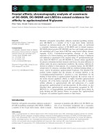
Fig. 1 Dendritic cells control immune response to cancer. The immune system is
endowed with the ability to recognize a universe of diverse molecules called antigens,
including cancer antigens, and to generate responses specific to the recognized antigens. Lymphocytes (T, B, NK, and NKT cells) and their products are under the control
of DCs. DCs reside in peripheral tissues where they are poised to capture antigens.
Antigen-loaded migratory DCs travel from tissues through the afferent lymphatics
into the draining lymph nodes. There, they present processed protein and lipid Ags
to T cells via both classical (MHC class I and class II) and non-classical (CD1 family) antigen presenting molecules. The soluble antigens also reach the draining lymph nodes
through lymphatics and conduits where they are captured, processed, and presented
by lymph-node resident DCs. Ag presentation by non-activated (immature) DCs leads to
tolerance and/or development of Tregs. Activated (mature), antigen-loaded DCs are
geared towards the launching of antigen-specific immunity leading to the T cell proliferation and differentiation into helper and effector cells with unique functions and cytokine profiles. DCs are also important in launching humoral immunity. Thus, DCs are at
the center of anti-cancer immunity.
2.1 Dendritic cell maturation as checkpoint of tolerance
and immunity
One of DC vulnerabilities in the context of cancer is the direct link between
their maturation and function as measured by the induction of T cell
response (Steinman, 2011). To this end, in the steady state, non-activated
(immature) DCs present antigens (including self-antigens) to T cells, thereby
inducing tolerance either through T cell deletion or through differentiation
of regulatory/suppressor T cells (Fig. 1). These immature DCs can be
ARTICLE IN PRESS
6
Jan Martinek et al.
considered “immunological sensors,” alert for potentially dangerous
microbes but also for the alterations of tissue homeostasis and sterile inflammation, and are capable of decoding and integrating such signals ( Janeway
Jr. and Medzhitov, 2002; Kawai and Akira, 2006; Pulendran et al., 2001).
Immature DCs have special characteristics including: (1) expression of a
specific set of damage sensing pathways (Banchereau et al., 2000; Steinman,
2011); (2) ability to efficiently capture Ags (Banchereau et al., 2000); (3) accumulation of MHC class II molecules in the late endosome-lysosomal compartment enabling loading of the peptide and assembly of peptide-MHC
complexes that can then be transferred to cell surface (Steinman, 2011);
and (4) low expression of costimulatory molecules (Steinman, 2011). These
properties can be harnessed by cancers to generate Tregs rather than T cells
able to reject tumors (Fehervari and Sakaguchi, 2004; Idoyaga et al., 2013;
Melief, 2008; Tanchot et al., 2012; Yamazaki et al., 2006). For example, in
a mouse model of melanoma the mere increase of DC infiltrate by their
mobilization with FLT3L was not sufficient for tumor rejection even in
the presence of PD-1 blockade (Salmon et al., 2016a). Yet, addition of DC
activator such as TLR-3 ligand poly IC facilitated tumor regression
(Salmon et al., 2016a).
Mature Ag-loaded DCs can launch differentiation of Ag-specific T cells
into effector cells (reviewed in Banchereau et al., 2000) (Fig. 1). DC maturation is associated with: (1) down-regulation of Ag-capture activity
(Trombetta and Mellman, 2005); (2) surface expression of CCR7 enabling
migration of DCs into draining lymph nodes (Dieu-Nosjean et al., 1999;
Forster et al., 1999); (3) translocation of peptide-MHC (pMHC) complexes
to cell surface together with co-stimulatory molecules (Lanzavecchia and
Sallusto, 2001); and (4) ability to secrete cytokines such as IL-12 (Veglia
et al., 2019) and IL-15 (Waldmann and Tagaya, 1999) supporting T cell differentiation. The ligation of the co-stimulatory receptor CD40 is an essential
signal for the final differentiation into fully mature DCs (Banchereau et al.,
1994). However, DC maturation alone does not result in a unique DC
phenotype. Instead, the different signals that are provided either directly
or through the surrounding cells that respond to damage induce DCs to
acquire distinct phenotypes that eventually contribute to different immune
responses (Pulendran et al., 2001) (Fig. 2). For example, γδ-T cells and NK
cells release IFN-γ, mast cells release pre-formed IL-4 and tumor necrosis
factor (TNF), plasmacytoid (p)DCs secrete IFN-α, stromal cells secrete
IL-15 and thymic stromal lymphopoietin (TSLP) while neutrophils provide
immunostimulatory DNA. This plasticity in response to external signals
ARTICLE IN PRESS
7
Interplay between dendritic cells and cancer cells
Cancer cell
Dendritic cell
Stromal cells
NK cells
Neutrophils
pDCs
Mast cells
MDSCs
Macrophages
T regs
CD8 T cells
CD4 T cells
Th2s
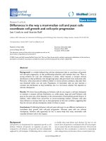
Fig. 2 Interplay of dendritic cells and cancer: contribution of other cells. DCs represent
the link between the innate and adaptive immunity and as such they integrate signals
from surrounding cells as well as are engaged in cross-talk with stromal cells other
leukocytes and cancer cells. Cancer cells and dendritic cells can impact each other indirectly by modulating intermediate cells, which is represented by the arrows in the
figure. This is driven by a variety of cell types, multiple surface bound and soluble factors
too numerous to be represented here but several examples includes: secretion of
stem cell factor (SCF) by numerous mouse and human cancer cell lines supported
c-KIT expressing mast cells, which in turn secreted multiple cytokines such as IL-6,
TNF-a VEGF, iNOS and CCL2, inducing tumor remodeling and altering DCs maturation/activation in TEM (Huang et al., 2008). Another example comes from TSLP production by cancer cells and stromal cells in breast and pancreatic cancers (De Monte et al.,
2011; Kuan and Ziegler, 2018; Olkhanud et al., 2011b; Pedroza-Gonzalez et al., 2011b).
TSLP drives DC maturation leading to expression among others of OX40-L, which
enables priming of IL-4/IL-13 secreting Th2 T cells. In turn, IL-4 and IL-13 modulate
TEM by promoting development of suppressive macrophages producing EGF that
supports cancer cell growth as well as directly impacting cancer cells by inhibition of
apoptosis. ITL7/BST2 mediated interaction between pDCs and cancer cells will suppress
IFN-α and TNF-a production in pDCs (Cao et al., 2009), while cancer derived PGE2
and TGF-β synergistically leads to production of IL-6/IL-8 by pDCs (Bekeredjian-Ding
et al., 2009). This in turn will have broad impact on both the innate (monocyte differentiation to macrophages and attraction of neutrophils) and adaptive (up regulation
of OX40-L and ICOS-L on pDC upon maturation, resulting in T regulatory and Th2
T cells activation) immunity in TEM. Last but not least, by secreting IL-12, DCs can
directly support CD8+ CTL and NK cells (Mittal et al., 2017). In turn CD8+ T cells and
NK cells will secrete IFN-y, which will stimulate CXCL9/10 production from DCs resulting
in an influx of effector T cells to the tumor environment (Mikucki et al., 2015). Additionally, NK cells can produce FLT3L, a growth factor promoting pre-DCs differentiation and
DCs survival (Barry et al., 2018).
ARTICLE IN PRESS
8
Jan Martinek et al.
represents another vulnerability that can be exploited by cancers to escape
immune-mediated elimination. Whereas some of the mechanisms dictating
DC maturation are differentially regulated in distinct DC subsets, many of
the principles are shared.
2.2 Antigen capture and modulation of DC maturation
DCs are scarce in tumor tissues when compared to other myeloid cells
such as macrophages and must therefore compete for both Ag capture
and T cell access. Ag capture is a critical step controlling the acquisition
and subsequent presentation of cancer Ags (Durand and Segura, 2015;
Mellman and Steinman, 2001; Trombetta and Mellman, 2005) and is often
linked with modulation of DC maturation (Fig. 3).
Acquisition of cancer Ags can be mediated via several pathways including
phagocytosis (Guermonprez and Amigorena, 2005); receptor mediated
endocytosis (as, for example, with the lectin Clec9A (Schreibelt et al.,
2012)); capture of IgG-Ags immune complexes (Liu et al., 2006;
Nimmerjahn and Ravetch, 2006; Rovere et al., 1998); pinocytosis enabling
capture of soluble molecules (de Baey and Lanzavecchia, 2000); nibbling
enabling capture of cell membrane fragments from live cells (Harshyne
et al., 2003); capture of extracellular vesicles (Muller et al., 2016; Wolfers
et al., 2001a); and capture of pre-formed peptides (cross-dressing)
(Wakim and Bevan, 2011). Hereunder, we will expand on some of these
mechanisms to illustrate the potential vulnerabilities that can be exploited
by cancer cells to escape immune elimination.
2.2.1 Phagocytosis
Phagocytosis is an active process of ingestion of particulate Ags that is essential for: (1) the clearance of apoptotic bodies from dying cells; and (2) for
the efficient uptake of pathogens and Ags from dying infected and/or cancer
cells (Aderem and Underhill, 1999; Platt et al., 1998). When captured by
macrophages, apoptotic bodies are degraded ( Jutras and Desjardins,
2005). However, when captured by DCs, their antigenic material can be
cross-presented to T cells to elicit Ag-specific CD4+ (Inaba et al., 1998)
and CD8+ T cell responses (Albert et al., 1998a; Berard et al., 2000a). Studies
pioneered by Zitvogel and Kroemer labs demonstrated the links between
phagocytosis and so-called immunogenic cell death in response to chemotherapy (Obeid et al., 2007). The critical mechanism involves the recognition of calreticulin translocated to the surface of apoptotic bodies from
cancer cells and its availability for recognition by receptors on DCs
ARTICLE IN PRESS
9
Interplay between dendritic cells and cancer cells
IL-10, CCL4,
CCL2, CXCL1,
CXCL5, PGE2,
ATP, Arginase,
TSLP,TGF-β,
HMGB1
TIM-3 Phosphatidylserine
TGF- R Activated TGFIntegrins E-cadherin
EpCAM CD44
LFA-1 ICAM-1
Fc Receptor
IL-1β, IL-6,
IL-8, IL-15, IL-10,
IFN-α, TNF-α
DNA
MHC-I
Vesicles/
Exosomes
RNA
MHC-II
Cancer
antigen
Dexosome
Apoptotic
bodies
1. Endocytosis
2. Fusion
STING
RIG-1
TLRs
Fc Receptors
CR1/CR2/CR3
SIRP
DEC205, CD207,
CD206, CD209,
DCIR, AXL, TIM-3
3. Phagocytosis
Fig. 3 Interplay of dendritic cells and cancer: direct interactions. (A) Interactions
between DCs and intact cancer cells driven by secreted and surface molecules: Here
we illustrate with few examples surface bound receptors and ligands, as well as also
secreted chemokine, cytokines, and metabolites involved in the interplay between
DCs and intact cancer cells. In addition to molecular pathways discussed in the
text and other legends, cancer cells can secrete CCL2 and CCL20, attracting CCR2+
tumor promoting monocytes and macrophages but also tolerogenic immature DCs
(Nagarsheth et al., 2017). IL-6 and IL-8 (CXCL8) secreted by DCs have been shown to
support tumor growth, survival and invasiveness in multiple types of cancer (Ara
et al., 2009; Araki et al., 2007; Yao et al., 2007). Through the secretion of IL-10, TGF-β
and other factors, cancer cells induce T cell Ig and mucin domain 3 (TIM-3) up regulation
(Continued)
ARTICLE IN PRESS
10
Jan Martinek et al.
(Obeid et al., 2007). This discovery showed that cancer cell apoptosis could
efficiently activate an immune response if the correct combination of “eatme” signals is present on the surface of dying cells. However, the putative
DC calreticulin receptors that are important for the sensing of immunogenic
cell death remain to be uncovered (Martins et al., 2010).
Another important mechanism of cancer cell phagocytosis is mediated by
CD47-SIRPα interactions (Chao et al., 2010; Jaiswal et al., 2009; Majeti
et al., 2009) and has been recently shown to contribute to immune escape
by delivering a “don’t’-eat-me” signal. Indeed, CD47, a "don’t eat me" signal for phagocytic cells including DCs, is overexpressed on cancer cells as
compared to matched adjacent normal (nontumor) tissue (Chao et al.,
2010; Jaiswal et al., 2009; Majeti et al., 2009). In vitro, blockade of
CD47 signaling using monoclonal antibodies enabled phagocytosis of cancer
cells that were otherwise protected (Willingham et al., 2012). Furthermore,
this pathway plays a role in DC maturation as SIRP-α engagement by
CD47-Fc leads to immature DC phenotype, decreased cytokine production, and low IFN-γ production by T cells after priming linked with an
impaired development of a T helper (Th)1 response (Hagnerud et al.,
2006; Latour et al., 2001).
The CD47-SIRPα axis appears to also dictate the fate of captured DNA
as blocking the interaction of SIRPα with CD47 preferentially increased the
sensing of captured DNA in DCs but not in macrophages (Xu et al., 2017).
Fig. 3—Cont’d by DCs. TIM-3 senses danger signals such as tumor derived nucleic
acid it can also sense phosphatidylserine (PS). PS serves as an “eat me” signal and is
exported to the outer membrane layer under oxidative stress to facilitate phagocytosis
(Birge et al., 2016). Cancer derived HMGB1 can interact with TIM-3 and inhibit its function (Chiba et al., 2012). ICs can be internalized by DCs and deliver Ags for presentation
to T cells (Amigorena et al., 1992; Geissmann et al., 2001). (B) Release and capture of
cancer antigen. Here we illustrate how intact and dying cancer cells release cancer antigens for capture by DCs. We show examples related to extracellular vesicles and apoptotic bodies derived from cancer cells and pathways through which DCs can capture
them. Cancer cells can produce extracellular vesicles by “blebbing” of the cell membrane and encapsulating parts of the cytosol or by formation of exosomes from the multivesicular endosome. These vesicles contain genetic material, cancer antigens as well as
immunosuppressive proteins such as PD-L1 and Fas-L (Bobrie and Thery, 2013; Sansone
et al., 2017; Schuler et al., 2014; Wolfers et al., 2001b). Nucleic acids, contained within
extracellular vesicles and apoptotic bodies can trigger intracellular danger associated
receptors/pathways such as TLRs, STING and RIG-I in DCs. ICAM-1 positive vesicles
can be endocytosed after binding to DC surface via LFA-1 interaction (Chiba et al.,
2012), they can also fuse with DCs membrane via fusion molecules such as flotillin or
GTPases (Subra et al., 2010).
ARTICLE IN PRESS
Interplay between dendritic cells and cancer cells
11
Mechanistically, CD47 blockade leads to reduced degradation of tumor
mitochondrial DNA (mtDNA) in DCs. mtDNA is then recognized by
cyclic-GMP-AMP synthase (cGAS) in the DC cytosol, contributing to
type I IFN production and generation of anti-cancer adaptive immunity
(Xu et al., 2017). Indeed, the innate immune sensing of tumors largely
occurs through the host STING pathway, which leads to type I IFN production, DC activation, cross-presentation of cancer antigens to CD8+
T cells, and eventually T cell recruitment into the TME (Woo et al., 2014).
2.2.2 Capture of immune complexes
Cancer cell membrane-bound antibodies and or soluble Ags can be bound
by immunoglobulins and form immune complexes (ICs). These can be
sensed by Fc and complement receptors on DCs (Sancho and Reis e,
2013). In many cancers, IgG antibodies are produced that recognize cancer
cells, form immune complexes and activate Fcγ receptors (Sancho and Reis
e, 2013). ICs also appear to play a potent role in priming CD8+ T cell immunity in response to therapeutic antibodies such as Trastuzumab (Gall et al.,
2017) or antibodies targeting EGFR (Banerjee et al., 2008). Fcγ receptors
family comprise two kind of receptors, type I (e.g., “classical” FcγRs) and
type II (e.g., non-classical FcR: FcRn, TRIM21) receptors (Bruhns and
J€
onsson, 2015; Pincetic et al., 2014). The "classical" FcγRs include the activating receptors FcγRI, FcγRIIA, FcγRIIC, FcγRIIIA FcγRIIIb and
the inhibitory receptor FcγRIIIb. The balance of activating and inhibitory
signaling is a key regulatory process controlling DC activity (Bournazos
et al., 2016; Guilliams et al., 2014; Platzer et al., 2014). For example, DC
maturation is linked with down-regulation of FcγRs consistent with
decrease in phagocytic function and gain of Ag-presenting function. In
mouse, antigen targeted to select activating and the inhibitory FcγR, results
under steady state in a short-term expansion of antigen-specific T cells,
whereas under inflammatory conditions especially, the activating FcγRIV
is able to induce superior CD4+ and CD8+ T cell responses (Lehmann
et al., 2017).
TRIM21 is an intracellular receptor that interacts with IgG-virus
complexes to neutralize viruses prior to replication and initiate degradation
in the proteasome (Keeble et al., 2008). It can also participate in Ag crosspresentation by DCs, (Ng et al., 2019). FcRn regulates IgG recapture, participates in cross-presentation (Baker et al., 2011), and was implicated
in anti-cancer immune response triggered by DCs in colorectal cancer
(Baker et al., 2013; Swiercz et al., 2016).
ARTICLE IN PRESS
12
Jan Martinek et al.
2.2.3 Other pathways of DC modulation
Tumor-secreted extracellular vesicles (EVs) are critical mediators of
intercellular communication between tumor cells and stromal cells in local
and distant microenvironments (Becker et al., 2016). EVs can be captured by
APCs in TEM and increasing evidence suggests this could be an important
regulatory pathway controlling the launching of immune responses. EVs
can impact Ag-independent interactions such as switching monocyte and
T cell differentiation towards a tolerogenic phenotype (Mangino et al.,
2017). Antigenic material transferred by exosomes derived from cancers
can be processed and presented by recipient DCs (Becker et al., 2016).
Furthermore, DCs themselves can produce EVs that directly activate
antigen-specific effector T cells (Tkach et al., 2017).
Another important component of the TME with a substantial impact
on DC function are lipids (Schupp et al., 2017). Indeed, lipid accumulation
in DCs can contribute to altered DC function via impaired antigen
cross-presentation, leading to inadequate CD8+ T cell activation.
2.3 Dendritic cell subsets in cancer
As DC subsets are discussed in another chapter, we will only focus on the
aspects most relevant to DC interplay with cancer cells.
Human blood DC subsets can be distinguished by differential expression
of three surface molecules: CD303 (BDCA-2), CD1c (BDCA-1), and
CD141 (BDCA3) (Chiodoni et al., 1999). CD303+ pDCs represent a
front-line of anti-viral immunity through their ability to secrete large quantities of type I IFN in response to viral encounter (Siegal et al., 1999). pDCderived type I IFN may promote maturation of other DC populations
(YJ, 2005) therefore helping to activate novel T cell clones. In their resting
state, pDCs are considered to play an important role in tolerance (YJ, 2005).
The other two subsets are termed conventional DCs and include
cDC1, which are Clec9A+ XCR1+ CD141+ CD1cÀ and cDC2, which
are SIRPα+ CD11b+ CD141–CD1c+. In mice cDC1 encompass lymphoid
tissue resident CD8a+ DCs and peripheral tissue resident CD103+ CD11b–
DCs (Siegal et al., 1999). cDC1 are under the transcriptional regulation of
IRF8, Id2 and Batf3 (YJ, 2005) while cDC2 are regulated by IRF4 (Bajan˜a
et al., 2016). cDC1 uniquely express TLR3, produce IL-12, and efficiently
cross-prime CD8+ T cells when activated by the TLR3 ligand, poly I:C
(Banchereau et al., 2000; Fehervari and Sakaguchi, 2004; Janeway Jr. and
Medzhitov, 2002; Kawai and Akira, 2006; Melief, 2008; Mellman
ARTICLE IN PRESS
Interplay between dendritic cells and cancer cells
13
and Steinman, 2001; Pulendran et al., 2001); however, human cDC2 also
cross-present antigens to CD8+ T cells, ( Jongbloed et al., 2010; Mittag
et al., 2011; Poulin et al., 2010; Yu et al., 2013). Furthermore, cDC2 are
uniquely able to drive differentiation of CD8+ T cells expressing CD103
(Yu et al., 2013), which binds to E-cadherin resulting in tumor cell rejection
(Banchereau et al., 2000). Indeed, mucosal homing and retention of CD8+
T cells contribute to mucosal cancer vaccine efficacy (Sandoval et al., 2013).
These results highlight the critical role tissue DCs play in imprinting the
trafficking patterns of elicited T cells (Mora et al., 2003).
The human skin hosts LCs in the epidermis and interstitial DCs (dermal
DCs). The dermal DCs can be further subdivided into CD1a+ DCs and
CD14+ DCs. Many studies of human cutaneous DCs demonstrated their
phenotypic and functional heterogeneity with regards to cellular immunity
and priming of highly efficient CTLs ( Joffre et al., 2012). The deeper understanding of their biology was made possible by the discovery of TNF as a
critical factor for in vitro LC differentiation from human progenitor cells
(Caux et al., 1997). These in vitro methods paved the way for the dissection
of molecular and cellular mechanisms regulating differentiation of LCs and
CD14+ interstitial DCs (Caux et al., 1997). Our studies concluded that
human CD14+ DCs can directly help activated B cells, as well as induce
naı¨ve T cells to differentiate into cells with properties of T follicular helper
cells (Tfh) (Klechevsky et al., 2008), thus, they may be specialized for development of humoral responses (Klechevsky et al., 2008). They however
induce CD8+ T cells, which are unable to kill cancer cells in a process mediated by ILT-4 and IL-10. On the contrary, LCs are more efficient in crosspresenting peptides from protein Ags to CD8+ T cells, and in priming CD8+
T cells in becoming potent CTLs partly through their ability to produce
IL-15 (Klechevsky et al., 2008). Monocytes can give rise to various forms
of inflammatory DCs (Klechevsky et al., 2008). Under certain conditions,
monocyte-derived DCs (Mo-DCs) are also capable of cross-priming
CD8+ T cells in response to cell-associated antigens both in mouse and
human (Albert et al., 1998b), (Berard et al., 2000b; Marigo et al., 2016).
Thus, in the human, several DC subsets are able to cross-prime CD8+
T cells to cancer Ags, while the experimental evidence in mouse supports
a leading role for cDC1s in this.
Further unraveling of the mechanisms regulating DC cross-presentation
capacity and response to TME-derived factors might open novel avenues for
intervention. Single cell RNAseq (scRNAseq) led to transcriptional definition of several DC types, including: a new DC subset that shares properties
ARTICLE IN PRESS
14
Jan Martinek et al.
with pDCs but potently activates T cells (Villani et al., 2017), thereby corroborating earlier studies suggesting the presence of such cells (Matsui et al.,
2009); a new subdivision within cDC2s; and circulating progenitors of cDCs
(Collin and Bigley, 2018; See et al., 2017). Further studies are needed to
better characterize the role of these redefined blood DC subsets in cancer.
When applied to tissue, single-cell approaches, although biased by inevitable
cell loss due to tissue digestion and preparation of single-cell suspensions,
offers a means to characterize and possibly redefine the composition of
the TME at different stages of tumor development. To this end, the Merad
lab simultaneously mapped early lung cancer as well as non-involved tissue
and blood from the same patient. Such paired analysis revealed depletion
of cDC1s (CD141+ DCs) and enrichment of macrophages even at the early
stages of disease (Lavin et al., 2017; Zilionis et al., 2019). Analysis of transcriptional profiles of DC subsets purified from primary breast cancers in
patients revealed DC subset-specific programming, suggestive of complex
interplay between ontogeny and tissue imprinting in conditioning DC
diversity in the TME (Michea et al., 2018).
Under steady state, DCs that reside in tissues are dependent upon FLT3
(fms-related tyrosine kinase receptor 3) and macrophage-colony stimulating
factor receptor (MCSF-R) (Helft et al., 2010). However, inflammatory processes such as those initiated by developing cancers might substantially alter
DC compartments. For example, in mouse models of cancer, the GM-CSF
produced in the TME facilitates the attraction of CD11b+ DCs that might
not be as efficient as other DC subsets in the induction of CD8+ T cell
immunity (Broz et al., 2014a). While the origin of each and all DCs recruited to sites of inflammation is still under investigation, monocytes can
give rise to inflammatory DCs, which have been characterized in psoriatic
skin (Knutson et al., 2001; Park et al., 2008) ascites fluid of ovarian cancer,
and synovial fluid of rheumatoid arthritis (Disis and Sciffman, 2001). Recent
studies in mice showed that a subset of monocytes can differentiate under
the regulation of GM-CSF and IL-4 into monocyte-derived DCs capable
of efficient cross-priming, and that the transcription factor IRF4, but
not BATF3, was critical (Brisen˜o et al., 2016). Indeed, in the absence of
IRF4 monocytes differentiated into macrophages even in the presence
of GM-CSF and IL-4. In this way, we have begun to unravel the transcriptional regulation of antigen presentation and plasticity of DCs, which is
expected to have a profound impact on reprogramming of the TME. Even
so, more work is needed to understand the precise roles that various DC
subsets play in the regulation of anti-cancer immunity.
ARTICLE IN PRESS
Interplay between dendritic cells and cancer cells
15
3. Dendritic cells dictate the outcome of immune
response to cancer
3.1 DCs control anti-tumor immune response
The hypothesis of “cancer immunosurveillance” was formulated by Burnet
and Thomas more than 50 years ago, who predicted that lymphocytes could
act as sentinels in recognizing and eliminating arising nascent and lowabundance neoplastic cells (Burnet, 1957). Indeed, studies in mice in the
1950s showed that chemically-induced tumors were immunogenic and
could elicit therapeutic immunity. While numerous teams have worked
on the mechanisms of these responses, R. Schreiber has been leading the
concept of elimination phase by primed immunity; equilibrium phase during which immune system exerts pressure on surviving cancer cells which
eventually leads to escape phase during which the immune system is unable
to control cancer (Dunn et al., 2004). It is thought that, in the initial phase of
immunogenicity, the mutated cells can be recognized by components of the
innate immune system. Indeed, immunodeficient mice (lacking IFN-γ and
recombination-activating gene 2) cannot control tumors as evidenced by
an increased incidence of cancer (Diamond et al., 2011). The role of DCs
in spontaneous tumor rejection has been implied in Batf3–/– mice, where
rejection of highly immunogenic syngeneic tumors was impaired possibly
due to defective DC cross-presentation capacity (Hildner et al., 2008).
NK cells can control the process of rejection, and cDC1 accumulation in
mouse tumors often depends on CCL5, XCL1 and FLT3L, all of which
can be produced by NK cells (Barry et al., 2018; B€
ottcher et al., 2018). Interestingly, in human cancers, CCL5, XCL1, and XCL2 transcripts closely
correlate with gene signatures of both NK cells and cDC1s and are associated
with increased overall patient survival (B€
ottcher et al., 2018). Cancerderived prostaglandin E2 (PGE2) can alter this axis (B€
ottcher et al., 2018).
However, in some mouse tumor models, NK cells inhibited the expansion
of tumor-specific CD8+ T cells during the priming phase and controlled the
frequency of CD8+ effector memory T cells (TEMs), leading to a diminished
recall response and reduced tumor control after a secondary tumor challenge
(Iraolagoitia et al., 2016). The underlying mechanism involved the regulation of DC maturation through PD-L1-expressing NK cells that emerged
during tumor growth (Iraolagoitia et al., 2016).
The cDC1 protein WDFY4 (WD repeat- and FYVE domain–containing
protein 4) is critical in tumor rejection by regulating cross-presentation
ARTICLE IN PRESS
16
Jan Martinek et al.
(Theisen et al., 2018). Wdfy4À/À mice cannot reject a highly immunogenic
fibrosarcoma or mount antitumor CD8+ T cell responses despite intact tumor
infiltration by cDC1s (Theisen et al., 2018).
Perhaps the most striking example of naturally occurring tumor immunity in humans is that observed in paraneoplastic diseases, some of which are
neurological disorders that arise as a consequence of anti-tumor immune
responses (Albert et al., 1998b; Darnell et al., 2000). Onconeural antigens
(like cdr2), normally expressed on neurons, can also be expressed on breast
cancer cells (Darnell et al., 2000); some patients develop a strong antigenspecific CD8+ T cell-mediated response against their breast cancer, resulting
in autoimmune cerebellar degeneration and severe neurological dysfunction, (Darnell et al., 2000). The presence of naturally occurring immunity
against a broad range of tumor-associated antigens including HER-2/
neu, MUC1, cyclin B1 and survivin has now been documented in patients
with breast cancer (Finn, 2008). Indeed, some early-stage clinical studies are
attempting to augment this intrinsic immunity in patients at high risk for
disease recurrence (Disis and Sciffman, 2001; Knutson et al., 2001; Park
et al., 2008). However, the native immune response to cancer co-exists with
the cancer and is therefore not protective, either because of tumor escape,
for example, through clonal evolution, or because the response might have
been generated by corrupted DCs.
3.2 The central role of type I IFN in tumor rejection
Pioneering studies by Marincola et al. analyzed, using DNA microarray
platforms, the question of genomic predisposition of response to therapy
at different metastatic sites in melanoma patients treated with antigenspecific vaccination plus systemic administration of r-IL2 (Galon et al.,
2013; Wang et al., 2002). The response was associated with the expression
in pretreatment metastases of genes related to chronic inflammation,
including numerous IRFs, consistent with a type I IFN signature. Similar
findings were made in the context of DC-based vaccination, suggesting
that responsiveness is in part dependent upon the genetic predisposition of
tumors to be susceptible to immune manipulation (Gajewski et al., 2009).
This type I IFN signature is found in a broader range of immune-mediated
tissue-specific destruction including tumor rejection in response to immunotherapy, allograft rejection, GVHD, flares of autoimmunity (Pascual
et al., 2010), and clearance of pathogen during acute infection (Wang
et al., 2008) .
ARTICLE IN PRESS
Interplay between dendritic cells and cancer cells
17
In pre-clinical models, mice lacking IFNAR1 (IFN-α/β receptor 1) in
DCs, cannot reject highly immunogenic tumor cells (Diamond et al., 2011)
and cDC1s from these mice display defects in antigen cross-presentation to
CD8+ T cells. In contrast, mice that lack IFNAR1 in granulocytes and macrophages are able to reject these tumors (Fuertes et al., 2011). Type I IFN is
involved not only in the induction phase of the cancer-immunity cycle but
also in the attraction of effector CD8+ T cells to tumor sites. This is due to
CXCL9 and CXCL10 chemokine expression by APCs that is induced upon
type I IFN production by APCs (Fuertes et al., 2013; Padovan et al., 2002).
Thus, type I IFN plays a central role in orchestrating tumor immune responsiveness via DCs. Furthermore, local secretion of type I IFN might switch
the differentiation of monocytes, leading to generation of monocytederived DCs as we found in systemic lupus erythematosus (Blanco et al.,
2001). Yet, chronic IFN signaling might fuel immune therapy resistance
in cancer. Indeed, prolonged IFN signaling (both type I and type II) allows
tumors to acquire STAT1-regulated epigenomic changes and augments
expression of Interferon Stimulated Genes (ISGs) and ligands for multiple
T cell inhibitory receptors (Benci et al., 2016).
3.3 DCs in pro-tumor immunity and response to treatment
Under the evolutionary pressure of the immune system, tumors fight back to
ensure survival of the fittest mutated cell(s). In this context, tumors adopt
multiple different strategies to suppress and/or corrupt DCs at all stages of
DC differentiation, maturation and function.
3.3.1 Studies in the human
As opposed to tumor rejection studies, most of which come from mouse
models, the concept of the role of DCs in tumor progression originated from
pathologists’ observations of human tumors. Many of these studies were performed in the 80s and 90s in patients with melanoma, renal cancer or breast
cancer (Enk et al., 1997; Gabrilovich et al., 1997; Steinbrink et al., 1999;
Thurnher et al., 1996). Indeed, in melanoma tumors, IL-10 suppresses
DC function and their capacity to trigger mixed lymphocyte reaction and
T cell proliferation (Enk et al., 1997; Steinbrink et al., 1999). While several
groups described the presence of intratumoral DCs in breast cancer (Bell
et al., 1999), their clinical impact was not resolved. We have found that
breast cancers are infiltrated with immature DCs present in tumor beds
(some of which express markers of LCs such as Langerin) and mature
CD83+ DCs in the stroma (Bell et al., 1999). Further studies revealed the
ARTICLE IN PRESS
18
Jan Martinek et al.
infiltration of breast cancers with inflammatory Th2 (iTh2) cells, which
coexpress interleukin (IL)-4/IL-13 and tumor necrosis factor (TNF)-α,
but not IL-10. iTh2 cells accelerate tumor development in humanized
mouse models of breast cancer through the activity of IL-13 (Aspord
et al., 2007). In genetically engineered mouse models of mammary cancer,
iTh2 cells accelerate the development of pulmonary metastasis via IL-4
(DeNardo et al., 2009). Generation of iTh2 cells in breast cancer depends
on the presence of mature tumor-infiltrating OX40L+ DCs (PedrozaGonzalez et al., 2011a). In experimental models of breast cancer, this DC
phenotype is driven by cancer-derived thymic stromal lymphopoietin
(TSLP) (Pedroza-Gonzalez et al., 2011a) (Olkhanud et al., 2011a). This
pro-tumor response can be reprogrammed by targeting dectin-1, an innate
immune receptor with activating motifs (ITAM), which can direct DCs
from inducing Th2 responses into Th1 responses (Baran et al., 2007;
Gringhuis et al., 2009; Wu et al., 2014). More recently, we showed that
production of IL1β in primary breast cancer tumors is linked with advanced
disease and originates from tumor-infiltrating CD11c+ myeloid cells
(Wu et al., 2018). IL1β production is triggered by cancer cell membranederived TGFβ. Neutralizing TGFβ or IL1 receptor prevents breast cancer
progression in a humanized mouse model, and this is associated with a
decrease in iTh2 cells and inhibition of OX40L+ DCs (Wu et al., 2018).
Thus, our work has challenged the notion that the only outcome of
cancer-DC interactions is suppression of DC function and showed that
they can be immunogenic, active yet corrupted, and can generate a
tumor-promoting response.
Cancers are not alike when it comes to mechanisms by which they modulate DCs. For example, in non-small cell lung cancer, the DCs isolated from
tumors are immature, suggesting blockade of DC maturation by lung cancer
cells (Perrot et al., 2007). Their antigen presentation ability and cytokine production are diminished and cannot be rescued even with TLR stimulation
(Yang et al., 2018). Mechanistically, lung tumor-isolated DCs express high
levels of co-inhibitory molecule B7-H3 and secrete more IL-10 and less
IL-12p70, which together lead to low T cell stimulatory capacity (Schneider
et al., 2011). Another example of a strategy employed by cancer cells comes
from colorectal cancer where tumor-derived mucins lead to increased DC
apoptotic death in vitro by engaging Siglec-3 (Rughetti et al., 2005).
3.3.2 Mouse models
Animal models of tumors permit elegant mechanistic studies, with the tradeoff that they do not reflect all of the features of genetic heterogeneity,
ARTICLE IN PRESS
Interplay between dendritic cells and cancer cells
19
complex tissue architecture, and immune microenvironment of human cancer. In particular, there is transcriptional divergence and distinct promoter
structures of the genes coding cytokines and chemokines across species
and variability in expression among innate immune cells (Hagai et al.,
2018; Mestas and Hughes, 2004). This might account for at times different
biological outcomes between human ex vivo and in vitro studies and preclinical models. Another important consideration is that, in the human,
the nature of cancer-DC-immune system interactions is chronic, thereby
leading to accumulation of events as well as chronic tissue remodeling processes. Most of the current mouse models focus on acute responses, possibly
accounting for challenges in extrapolating results from mouse to human.
Nevertheless, studies in Batf3À/À mice led to the identification of a crucial
role for CD103+ cDC1s in tumor response to checkpoint blockade (Salmon
et al., 2016b) (Sa´nchez-Paulete et al., 2016). Salmon et al. found that human
melanomas contain very few infiltrating DCs (Salmon et al., 2016b).
Similarly, tumors in two independent mouse models of melanoma, transplantable B16 tumors and a BRAF-driven genetically-engineered mouse
model, showed limited accumulation of DCs. As discussed earlier, mobilization of DCs with FLT3L and their maturation with poly IC enables priming of anti-cancer immune responses and are essential for response to
anti-PD1. This mechanism applies to the check-point inhibitor response
as well as to response to immunostimulatory antibodies such as CD137 agonists (Salmon et al., 2016b). Furthermore, effective antitumor responses to
anti-PD1 required IL-12 produced by tumor-infiltrating DCs (Garris et al.,
2018), which was driven by sensing of IFN-γ released from neighboring
T cells following PD-1 blockade (Garris et al., 2018). IL-12 production
can also be liberated by blockade of IL10 as shown in the MMTV-PyMT
mammary tumor model (Ruffell and Coussens, 2015). In this model,
both paclitaxel and reprogramming of the immunosuppressive tumor
microenvironment through depletion of IL-10-expressing macrophages
are necessary to reactivate DCs, enable IL-12 secretion, and generation of
anti-tumor immunity (Ruffell and Coussens, 2015). Tumor-residing
CD103+ DCs are also required for the recruitment of effector T cells into
the TME and are a major component of the establishment of the T cellinflamed tumor phenotype (Spranger et al., 2017). Thus, DCs fight for
T cell occupancy in the tumor by mobilizing specific subsets. The scarcity
of DCs in tumors that has been observed in multiple experimental and
human tumor types (Broz et al., 2014b; Ruffell and Coussens, 2015;
Spranger et al., 2015), may represent one of the major barriers to clinical
responses. Spranger et al. recently uncovered a role for oncogenes in the
ARTICLE IN PRESS
20
Jan Martinek et al.
regulation of DC recruitment to cancer (Spranger et al., 2017). The activation of cancer cell-intrinsic Wnt/β-catenin pathway was linked with the
lack of T cell priming against cancer-associated antigens in vivo due to defective recruitment of Batf3-dependent CD103+ DC into the tumor (Spranger
et al., 2017). Furthermore, pre-clinical models and a clinical trial in patients
with indolent non-Hodgkin’s lymphoma have indicated that DCs can be
recruited to non-responsive tumors for improved outcomes (Hammerich
et al., 2019). While lymphoma cells potently prime antigen-specific
T cells in vitro, direct priming is not sufficient to induce antitumor immunity
in vivo. There, a triple combination therapy regimen composed of Flt3L to
recruit intratumoral DCs and a local radiotherapy to release tumor antigens
for capture by DCs, together with poly-ICLC to induce DC maturation and
facilitate cross-presentation of released antigens to T cells, yielded substantial
tumor-specific CD8+ T cell response (Hammerich et al., 2019). The rate of
tumor rejection was further augmented by PD-1 blockade. These observations in mouse model reflected the clinical observations, where treated
patients displayed durable regressions of distant (untreated) tumors suggesting induction of systemic immunity (Hammerich et al., 2019). Thus, checkpoint blockade efficacy requires, and can be augmented through, induction
of antigen cross-priming by DCs.
3.4 Chronic inflammation promotes immune escape via DCs
Chronic inflammation, a hallmark of many cancers, is maintained by interplay of intrinsic (oncogenes and tumor suppressor genes) and extrinsic
(immune and stromal components) factors (Palucka and Coussens, 2016b)
(Mantovani et al., 2008) (Mantovani et al., 2019a) (Dougan et al., 2019a;
Pio et al., 2019). Acute and chronic inflammation can have distinct effects
on cancer and ultimately treatment outcomes (Palucka and Coussens,
2016b). Although signals that trigger acute inflammatory reactions often
stimulate DC maturation and antigen presentation, chronic inflammation
can be immunosuppressive as we discussed above. This antagonism between
inflammation and immunity also affects the outcome of cancer treatment
(Shalapour and Karin, 2015). As discussed above, chronic IFN signaling
can be a contributing factor to resistance. A similar concept can apply to
other cytokines such as acute response cytokines including IL-1, IL-6 and
TNF, and beta chain-signaling cytokines, especially GM-CSF.
GM-CSF is a critical cytokine for the generation of DCs (Caux et al.,
1992, 1996; Chapuis et al., 1997; Inaba et al., 1993; Santiago-Schwartz
et al., 1992; Zhou and Tedder, 1996). In vivo, the GM-CSF receptor acts
ARTICLE IN PRESS
Interplay between dendritic cells and cancer cells
21
in the steady state to promote the survival and homeostasis of nonlymphoid
tissue-resident CD103+ and CD11b+ DCs (Greter et al., 2012). In cancer,
GM-CSF has dual roles (Dougan et al., 2019b). Cancer cells themselves can
secrete GM-CSF to recruit macrophages, and promote differentiation of
myeloid-derived suppressor cells and the epithelial-to-mesenchymal transition (EMT). GM-CSF can also attract eosinophils and promote Th2 immunity (Dougan et al., 2019b). However, GM-CSF can also facilitate tumor
infiltration with DCs and Ag uptake and cross-presentation (Dougan
et al., 2019b). Furthermore, T cell-derived GM-CSF promotes maturation
and activation of APCs, which in turn potentiate T cell functions (Min et al.,
2010; Wada et al., 1997). The complexity behind the production of T cellderived GM-CSF suggests that it represents a powerful immune system
mediator (Ushach and Zlotnik, 2016).
Early in vitro experiments aimed at establishing the impact of cancerderived factors on human DC differentiation from CD34+ HPCs as well
as on their function showed that cancer-derived IL-6 and M-CSF play
inhibitory roles in renal cell carcinoma (Menetrier-Caux et al., 1998).
Mechanistically, the inhibition of DC differentiation was linked with
the induction of M-CSF receptor expression by IL-6 and a loss of
GM-CSF receptor expression at the surface of CD34+ HPCs by action
of M-CSF (Menetrier-Caux et al., 1998). The interplay between IL-6
and M-CSF also acts at the level of mature monocytes, where IL-6
switches monocyte differentiation to macrophages rather than DCs in
the presence of fibroblast and breast cancer cell lines (Menetrier-Caux
et al., 1998). Furthermore, IL-6 synergizes with TGFβ and GM-CSF to
direct blood monocyte differentiation into monocytic-MDSCs at the
expense of macrophages and DCs (Menetrier-Caux et al., 1998). Thus,
IL-6 is an essential factor in the molecular control of monocyte-derived
APC development. Interestingly, TNF, but not IL-1, induced monocytes
to become DCs despite the presence of fibroblasts and/or breast cancer
cells (Chomarat et al., 2003). TNF was found to decrease the expression
and internalization of the M-CSF receptor, thus overriding the IL-6/
M-CSF pathway (Chomarat et al., 2003). Thus, TNF facilitates the
induction of adaptive immunity by promoting monocyte-derived DCs
(Chomarat et al., 2003). These studies were further confirmed at the level
of transcriptional regulation where IRF4 and MAFB were shown to be
critical regulators of monocyte differentiation into DCs and macrophages,
respectively (Goudot et al., 2017). The aryl hydrocarbon receptor (AHR)
acts as a molecular switch for monocyte fate specification in response to
environmental factors (Goudot et al., 2017).
ARTICLE IN PRESS
22
Jan Martinek et al.
Many studies have indicated that IL1β might play a deleterious role in
modulating tumor-associated immunity (Curtis et al., 2012; Mantovani
et al., 2019b; Salcedo et al., 2013). For example, in syngeneic mouse models,
IL1β can counter antitumor effects, triggered by doxorubicin treatment, by
recruiting MDSCs that promote immunosuppression as well as promote
cancer invasiveness and metastasis (Zitvogel et al., 2012); and by conferring
a proliferative advantage to the cancer cells (reviewed in Elaraj et al., 2006).
We found that tumor infiltrating DCs in breast cancer display high levels of
IL-1β (Garris et al., 2018) and (Fig. 4). Treatment of patients with metastatic
breast cancer with the IL1 receptor antagonist anakinra eliminated a systemic
transcriptional signature of IL1-associated inflammation in blood cells.
Moreover, blood transcriptional profiles indicated that in vivo anakinra treatment effectively rescued immune cells’ cytotoxic programs that could contribute to enhanced antitumor activity. The link between TGFβ and IL1β
that we demonstrated in the context of breast cancer is of interest in view of
earlier studies reporting a suppressive role for TGFβ in immunosurveillance
against transplantable as well as spontaneous tumors in mice via NKT-cellderived IL13 (Ryan Kolb et al., 2016) (Mantovani et al., 2019a). In a mouse
model of orthotopically introduced 4T1 breast cancer cells, IL-1β-deficient
mice support spontaneous tumor regression (Kaplanov et al., 2019). This is
associated with low levels of macrophages, which favors the infiltrate of
CD11b+ DCs and changes the balance between IL-1- and IL-12 in the
TME, thereby supporting anti-tumor immunity. Treating WT mice first
with anti-IL-1β Abs followed by anti-PD-1 Abs abrogated early tumor progression (Kaplanov et al., 2019). Thus, DCs are involved in orchestrating
chronic inflammation at tumor sites. Unraveling the underlying mechanisms
as well as identifying contributing factors such as host genetics and the
microbiome response will likely lead to novel biomarkers and targets for
intervention.
4. Conclusions and future studies
Targeting the immune system has changed the landscape of cancer
treatment, both in the clinic and conceptually. Yet, many patients still do
not benefit from this paradigm shift. The determinants of treatment response
are likely multifactorial and include cancer cell-intrinsic factors; host environment as, for example, microbiome; host genetics dictating the response
to environment predisposing to certain type of response independently of
cancer; and last but not least the impact of these on the immune status in
ARTICLE IN PRESS
23
Interplay between dendritic cells and cancer cells
A
B
C
D
E
F
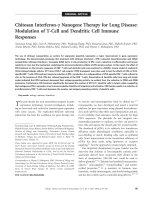
Fig. 4 DCs role in the Yin and Yang circle of cancer immune response. The Yang side of
the tumor immune response: Images illustrate different stages of anti-cancer immune
response leading to cancer rejection. (A) the capture of cancer antigen by tumor infiltrating DCs (Cytokeratin (CK) in green and CD11c+ DCs in red). (B) Activated DCs provide
co-stimulatory signal to T cells (CD3+ T cells (green) are in close contact with CD11c+
(not shown) DCs expressing the co-stimulatory molecule CD86 (red)). This process
can take place at the draining lymph node or, as shown here, at the tumor site with cancer
cells stained for CK (blue). (C) after Ag presentation along with adequate co-stimulation,
Ag-specific T cells will kill cancer cells via the action of granzymes and perforin (cytotoxic
CD3+ (not shown) CD8+ (red) T cell, killing a target cancer cell (blue) by perforin (green)
secretion. The Yin side of the tumor immune response: Images depicting how cancer cells
can corrupt DCs, reprogramming them into launching an immune response that will support cancer growth and progression. (D) Cancer cells can produce factors that will corrupt
DC maturation (CK+ cancer cells (blue) present surface bound TGF-β (red) to DCs, which
induces them to secrete IL-1β (green)). (E) Corrupted DCs, will secrete factors with an
impact on the whole TME. This is shown in E. where CD11c+ DCs (green) produce and
secrete IL-1β in proximity but also directly onto CK+ cancer cells (blue). (F) Corrupted
immune system will then support cancer progression (CK+ cancer cells (blue), based
on their KI67 staining (green), are highly proliferating despite being in close proximity
with tumor infiltrating CD11c+ DCs (red)).
the steady state and upon cancer challenge. Thus, progress will come
from basic studies and deep analysis of patient tissues linked with causative
studies in pre-clinical models. Next generation immunotherapies will
be based on cycles of interventions designed to boost and modulate
ARTICLE IN PRESS
24
Jan Martinek et al.
anti-cancer immunity. Eventually all patients will be treated with
checkpoint inhibitors, either directly or after interventions targeting
inflammation, by vaccination to boost T cell repertoires, or by adoptive
T cell transfer. The majority of patients will subsequently develop acquired resistance followed by immune escape; this will lead to the next
cycle of treatments incorporating multi-modal biomarkers. Despite rapid
progress in the field, much remains to be discovered and defined in
terms of biomarkers. The cancer-immunity cycle represents a framework
enabling uncovering of mechanisms operative at each step. We must fully
understand the rules of T cell priming in vivo in humans and develop
strategies for directing T cells to tumors. Last but not least, the role of
Tregs, so well established in murine cancer, will need to be redefined
in humans.
Recent studies place one DC subset, cDC1, at the center of regulation
of cancer immunity. How then do other DC subsets contribute to and
modulate anti-cancer immunity and by what mechanisms? Are the mechanisms regulating DC-T cell interactions at the tumor shared with those
operating in the draining lymph node? The studies on modulation of
DCs in lymph nodes draining tumors have only begun (Binnewies
et al., 2019) and this line of investigation is likely to enhance our understanding of how the new T cell repertoire can be primed in the context of
cancer environment. Metabolic regulation of DCs creates another layer
of control by the TEM that will need to be explored (Sinclair et al.,
2017; Wculek et al., 2019). Another important question is how the differences in the phenotype of human DC subsets between individuals and
tissues (Alcantara-Hernandez et al., 2017) impact the launching of anticancer immunity and response to check point inhibitors. By analogy to
its role in autoimmune diseases, host genetic variation is likely to have a
significant contribution to DC-cancer interactions (Hafler and Jager,
2005; Ye et al., 2014). Genome-wide association studies (GWAS) have
identified more than three hundred susceptibility loci predisposed to the
development of autoimmune diseases. These studies of patients affected
by severe autoimmune or immunodeficiency syndromes have led to the
discovery of several causative variants (Gutierrez-Arcelus et al., 2016).
Polymorphisms of Human Leukocyte Antigen (HLA) molecules have
been associated with development of virally-induced tumors such as head
and neck, cervical, and nasopharyngeal cancers (Brodin et al., 2015;
Brodin and Davis, 2017; Mangino et al., 2017). Resolving all this will
keep busy for a while!
ARTICLE IN PRESS
Interplay between dendritic cells and cancer cells
25
Acknowledgments
We thank patients and healthy donors for participation in our studies; our current and former
lab members and collaborators; Dr. Taneli Helenius for editing the manuscript, the JAX
creative services and the Imaging sciences services at the Jackson Laboratory for expert
assistance with this publication. Due to space limitations we could cite only selected
papers. Supported by The Jackson Laboratory; R01 CA219880 (KP); U01 AI124297
(JB); and P30CA034196 (Research reported in this publication was partially supported by
the National Cancer Institute of the National Institutes of Health under Award Number
P30CA034196. The content is solely the responsibility of the authors and does not
necessarily represent the official views of the National Institutes of Health).
References
Abbas, A.K., Lichtman, A.H., 2003. Cellular and molecular immunology, fifth ed. Saunders,
Philadelphia, p. 562.
Aderem, A., Underhill, D.M., 1999. Mechanisms of phagocytosis in macrophages. Annu.
Rev. Immunol. 17, 593–623.
Albert, M.L., Sauter, B., Bhardwaj, N., 1998a. Dendritic cells acquire antigen from apoptotic
cells and induce class I- restricted CTLs. Nature 392 (6671), 86–89.
Albert, M.L., Darnell, J.C., Bender, A., Francisco, L.M., Bhardwaj, N., Darnell, R.B.,
1998b. Tumor-specific killer cells in paraneoplastic cerebellar degeneration. Nat.
Med. 4, 1321–1324.
Alcantara-Hernandez, M., et al., 2017. High-dimensional phenotypic mapping of human
dendritic cells reveals interindividual variation and tissue specialization. Immunity
47 (6), 1037–1050. e6.
Amigorena, S., et al., 1992. Tyrosine-containing motif that transduces cell activation signals
also determines internalization and antigen presentation via type III receptors for IgG.
Nature 358 (6384), 337–341.
Appay, V., Douek, D.C., Price, D.A., 2008. CD8+ T cell efficacy in vaccination and disease.
Nat. Med. 14 (6), 623–628.
Ara, T., et al., 2009. Interleukin-6 in the bone marrow microenvironment promotes the
growth and survival of neuroblastoma cells. Cancer Res. 69 (1), 329–337.
Araki, S., et al., 2007. Interleukin-8 is a molecular determinant of androgen independence
and progression in prostate cancer. Cancer Res. 67 (14), 6854–6862.
Aspord, C., Pedroza-Gonalez, A., Gallegos, M., Tindle, S., Burton, E.C., Su, D.,
Marches, F., Banchereau, J., Palucka, A.K., 2007. Breast cancer instructs dendritic cells
to prime interleukin 13-secreting CD4 + T cells that facilitate tumor development.
J. Exp. Med. 204 (5), 1037–1047.
Bajan˜a, S., Turner, S., Paul, J., Ainsua-Enrich, E., Kovats, S., 2016. IRF4 and IRF8 Act in
CD11c+ cells to regulate terminal differentiation of lung tissue dendritic cells.
J. Immunol. 196 (4), 1666–1677.
Baker, K., Qiao, S.-W., Kuo, T.T., Aveson, V.G., Platzer, B., Andersen, J.-T., Sandlie, I.,
et al., 2011. Neonatal Fc receptor for IgG (FcRn) regulates cross-presentation of IgG
immune complexes by CD8 ÀCD11b + dendritic Cells. Proc. Natl. Acad. Sci. U. S.
A. 108 (24), 9927–9932.
Baker, K., Rath, T., Flak, M.B., Arthur, J.C., Chen, Z., Glickman, J.N., Zlobec, I., et al.,
2013. Neonatal Fc receptor expression in dendritic cells mediates protective immunity
against colorectal cancer. Immunity 39 (6), 1095–1107.
Banchereau, J., Steinman, R.M., 1998. Dendritic cells and the control of immunity. Nature
392 (6673), 245–252.