Epigenetic regulation by BAF (mSWI SNF) chromatin remodeling complexes in late cortical development and beyond
Bạn đang xem bản rút gọn của tài liệu. Xem và tải ngay bản đầy đủ của tài liệu tại đây (5.69 MB, 107 trang )
Epigenetic regulation by BAF (mSWI/SNF) chromatin
remodeling complexes in late cortical
development and beyond
Dissertation
for the award of the degree
‘‘Doctor of Philosophy’’ (Ph.D.)
of the Georg-August-University of Goettingen
within the doctoral program
of the Georg-August University School of Science (GAUSS)
Submitted by
Huong Nguyen
from Bac Giang, Vietnam
Goettingen, 2019
Thesis Committee
Prof. Dr. Jochen Staiger
Department of Neuroanatomy,
University Medical Center Goettingen
Prof. Dr. Gerhard Braus
Department of Molecular Microbiology and Genetics,
University of Goettingen
Prof. Dr. Thomas Dresbach
Department of Anatomy and Embryology,
University of Goettingen
Members of the Examination Board:
Prof. Dr. Jochen Staiger
Department of Neuroanatomy,
University Medical Center Goettingen
Prof. Dr. Gerhard Braus
Department of Molecular Microbiology and Genetics,
University of Goettingen
Prof. Dr. Thomas Dresbach
Department of Anatomy and Embryology,
University of Goettingen
Further members of the Examination Board:
Prof. Gregor Eichele,
Max Planck Institute for Biophysical Chemistry, Goettingen
Prof. Anastassia Stoykova
Max Planck Institute for Biophysical Chemistry, Goettingen
Prof. Dr. André Fiala
Department of Molecular Neurobiology of Behavior
Date of the oral examination: 03.07.2019
Affidavit
I herewith declare that the PhD thesis entitled ‘‘Epigenetic regulation by BAF
(mSWI/SNF) chromatin remodeling complexes in late cortical development and
beyond’’ was written independently, with no other sources and aids than quoted.
Goettingen, May 22th, 2019
Huong Nguyen
Acknowledgements
First of all, I would like to thank Prof. Staiger for giving me opportunity to work in
his institute and supporting me during my PhD time.
I would like to thank Dr. Tuoc Tran for giving me the chance to work in his
research group. I am very thankful for being always available for discussions,
answering questions and for always being positive.
I owe many thanks to the members of my thesis committee, Prof. Staiger,
Prof. Braus and Prof. Dresbach for their scientific advice during my PhD period.
I would like to thank members of
my Molecular Neurobiology Group:
Godwin Sokpor for his collegiality, cooperation and great scientific discussion.
Many thanks go especially to our group assistants Linh Pham for her technical helps.
Furthermore, I want to extend my thanks to members of the institute for
Neuroanatomy lab for their direct or indirect contribution to my project.
I would also like to thank my husband, my son, my parents and the rest of
my family for their enormous support during my studies, and for making my life happy!
Table of Contents
Chapter 1: General Introduction ............................................................................. 1
1.1.
Epigenetic modifications in cell biological processes ........................................ 1
1.2.
ATP-dependent chromatin modifiers ................................................................ 2
1.3.
Biochemical features of the SWI/SNF (BAF) Complex ..................................... 3
1.4.
Regulation of cortical development by the mammalian SWI/SNF (BAF)
complex ............................................................................................................ 4
Chapter 2: Epigenetic regulation by BAF (mSWI/SNF) chromatin remodeling
complexes is indispensable for embryonic development ..................................... 8
2.1. Abstract ............................................................................................................... 8
2.2. Introduction........................................................................................................... 9
2.3. Results and Discussion ...................................................................................... 11
2.3.1. BAF155 and BAF170 are indispensable for brain development and
embryogenesis .......................................................................................................... 11
2.3.2. BAF155 and BAF170 control the stability of BAF complexes in both cultured cells
and embryos.............................................................................................................. 13
2.3.3. The loss of BAF complexes induces the accumulation of H3K27me2/3-marked
heterochromatin ....................................................................................................... 16
2.4. Conclusion.......................................................................................................... 20
2.5. Materials and Methods ....................................................................................... 20
2.5.1. Transgenic mice .............................................................................................. 20
2.5.2. Immunohistochemistry (IHC) and Western blotting (WB) ................................ 20
2.5.3. Imaging and quantitative and statistical analyses ............................................ 21
Chapter 3: Epigenetic Regulation by BAF Complexes Limits Neural Stem Cell
Proliferation by Suppressing Wnt Signaling in Late Embryonic Development . 22
3.1. Summary ............................................................................................................ 22
3.2. Introduction......................................................................................................... 23
3.3. Results ............................................................................................................... 25
3.3.1. Loss of BAF complexes causes a genome-wide increase in the level of both
active and repressive epigenetic marks at distinct loci in the developing pallium during
late neurogenesis. .................................................................................................... 25
3.3.2. Conditional inactivation of BAF complexes during late cortical development
impairs neurogenesis of upper cortical layer neurons and the hippocampus. ........... 28
3.3.3. The NSC pool is increased at late development stages in the dcKO pallium .. 33
3.3.4. RGs acquire a NE-like identity in the BAF155/BAF170-deficient pallium. ....... 37
3.3.5. Change in spindle orientation, and increased proliferative capacity of NSCs in
the BAF155/BAF170-deficient pallium. ..................................................................... 40
3.3.6. Elimination of BAF155 and BAF170 de-represses Wnt signaling in late
corticogenesis. .......................................................................................................... 42
3.4. Discussion .......................................................................................................... 47
3.4.1. BAF155/BAF170-dependent maintenance of RG cell fate during late cortical
neurogenesis. ............................................................................................................ 48
3.4.2. BAF complexes control NSC proliferation and differentiation in early and late embryonic
stages via distinct epigenetic mechanisms. ......................................................................... 49
3.4.3. BAF complexes suppress Wnt signaling activity
50
3.5. Materials and Methods ....................................................................................... 51
3.5.1. Materials .......................................................................................................... 51
3.5.2. Methods........................................................................................................... 52
Chapter 4: General discussion ............................................................................... 72
Summary .................................................................................................................. 75
References ............................................................................................................... 76
List of figures........................................................................................................... 92
Abbreviations .......................................................................................................... 94
Curriculum Vitae ...................................................................................................... 97
Chapter 1
Chapter 1: General Introduction
1.1. Epigenetic modifications in cell biological processes
Epigenetic modifications are defined as mechanisms that regulate gene
expression without changes in the underlying DNA sequence (Bernstein et al., 2007;
Bird, 2007). In the mammalian cells, epigenetic modifiers can alter chromatin
architecture and genomic function through different processes, including DNA, RNA or
histone modifications, and activity of non-coding RNAs (Strahl & Allis, 2000;
Goldberg et al., 2007; Kouzarides, 2007).
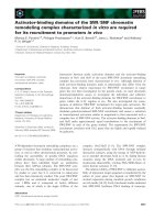
Figure 1.1 Chromatin remodeling BAF (mSWI/SNF) complex in neural development.
The BAF complex, epigenetic factors and transcription factors (TF) control gene expression.
TFs and ncRNAs bind to specific DNA sequences. The recruitment of BAF complexes and
other epigenetic factors on the genome leads to altered epigenetic marks (e.g., histone
acetylation, Ac; histone methylation, Me) and chromatin structure in order to activate or repress
a specific gene expression program in cell lineages. This figure taken from Sokpor et al. (2017).
Normally, epigenetic modifiers that target chromatin work as a complex
machinery to modulate higher-level chromatin configuration to impact many biological
processes, including cell renewal, differentiation, motility, maturation, survival and
1
Chapter 1
reprogramming (Figure 1.1) (Reik, 2007; Boland et al., 2014; Sokpor et al., 2017;
Hanna et al., 2018). The outcome of various epigenetic modifications broadly
converges on either gene repression or activation. Generally, epigenetic regulators
that promote gene expression activation remodel compact chromatin structure to an
open or relaxed chromatin. The relaxed chromatin is known to be transcriptionally
active because of related increase accessibility by transcription factors (Hirabayashi &
Gotoh, 2010; Juliandi et al., 2010; Coskun et al., 2012; Ronan et al., 2013;
Yao et al., 2016; Watson & Tsai, 2017). The converse is true for transcription
repression being caused by chromatin modifiers that render the chromatin compact.
The epigenetic regulators of chromatin structure can be categorized into: covalent
and non-covalent chromatin modifiers. Covalent modifiers regulate chromatin via
processes including methylation, acetylation, phosphorylation and ubiquitination,
whereas non-covalent chromatin modification includes ATP-dependent chromatin
remodelers which have been implicated in regulating many developmental
processes, including neurodevelopment (Strahl & Allis, 2000; Neilson et al., 2006;
Goldberg
et
al.,
2007;
Tran
et
al.,
2013;
Narayanan
et
al.,
2015a;
Bachmann et al., 2016b; Nguyen et al., 2016; Nguyen et al., 2018).
1.2. ATP-dependent chromatin modifiers
The ATP-dependent chromatin remodeling factors are multi-subunits complexes
that depend on energy obtained from ATP breakdown to orchestrate detectable
alterations in DNA-histone interactions that frequently translate in transcriptional
changes to influence cellular developmental processes (Hirabayashi et al., 2009;
Yoo & Crabtree, 2009; Hirabayashi & Gotoh, 2010; Ho & Crabtree, 2010;
Yao et al., 2016; Albert et al., 2017; Sokpor et al., 2017). Mechanistically, chromatin
remodeling involves nucleosomal mobilization that enhances the accessibility of DNA
sequences to regulatory proteins that target genomic loci (Reinke & Hörz, 2003;
Bailey et al., 2011).
ATP-dependent chromatin remodeling complexes typically have ATPase
subunits that allow them to hydrolyze ATP and to use the generated energy in order to
remodel the chromatin structure. The mobilization of chromatin domains to alter DNA
access is considered as a general mechanism that defines all ATP-dependent
2
Chapter 1
chromatin remodelers (Clapier et al., 2017). Based on similarities and differences in
their ATPase domains and related subunits, the chromatin remodelers can be further
classified into four categories of complexes: INO80/SWR, imitation switch (ISWI),
chromodomain helicase DNA-binding (CHD)/Nucleosome Remodeling Deacetylase
(NuRD), and switch/sucrose non-fermentable (SWI/SNF) (Flaus et al., 2006).
My study focused on the SWI/SNF complex that have been shown to play
indispensable role in embryonic development including neurodevelopment and
neuropsychiatric disorders (Sokpor et al., 2017).
1.3. Biochemical features of the SWI/SNF (BAF) Complex
The SWI/SNF complex was first identified in yeast to be composed of few
subunits (Neigeborn & Carlson, 1984; Wang et al., 1996a). However, the mammalian
orthologs, mSWI/SNF, or the Brg1/Brm associated factor (BAF) complex is made up
of about 15 subunits totaling about 2 Megadalton (MDa) in size (Lessard et al., 2007;
Wu et al., 2007).
The BAF complex is typically found around gene promoters and enhancers,
thus making them participate in gene expression programs that orchestrate cell
biological processes including cell renewal, specification, differentiation and migration.
Like other ATP-dependent chromatin remodelers, the BAF complex is composed of
exchangeable ATPase catalytic core(s): either BRM/SWI2 related gene 1 (BRG1) or
Brahma
(BRM)
depending
on
cell
lineage
(Neigeborn
&
Carlson,
1984;
Wang et al., 1996a; Lessard et al., 2007; Wu et al., 2007; Kadoch et al., 2013).
The BAF complex also contains other core subunits, including BAF155, BAF170 and
BAF47 and variant subunits such as BAF60, BAF100, and BAF 250 that are
ubiquitously expressed in the mammalian cell (Phelan et al., 1999; Sokpor et al., 2018).
Some of variant subunits are expressed specifically in certain cell lineages such as
BAF45A, BAF53A in neural stem cells and BAF45B, BAF53B in neurons
(Bachmann, 2016; Lessard, 2007).
3
Chapter 1
Mechanistically, BAF complex is able to convert condensed chromatin
(heterochromatin) to transcriptionally active euchromatin via histone dimer exchanges
or nucleosomal mobilization, ejection, and unwrapping (Phelan et al., 1999;
Whitehouse et al., 1999; Saha et al., 2002; Gutiérrez et al., 2007; Tang et al., 2010).
Many BAF subunits contain binding domains that allow the BAF complex to
interact with DNA and/or histone and regulate gene expression in cell lineage restricted
manner. The BAF complex displays variability and specificity in vivo due to
combinatorial assembly and switch of its subunits to form complexes with specific
remodeling outcomes and gene expression effects on cell fate (Lessard et al., 2007;
Wu et al., 2007; Kadoch et al., 2013; Tran et al., 2013; Bachmann et al., 2016a).
1.4. Regulation of cortical development by the mammalian
SWI/SNF (BAF) complex
During early development of the cerebral cortex, neuroepithelial (NE) cells which
initially predominate the germinative zone of the presumptive cortex undergo
proliferative (symmetric) division to increase their pool and subsequently switch to
differentiative (asymmetric) division to produce the more specialized apical progenitors
(radial glial [RG] cells) and pioneer neurons (Martínez-Cerdeño et al., 2006;
Kriegstein
&
Alvarez-Buylla
2009).
The downregulation
of
tight
junctional
complexes and the adoption of astroglial fate are characteristic changes that
occur during such transformation of NE into RG cells (Mollgøard & Saunders, 1975;
Aaku-Saraste et al., 1997; Hartfuss et al., 2001; Malatesta et al., 2003). Majority of
NE cells differentiate to RG cells around embryonic day 12.5 (E12.5) of mouse
cortical development (Kriegstein & Alvarez-Buylla, 2009; Sahara & O'Leary, 2009).
The parent RG cells also known referred to as apical RG cells actively proliferate to
increase their population and later exit the cell cycle as other subtypes of apical RG
cells or basal progenitors, or as neurons that migrate to make the nascent cortical
plate (Florio & Huttner, 2014). By mid-corticogenesis the developing cortex
is populated by diverse neural precursor cells that produce majority of the neurons
4
Chapter 1
that form the laminae of the cortical plate. The RG cells in the ventricular zone of
the developing cortex switch from neurogenic fate to astrocytic progenitor fate to
produce astrocytes and in the mouse cortex it starts from E17.5 (Morest, 1970;
Schmechel & Rakic, 1979; Misson et al., 1991).
Many transcriptional and epigenetic factors have been identified to regulate
various discrete cortical developmental process including neural progenitor
cell
specification,
proliferation,
differentiation,
migration
and
maturation
(Sokpor et al., 2017; Elsen et al., 2018). The BAF complex plays critical role in many
aspects brain development and function. Specific subunits of the BAF complex have
been associate to neurodevelopmental processes, including progenitor proliferation
and differentiation, and neuronal migration, maturation and synaptogenesis.
As a result, malfunction of the BAF complex have been linked to several
neurodevelopmental and neuropsychiatric disorders (Sokpor et al., 2017).
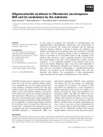
Figure 1.2. Model about the degradation of BAF complexes. Deletion of BAF complex lead
to dissociation of the other subunits and their degradation by the protein destruction system. This
figure taken from (Narayanan et al. 2015).
In the studies presented here, we developed mouse models to inactivate
BAF complex globally in the developing embryo and conditionally in the dorsal
telencephalon. The ablation of BAF complex was achieved by deletion of BAF155 and
BAF170, leading to dissociation of the other subunits and their degradation by
the protein destruction system (Figure 1.2) (Narayanan et al., 2015a). That way,
the chromatin remodeling function of the BAF complex is lost in cells with constitutional
5
Chapter 1
deletion of BAF155 and BAF170. Upon analyzing the BAF complex-deficient mouse
embryo, we identified that the Brg1/Brm associated factor plays critical roles in
embryogenesis and organ development (Nguyen et al., 2016). Furthermore, we found
evidence implicating the regulatory influence of BAF complex on cortical, hippocampal
and olfactory epithelium morphogenesis through regulation of neural progenitor
proliferation and differentiation (Tran et al., 2013; Bachmann et al., 2016b;
Nguyen et al., 2016; Tran et al., 2017; Nguyen et al., 2018).
Aims and general results of the studies
The studies aimed to clarify the role of BAF complexes in late cortical
development and beyond. The studies addressed two major questions: (i) the in vivo
validity and reproducibility of the mouse model of inactive BAF complex, and
(ii) the implication of loss of BAF complex on cortical organogenesis. To answer these
questions we first investigated the role of BAF155 and BAF170 in maintaining
the stability of the BAF complex in the entire mouse embryo and specifically in
the developing mouse forebrain. Second, we dived into how the BAF complex regulate
neurogenesis during late cortical development. Our generated BAF complex mutant
model provided a novel and investigative tool to probe into the above mention question
in order to confirm our understanding of how the epigenetic regulation by the BAF
complex influence cortical development.
The published findings presented in chapter 2, we identified an indispensable
BAF complex function in directing general development of the mouse embryo and
profoundly in the early development of the forebrain. Globally, the BAF complex
controls the installing of the transcription repressing heterochromatin marks
H3K27me2 and H3K27me3 via modulation of the H3K27 demethylases (UTX and
JMJD3) enzymatic activity know to control the dynamics of such transcription
repression marks (Nguyen et al. 2018; Nguyen et al. 2016; Narayanan et al. 2015). As
a result, deletion of the BAF complex resulted in marked upregulation of H3K27me2
6
Chapter 1
and H3K27me3 in many organs in the mouse embryo, including the brain leading to
developmental disturbance.
In chapter 3, we showed the mechanistic details of how the BAF complex regulate
cortical development. We found that the BAF complex functions as both repressors
and activators to control the epigenetic landscape and related corticogenic gene
expression programs in late cortical development. Specifically, BAF complexes
ablation led to H3K27me3-linked repression of neuronal differentiation-associated
genes, with simultaneous H3K4me2-mediated enhancement of proliferation-related
genes through Wnt signaling de-repression. Interestingly, loss of BAF complex
encourage proliferation of NE-like neural stem cells and apparently prolonged their
transformation into RC cells. Altogether, loss of BAF complex functionality resulted in
impaired neural progenitor proliferation and differentiation, and had a Wnt-dependent
impact on proper cerebral cortex (neocortex and hippocampus) development.
7
Chapter 2
Chapter 2: Epigenetic regulation by BAF (mSWI/SNF)
chromatin remodeling complexes is indispensable for
embryonic development
Huong Nguyen1#, Godwin Sokpor1#, Linh Pham1, Joachim Rosenbusch1,
Anastassia Stoykova2,3, Jochen F. Staiger1,3, and Tuoc Tran 1,3
Personal contributions: I and G.S. were involved in characterization of dcKO
phenotypes, data analysis and preparation of the manuscript. L.P. and J.R. contributed
to histological analyses; T.T. supervised, and wrote the manuscript; J.F.S., A.S. offered
suggestions for the study.
# Equally contributed authors
2.1. Abstract
The multi-subunit chromatin-remodeling SWI/SNF (known as BAF for
Brg/Brm-associated factor) complexes play essential roles in development.
Studies have shown that the loss of individual BAF subunits often affects local
chromatin structure and specific transcriptional programs. However, we do not fully
understand how BAF complexes function in development because no animal mutant
had been engineered to lack entire multi-subunit BAF complexes. Importantly, we
recently reported that double conditional knock-out (dcKO) of the BAF155 and BAF170
core subunits in mice abolished the presence of the other BAF subunits in
the developing cortex. The generated dcKO mutant provides a novel and powerful tool
for
investigating
how
entire
BAF
complexes
affect
cortical
development.
Using this model, we found that BAF complexes globally control the key
heterochromatin marks, H3K27me2 and -3, by directly modulating the enzymatic
activity of the H3K27 demethylases, Utx and Jmjd3. Here, we present further insights
into how the scaffolding ability of the BAF155 and BAF170 core subunits maintains
the stability of BAF complexes in the forebrain and throughout the embryo
during development. Furthermore, we show that the loss of BAF complexes in the
above-described model up-regulates H3K27me3 and impairs forebrain development
and embryogenesis. These findings improve our understanding of epigenetic
8
Chapter 2
mechanisms and their modulation by the chromatin-remodeling SWI/SNF complexes
that control embryonic development.
2.2. Introduction
Embryogenesis and organogenesis are determined by the combined effects of
myriad developmental events. In recent years, we have made substantial advances in
understanding how embryonic development is regulated (Ho & Crabtree, 2011;
Kojima et al., 2014). The early development are coordinated by different molecular
programs, in which epigenetic and chromatin-related controls are known to play crucial
roles (Ho & Crabtree, 2011).
Epigenetic
regulation,
which
modulates
the chromatin structure without altering the DNA sequence, has profoundly heritable
influences on transcriptional programs (Heard & Martienssen, 2014). These changes
in chromatin organization activate or repress gene expression programs either globally
or locally, and may thus shape specific developmental events. Epigenetic mechanisms
and chromatin regulation influence the ability of transcription factors (TFs) to access
regulatory elements in their target genes. This occurs primarily via histone modification
(Goldberg et al., 2007) or the action of ATP-dependent chromatin remodeling
complexes, such as SWI/SNF (BAF) complexes (Wen et al., 2009; MuhChyi et al., 2013;
Narlikar et al., 2013; Ronan et al., 2013). In addition, recent studies have shown that
DNA methylation (Wu & Zhang, 2014) and long non-coding RNA (lncRNA)-based
mechanisms (Bohmdorfer & Wierzbicki, 2015) also contribute to the complexity of
epigenetic regulation during development.
The types of covalent histone modification include histone acetylation,
methylation,
ubiquitination
and
phosphorylation
(Strahl
&
Allis,
2000;
Goldberg et al., 2007). Histone modification (epigenetic marks) is catalyzed by two
enzyme classes: histone writers (e.g., histone acetyltransferases, methyltransferases,
kinases, and ubiquitin ligases) and histone erasers (e.g., histone deacetylases,
demethylases, phosphatases, and deubiquitinases). The mis-regulation of histone
writers and erasers will typically alter the epigenetic program and have profound effects
on development (Strahl & Allis, 2000; Goldberg et al., 2007).
9
Chapter 2
A number of non-covalent, energy-dependent chromatin remodeling complexes
modulate
the
dynamicity
of
chromatin
structures.
Among
them,
the SWI/SNF complexes are the best characterized in both development and disease.
Mammalian SWI/SNF (BAF) complexes are made up of two switchable ATPase
subunits (Brg1 or Brm), core subunits (BAF47, BAF155, and BAF170) and a variety of
lineage-specific subunits (Lessard et al., 2007; Ho et al., 2009b; Kadoch et al., 2013;
Ronan et al., 2013). The Brg1 and Brm ATPases hydrolyze adenosine triphosphate
(ATP) and utilize the obtained energy to alter chromatin (nucleosome) structures,
thereby
modulating
cellular
processes
such
as
gene
expression
(Hirschhorn et al., 1992; Laurent et al., 1993; Phelan et al., 1999). The various subunits
(at least 15 have been identified) are capable of undergoing combinatorial assembly
(Wang et al., 1996b; Ronan et al., 2013), yielding hundreds of distinct BAF complexes
that can direct specific transcriptional events during development in vivo.
The exceptional diversity of BAF complexes allows them to have functional specificity
in biological processes. To investigate the roles of BAF complexes in development,
researchers have focused on phenotypic analyses of model animals harboring
mutations in single BAF subunits (Ko et al., 2008; Ho & Crabtree, 2011;
Narayanan & Tran, 2014). However, although BAF complexes are known to play
essential roles in development, studies using knock-out mouse models for individual
BAF subunits have yielded incomplete information regarding the functions of
these complexes.
While the epigenetic machinery and chromatin-remodeling complexes are
known to play essential roles in development, we know little about how they interact to
coordinate developmental processes during embryogenesis and organogenesis.
Recently, our group developed cortex-specific BAF155/BAF170cKO mouse mutant
and showed that BAF complexes did not form in the cortices of these mice. We further
showed that the known BAF subunits undergo proteasome-mediated degradation in
the developing cortices of these mutants. Finally, we found that, during corticogenesis,
BAF complexes globally control key heterochromatin marks (H3K27me2/3) by directly
interacting with and modulating the enzymatic activity of the H3K27 demethylases.
Here, we discuss these recent discoveries (Narayanan et al., 2015) and present
10
Chapter 2
additional evidence suggesting that the BAF155 and BAF170 core subunits cooperate
to stabilize the BAF complex and maintain the global level of H3K27me3 both in
the developing forebrain and throughout the embryo. Our new findings indicate that
BAF complexes act as key regulators of embryogenesis.
2.3. Results and Discussion
2.3.1. BAF155 and BAF170 are indispensable for brain development and
embryogenesis
By employing cortex-specific conditional mouse mutagenesis, we showed that
the dual loss of the BAF155 and BAF170 subunits in double conditional knock-out
(dcKO) mutants severely perturbed the growth of cortical structures, blocked
the proliferation, differentiation and cell-cycle progression of cortical progenitors, and
triggered a massive increase in the number of apoptotic cells (Narayanan et al., 2015).
To further investigate how the loss of both BAF155 and BAF170 affects
forebrain
development,
we
generated
forebrain-specific
BAF155
and
BAF170 dcKO mice by crossing mice floxed for BAF155 (Choi et al., 2012) and
BAF170 (Tran et al., 2013) (BAF155fl/fl, BAF170fl/fl) with a FoxG1-Cre line
(Hebert & McConnell, 2000). In FoxG1-Cre mice, the Cre-recombinase is driven in all
telencephalic cells [including those of the cortex (Cx) and basal ganglia (BG)], but not
in other parts of the brain [e.g., in the diencephalon (Di)] (Hebert & McConnell, 2000).
Remarkably, we found that the dcKO_FoxG1-Cre mutants completely lacked all
telencephalic structures at E16.5 (Narayanan et al., 2015). This indicated that
the expressions of BAF155 and BAF170 are required for brain development.
11
Chapter 2
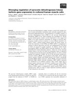
Figure 2.1. The expressions of BAF155 and BAF170 are indispensable for embryonic
development: dcKO_CAG-Cre embryos treated with TAM at E9.5 remained alive and showed
roughly preserved morphology at E13.5, but thereafter died between E14.5 and E15.5.
Scale bars = 1000 m.
To address whether BAF155 and BAF170 are essential for embryogenesis,
we generated and analyzed a line harboring a full dcKO_CAG-Cre mutant with
the tamoxifen (TAM)-inducible ubiquitous deleter, CAG-Cre line (Hayashi & McMahon,
2002) (Figure 2.1). The dcKO_CAG-Cre mutants were injected with either TAM or
corn oil (vehicle solution, control) at E9.5. Following TAM induction, we observed
Cre-recombinase activation in all cells of the body (Hayashi & McMahon, 2002).
12
Chapter 2
The mutants died between E14.5-E15.5, and exhibited a severe developmental
retardation (Figure 2.1). Together, these results show that the expressions of BAF155
and BAF170 are critical for determining overall embryogenesis,
including
the formations of the forebrain and cortex.
2.3.2. BAF155 and BAF170 control the stability of BAF complexes in both
cultured cells and embryos
Hundreds of distinct BAF complexes are predicted to form in vivo by
the
combinatorial
assembly
of
at
least
15
identified
BAF
subunits
(Ho & Crabtree, 2011). The functional specificity of a BAF complex is believed to reflect
the composite surfaces of its integrated subunits, which are essential for the ability of
these complexes to target the genome and interact with transcriptional factors (TFs),
co-activators, co-repressors, and signaling pathways (Ho & Crabtree, 2011).
We recently reported that BAF155 and BAF170 act as scaffolding subunits and are
required to ensure the stability of the entire BAF complex in the developing cortex
(Narayanan et al., 2015). The loss of BAF155 and BAF170 in cortex-specific dcKO
mutants leads to the dissociation of all other BAF subunits from the complex.
The free BAF subunits are subsequently ubiquitinated and degraded by
the proteasome system.
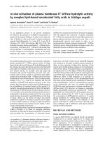
In an effort to extend our analysis to other parts of the brain, we examined
the expression levels of various BAF subunits (e.g., Brg1, Brm, BAF47, BAF60, and
BAF250) following the loss of BAF155/BAF170 in telencephalon of dcKO_FoxG1-Cre
embryos (Figure 2.2). Consistent with the Cre-recombinase activity in the Cx and BG
of dcKO_FoxG1-Cre mice, there was no detectable expression of BAF155 or BAF170
in these structures. In contrast, their expression levels were preserved in the Di,
where Cre is inactive (Figure 2.2 A, B). Similar to the reported effects in cortical tissues
(Narayanan et al., 2015), the loss of BAF155 and BAF170 in the telencephalon
abrogated the expression of all BAF subunits throughout this structure, including in the
BG (Figure 2.2C-G). To investigate whether both BAF155 and BAF170 are required to
stabilize BAF complexes throughout the embryo, the expression of BAF subunits was
examined in ubiquitously inducible dcKO_CAG-Cre embryos with global loss of
BAF155/BAF170 (Figure 2.3). These dcKO_CAG-Cre mutants were injected with
either TAM or corn oil (vehicle solution, as control) at E9.5 and analyzed at E13.5,
when the mutants were still viable. Following treatment with TAM, the expression levels
of
BAF155
and
BAF170
were
considerably
13
ablated
(Figure
2.3A-D).
Chapter 2
Moreover, the expression levels of the tested BAF subunits (Brg1, Brm, BAF47, and
BAF250) were severely diminished throughout the dcKO_CAG-Cre embryos,
as compared to controls (Figure 2.3E-L). These findings suggest that BAF155 and
BAF170 are required to maintain the expression levels of BAF subunits in
living animals.
Figure 2.2. Expression of BAF subunits in telencephalon-specific dcKO_FoxG1-Cre
mutants. (A-G) Images show immunohistochemical (IHC) analyses for various core subunits
of BAF complexes, including BAF155 (A), BAF170 (B), Brg1 (C), Brm (D), BAF47 (E),
BAF60 (F), and BAF250 (G), in the forebrains of dcKO_FoxG1-Cre mutants at E11.5.
The indicated BAF subunits are not detected in the BAF155/BAF170-knockout telencephalon.
Scale bars = 500 m. Abbreviations: Cx, cortex; BG, basal ganglia; and Di, diencephalon.
14
Chapter 2
Figure 2.3. Expression of BAF subunits in embryos of TAM-inducible full dcKO_CAGCre mutants. (A/C/E/G/I/K) E13.5 dcKO_CAG-Cre mutant embryos were treated with TAM
at E9.5, and whole-embryo sections were immunostained with antibodies against BAF155 (A),
BAF170 (C), Brg1 (E), Brm (G), BAF47 (I), and BAF250a (K).(B/D/F/H/J/L) Quantifications of
fluorescent signal intensities obtained from the sections described (A/C/E/G/I/G) (see also
Table S1 for statistical analysis). The results revealed that the protein expression levels of
BAF155 and BAF170 were reduced throughout the TAM-treated dcKO_CAG-Cre mutant
embryos, confirming the double knockdown of BAF155/BAF170. The expression levels of
the other tested BAF subunits were also diminished in mutant embryos compared to controls.
Scale bars = 500 m.
15
Chapter 2
In different tissues and cell lineages, BAF155 is highly expressed in proliferating
stem/progenitor
cells
but
generally
down-regulated
upon
differentiation
(Yan et al., 2008; Ho et al., 2009a; Tran et al., 2013). Conversely, little BAF170
is expressed in stem/progenitor cells (e.g., embryonic stem cells, or ESCs) and
at higher levels in differentiated cells (e.g., neurons) (Yan et al., 2008; Ho et al., 2009a;
Tran et al., 2013). We hypothesized that although only low expression levels are
detected for BAF170 in proliferating ESCs and for BAF155 in post-mitotic neurons,
this expression is necessary and sufficient to stabilize the embryonic stem cell (es)BAF
and neuronal (n)BAF complexes. Indeed, when we derived ESC lines from blastocysts
and primary neurons from forebrains (both representing the dcKO_CAG-Cre
genotype), we found that the depletion of BAF155 and BAF170 in these cultured cells
led to the loss of BAF subunit expression at the protein level (Narayanan et al., 2015).
These results collectively indicate that the knockout of BAF155/BAF170 in dcKO
mutants eliminates the presence of known BAF complex subunits both in vitro and in
vivo. Thus, the dcKO mutants provide a potent tool for investigating the roles of entire
BAF complexes during development.
2.3.3. The loss of BAF complexes induces the accumulation of H3K27me2/3marked heterochromatin
Previous studies suggested that the loss of individual BAF subunits has a local
(not global) influence on chromatin marks (Ho et al., 2011; Tran et al., 2013).
However, when we examined epigenetic marks in cortex-specific dcKO_Emx1-Cre
mice, which lacked entire BAF complexes, we observed a global reduction in
euchromatin along with increased H3K27me2/3 and decreased H3K9Ac in
the developing cortex during both embryonic and perinatal stages, as assessed by
assays
such
as
ChIP-Seq,
immunohistochemistry,
and
western
blotting
(Narayanan et al., 2015). Thus, our data showed for the first time that the presence of
BAF complexes is needed to maintain the balance between global repression and local
activation of epigenetic programs during cortical development (Narayanan et al., 2015).
16
Chapter 2
The intriguing observation that BAF complexes are lost from the telencephalonspecific dcKO_FoxG1-Cre and inducible full dcKO_CAG-Cre mutants prompted us to
study how this BAF155/BAF170 loss-of-function affects the H3K27me3 repressive
mark. We performed western blotting (WB) on telencephalic tissue lysates from E11.5
dcKO_FoxG1-Cre mutants using an antibody against H3K27me3. Similar to our
observation in cortical tissues, we found that the loss of BAF155 and BAF170
increased
the
level
of
H3K27me3
in
telencephalon
(Figure
2.4A/C).
Likewise compared to control (non-injected) embryos, the H3K27me3 level was
augmented in E13.5 dcKO_CAG-Cre embryos that had been injected with TAM at E9.5
(Figure 2.4B/C).
H3K27me2 and -3 are chromatin modifications that have been linked to the
down-regulation of gene expression (Cao et al., 2002; Pereira et al., 2010).
Thus, the massive enhancement of H3K27me3 in the dcKO mutants would be
expected to trigger obvious repression of gene expression. Indeed, gene expression
profiling of developing cortices from dcKO mutants revealed that most of the transcripts
were down-regulated, with only a few showing up-regulation (Narayanan et al., 2015).
Remarkably, BAF complexes were found to positively regulate most of the genes that
are repressed by the H3K27 methyltransferase, Ezh2 (Pereira et al., 2010;
Narayanan et al., 2015).
17
Chapter 2
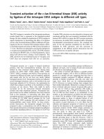
Figure 2.4. BAF complexes control the level of H3K27me3 in the brain and whole embryo
during development. (A) WB analysis of E11.5 telencephalons from telencephalon-specific
dcKO_FoxG1-Cre mutants revealed that the lost expressions of BAF155 and BAF170
elevated the level of H3K27me3. (B) dcKO_CAG-Cre embryos treated with TAM at E9.5
showed up-regulation of H3K27me3 at E13.5, compared to untreated control embryos.
(C) Densitometric quantification of the WB bands shown in (A and B; see also Table S2 for
statistical analysis). (D) Schematic indicating how altered levels of H3K27me2/3 demethylases
(UTX/Kdm6a and JMJD3/Kdm6b), BAF complexes, and the H3K27 methyltransferase
Ezh2 subunit of the PRC2 complex collectively modulate histone methylation, developmental
defects and diseases (e.g., tumorgenesis).
18