In vitro α-glucosidase inhibitory activity of compounds isolated from mangrove Lumnitzera littorea leaves
Bạn đang xem bản rút gọn của tài liệu. Xem và tải ngay bản đầy đủ của tài liệu tại đây (1.69 MB, 8 trang )
Science & Technology Development Journal, 22(1):106- 113
Original Research
In vitro α-glucosidase inhibitory activity of compounds isolated
from mangrove Lumnitzera littorea leaves
Nguyen Thi Le Thuy1 , Pham Thi Thuy2 , Poul Erik Hansen3 , Nguyen Kim Phi Phung2 ,∗
ABSTRACT
Introduction: Lumnitzera littorea grown at CanGio Mangrove Forest has been investigated. The
present study reports the isolation, characterization and evaluation of the α-glucosidase inhibitory
activity of isolated compounds from Lumnitzera littorea leaves. Methods: Their structures were
elucidated by spectroscopic methods (including MS, 1D and 2D–NMR) and comparison with values from the literature. From the n-hexane extract, nine compounds including lupeol (1), betulin (2), betulinic acid (3), oleanolic acid (4), corosolic acid (5), β -sitosterol (6), β -sitosterol 3-Oβ -D-glucopyranoside (7), stigmast-5-ene-3β -O-(6-O-hexadecanoyl-β -D-glucopyranoside) (8), and
stigmast-4-ene-3-one (9) were isolated and identified. Results: The results of the α-glucosidase
inhibitory activity showed thatcorosolic acid (5) and oleanolic acid (4) were the most potent, with
IC50 values of 17.86 ± 0.42 and 18.82 ± 0.59 µg/mL, respectively. Five of the other seven compounds exhibited inhibitory activity with IC50 values below 100 µg/mL, and higher than the positive control acarbose (127.64 ± 0.64 µg/mL).
Key words: Lumnizera littorea, mangrove plant, α-glucosidase inhibitory activity
1
Department of BioTechnology,
HoChiMinh city Open University,
Vietnam
2
Department of Chemistry, University of
Science, VNU-HCM
3
Department of Science and Enviroment,
Roskilde University, Denmark
Correspondence
Nguyen Kim Phi Phung, Department of
Chemistry, University of Science,
VNU-HCM
Email:
History
• Received: 2018-11-12
• Accepted: 2018-12-26
• Published: 2019-01-07
DOI :
/>
Copyright
© VNU-HCM Press. This is an openaccess article distributed under the
terms of the Creative Commons
Attribution 4.0 International license.
INTRODUCTION
METHODS
Diabetes is a chronic disease associated with unusually high levels of glucose in the blood. The goal of diabete therapy is the maintenance of normal blood glucose levels after a meal. Postprandial hyperglycemia
plays an important role in the development of type 2
diabetes and its complications. One of the therapeutic approaches for decreasing blood glucose rise after a
meal is to slow down the absorption of glucose by inhibition of carbohydrate hydrolyzing enzymes, such
as α-glucosidase. α-Glucosidase is an intestinal enzyme that breaks down α-1,4 linked polysaccharides
to α-glucose, which leads to the high blood sugar levels. The development of an α-glucosidase inhibitor
derived from natural products is an important contribution for the treatment of diabetes.
Lumnitzera littorea, a woody tree of the Combretaceae
family, grows at the Can Gio Mangrove Forest in
Vietnam. The antimicrobial activities of n-hexane,
ethyl acetate and methanol extracts of leaves of this
species were evaluated against six human pathogenic
microbes and the former extract was the most active 1 . Our published research showed that the αglucosidase inhibitory activity on all extracts and isolated flavonoids from the leaves of Lumnitzera littorea
were very strong 2 . The aim of this study was to isolate
phytoconstituents and evaluate the inhibition of αglucosidase activity of the compounds isolated from
the n-hexane extract of L. littorealeaves.
Plant materials
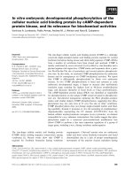
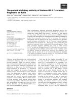
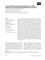
Leaves of Lumnitzera littorea (Jack) Voigt (Combretaceae) Figure 1were collected at Can Gio Mangrove
Forest of Ho Chi Minh city, Viet Nam in August of
2014. The scientific name of plant was authenticated
by Dr. Pham Van Ngot, Faculty of Biology, Ho Chi
Minh City University of Pedagogy. A voucher specimen (No US-B012) was deposited in the herbarium
of the Department of Organic Chemistry, University
of Science.
GENERAL EXPERIMENTAL
PROCEDURES
The NMR spectra were recorded on a Bruker Avance
III spectrometer at 500 MHz for 1 H NMR and 125
MHz for 13 C NMR spectra. ESI-MS were performed
on a Shimadzu +IDA TOF MS. TLC was performed
on silica gel 60 F254 (Merck, Darmstadt, Germany).
Gravity column chromatography was performed on
silica gel 60 (0.040–0.063 mm, Merck, Darmstadt,
Germany) and Sephadex LH-20 (GE Healthcare BioScience AB, Uppsala, Sweden). α-Glucosidase (EC
3.2.1.20) from Saccharomyces cerevisiae (750 UN) and
p-nitrophenyl-α-D-glucopyranoside were purchased
from Sigma Chemical Co. (St. Louis, MO, USA).
Acarbose and dimethyl sulfoxide were obtained from
Cite this article : Thuy N T L, Thuy P T, Hansen P E, Phung N K P. In vitro α-glucosidase inhibitory activity
of compounds isolated from mangrove Lumnitzera littorea leaves . Sci. Tech. Dev. J.; 22(1):106-113.
106
Science & Technology Development Journal, 22(1):106-113
Figure 1: Lumnitzera littorea (Jack) Voigt.
Merck (Darmstadt, Germany). Other chemicals were
of the highest grade available.
EXTRACTION AND ISOLATION
The fresh leaves were washed under running tap water
to remove all sandy particles and epiphytes and then
were dried and ground into fine powder. The powder (15,000 g) was exhaustively extracted with ethanol
at room temperature by the method of maceration.
After filtration, the ethanol solution was evaporated
to dryness under reduced pressure to yield a crude
ethanol residue (1,000 g). This crude was applied to a
silica gel solid phase extraction, eluted consecutively
with n-hexane, ethyl acetate, and finally with ethanol.
After evaporation under reduced pressure, three extracts were obtained: n-hexane (100 g), ethyl acetate
(250 g), and ethanol (550 g).
The n-hexane extract (100 g) was fractionated by silica gel column chromatography using a mixture of
n-hexane-ethyl acetate (98:2 to 0:100) to yield five
fractions (H1–H5). Fraction H2 (52.5 g) was applied to a silica gel column and eluted with chloroform:methanol (stepwise, 98:2 to 50:50) to give 6 subfractions (H21–H26). Subfraction H21 was rechromatographed on a silica gel column using chloroform:ethylacetate (stepwise 98:2 to 0:100), and then
purified by Sephadex LH–20 chloroform:methanol
(1:1) to obtain compound 1 (20 mg). Subfraction
H23 was further chromatographed on Sephadex LH–
20 chloroform:methanol (1:1) to give two compound
: compound 8 (10 mg) and 9 (15 mg).
Fraction H3 was further separated on a silica gel column and eluted with chloroform:methanol (stepwise,
107
9:1 to 0:100) to yield four fractions (H31–H34). Subfraction H31 was subjected to Sephadex LH–20 chloroform:methanol (1:1), then separated on a silica gel
Rp18 with water:methanol:acetone (2:3:5) to obtain
three compounds, such as compound 2 (10 mg), 3 (15
mg), and 4 (5 mg).
Fraction H4 was applied to a silica gel column and
eluted with chloroform:methanol:water (14:6:1) to
yield five fractions (H41–H45). Subfraction H41
was further separated on Sephadex LH–20 chloroform:methanol (1:1) to give compound 5 (15 mg) and
6 (25 mg). Subfraction H43 was rechromatographed
on a silica gel column with n-hexane:chloroform
(stepwise, 95:5 to 50:50) to obtain compound 7 (20
mg).
In vitro α-glucosidase inhibitory assay
The α-glucosidase inhibitory activity was evaluated
on all compounds according to the method of Apostolidis et al. 3 . A reaction mixture containing 60 µL of
100 mM phosphate buffer (pH 6.8), 20 µL of sample
(at the different concentrations), and 100 µL of 200
µM p-nitrophenyl-α-D-glucopyranoside solution (in
100 mM phosphate buffer) was incubated in 96-well
plates at 37 o C for 10 min. Then, 20 µL of 0.3 U/mL
α-glucosidase in the phosphate buffer was added to
the mixture. The reaction mixtures were incubated
at 37 o C for 10 min. Then, the reaction was stopped
by adding 20 µL of 50 mM NaOH. Absorbances were
recorded at 405 nm by a microplate reader and compared to a control which had 20 µL of buffer solution
in place of the sample. Acarbose was used as a positive
Science & Technology Development Journal, 22(1):106-113
control. The α-glucosidase inhibitory activity was expressed as % inhibition and was calculated as follows:
% Inhibition= [(Acontrol - Asample ) / Acontrol ]*100
The inhibitory concentration (IC50 ) for each sample
was calculated using a regression analysis from the
graph plotting scavenging activity against concentration. All experiments were carried out in triplicate
and the results were expressed as the mean ± SD of
three determinations.
Statistical analysis
All assays were conducted in triplicate. Statistical
analyses were performed with Statgraphics Plus Professional 16.0.03 for an analysis of variance (ANOVA),
followed by Duncan’s test. Differences at P<0.05 were
considered significant.
RESULTS
Structural elucidation
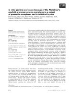
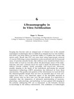
The phytochemical study of Lumnitzera littorea led to
the isolation and identification of nine compounds
whose structures are shown in Figure 2. The spectral properties of these known compounds, including
1
H-NMR and 13 C-NMR data, were identical to those
previously described in the literature.
Lupeol (1): white powder, ESI-MSm/z: 426.0 [M]+
for C30 H50 O. 1 H-NMR (500 MHz, CDCl3 ) d (ppm):
4.68 (brs, H-29a), 4.56 (brs, H-29b), 3.18 (dd, 11.5,
5.0 Hz, H-3), 1.68 (s, H-30), 1.03 (s, H-26), 0.96 (s,
H-23), 0.94 (s, H-27), 0.83 (s, H-25), 0.79 (s, H-28)
and 0.76 (s, H-24), 13 C-NMR (125 MHz, CDCl3 ) d
(ppm): 151.1 (C-20), 109.5 (C-29), 79.1 (C-3), 55.5
(C-5), 50.6 (C-9), 48.5 (C-18), 48.1 (C-19), 43.1 (C14), 43.0 (C-17), 41.0 (C-8), 40.2 (C-22), 39.0 (C-4),
38.9 (C-1), 38.2 (C-13), 37.3 (C-10), 35.7 (C-16), 34.4
(C-7), 30.0 (C-21), 28.1 (C-23), 27.6 (C-2, C-15), 25.3
(C-12), 21.1 (C-11), 19.5 (C-30), 18.5 (C-6), 18.2 (C28), 16.3 (C-25), 16.1 (C-26), 15.5 (C-24) and 14.7 (C27).
Betulin (2): white powder, ESI-MSm/z: 443.44
[M+H]+ for C30 H50 O2 . 1 H-NMR (500 MHz,
DMSO-d6 ) d (ppm): 4.68 (d, 2.0 Hz, H-29a), 4.58
(brs, H-29b), 3.80 (d, 10.5 Hz, H-28a), 3.33 (d, 11.0
Hz, H-28b), 3.18 (dd, 11.0, 4.0 Hz, H-3), 2.38 (m, H18), 1.68 (brs, H-30), 1.02 (s, H-25), 0.98 (s, H-27),
0.97 (s, H-23), 0.82 (s, H-26) and 0.76 (s, H-24), 13 CNMR (125 MHz, DMSO-d6 ) d (ppm): 150.6 (C-20),
109.8 (C-29), 79.1 (C-3), 60.7 (C-28), 55.5 (C-5), 50.6
(C-9), 48.9 (C-18), 47.9 (C-19, C-17), 42.9 (C-14),
41.1 (C-8), 39.0 (C-1), 38.9 (C-4), 37.5 (C-10), 37.3
(C-13), 34.4 (C-7), 34.1 (C-22), 29.9 (C-21), 29.3 (C16), 28.1 (C-23), 27.6 (C-2), 27.2 (C-15), 25.4 (C-12),
21.0 (C-11), 19.2 (C-30), 18.5 (C-6), 16.3 (C-25), 16.1
(C-26), 15.5 (C-24) and 14.9 (C-27).
Betulinic acid (3): white powder, ESI-MSm/z: 455.38
[M-H]− corresponding for C30 H48 O3 . 1 H-NMR
(500 MHz, CDCl3 ) d (ppm): 4.74 (brs, H-29a), 4.61
(brs, H-29b), 3.19 (dd, 11.0, 4.5 Hz, H-3), 3.00 (m, H19), 1.69 (s, H-30), 0.98 (s, H-26), 0.96 (s, H-27), 0.93
(s, H-23), 0.82 (s, H-25) and 0.75 (s, H-24), 13 C-NMR
(125 MHz, CDCl3 ) d (ppm): 180.5 (C-28), 150.6 (C20), 109.8 (C-29), 79.2 (C-3), 56.5 (C-17), 55.5 (C-5),
50.7 (C-9), 49.4 (C-19), 47.1 (C-18), 42.6 (C-14), 40.9
(C-8), 39.0 (C-4), 38.9 (C-1), 38.6 (C-13), 37.4 (C-10),
37.2 (C-22), 34.5 (C-7), 32.3 (C-16), 30.7 (C-15), 29.9
(C-21), 28.1 (C-23), 27.6 (C-2), 25.7 (C-12), 21.0 (C11), 19.5 (C-30), 18.4 (C-6), 16.3 (C-26), 16.2 (C-25),
15.5 (C-24) and 14.9 (C-27).
Oleanolic acid (4): white powder, C30 H48 O3 . 1 HNMR (500 MHz, CDCl3 ) d (ppm): 5.29 (t, 3.5 Hz, H12), 3.22 (dd, 11.5, 4.0 Hz, H-3), 1.14 (s, H-27), 0.99 (s,
H-29), 0.93 (s, H-30), 0.91 (s, H-23, H-25), 0.78 (s, H24) and 0.77 (s, H-26), 13 C-NMR (125 MHz, CDCl3 )
d (ppm): 177.8 (C-28), 143.7 (C-13), 122.9 (C-12),
79.2 (C-3), 55.4 (C-5), 47.8 (C-9), 46.6 (C-17), 46.1
(C-19), 41.9 (C-14), 41.3 (C-18), 39.5 (C-8), 38.9 (C1), 38.6 (C-4), 37.3 (C-10), 34.0 (C-21), 33.2 (C-29),
32.9 (C-22), 32.6 (C-7), 30.8 (C-20), 28.3 (C-23), 27.9
(C-15), 27.4 (C-2), 26.1 (C-27), 23.7 (C-30), 23.6 (C16), 23.2 (C-11), 18.5 (C-6), 17.2 (C-26), 15.7 (C-24)
and 15.5 (C-25).
Corosolic acid (5): white powder, ESI-MSm/z: 471.43
[M-H]− corresponding for C30 H48 O4 . 1 H-NMR
(500 MHz, CDCl3 ) d (ppm): 5.15 (d, 14.5 Hz, H12), 3.41 (m, H-2), 2.74 (d, 9.5 Hz, H-3), 2.11 (d, 11.5
Hz, H-18), 1.04 (s, H-27), 0.92 (s, H-23, H25), 0.91
(d, 7.0 Hz, H-30), 0.82 (d, 6.0 Hz, H-29), 0.75 (s, H26), 0.71 (s, H-24), 13 C-NMR (125 MHz, CDCl3 ) d
(ppm): 178.4 (C-28), 138.3 (C-13), 124.5 (C-12), 82.3
(C-3), 67.2 (C-2), 54.8 (C-5), 52.4 (C-18), 47.1 (C-17),
47.0 (C-9), 46.8 (C-1), 41.7 (C-14), 38.9 (C-8), 38.5
(C-4, C-19 & C-20), 37.6 (C-10), 36.3 (C-22), 32.6 (C7), 30.2 (C-21), 28.8 (C-23), 27.5 (C-15), 25.6 (C-16),
23.3 (C-27), 22.9 (C-11), 21.1 (C-30), 18.0 (C-6), 17.2
(C-29), 17.0 (C-26), 16.9 (C-25) and 16.4 (C-24).
β-Sitosterol (6): white powder, C29 H50 O. 1 H-NMR
(500 MHz, CDCl3 ) d (ppm): 5.35 (d, 5.0 Hz, H-6),
3.52 (m, H-3), 1.01 (s, H-18), 0.92 (d, 6.5 Hz, H-21),
0.85 (d, 7.5 Hz, H-29), 0.83 (d, 6.5 Hz, H-27), 0.81 (d,
7.0, Hz, H-26), 0.68 (s, H-19), 13 C-NMR (125 MHz,
CDCl3 ) d (ppm): 140.9 (C-5), 121.9 (C-6), 72.0 (C3), 56.9 (C-14), 56.2 (C-17), 50.3 (C-9), 46.0 (C-24),
42.5 (C-4 & C-13), 39.9 (C-12), 37.4 (C-1), 36.7 (C10), 36.3 (C-20), 34.1 (C-22), 32.1 (C-7 & C-8), 31.8
108
Science & Technology Development Journal, 22(1):106-113
Figure 2: The chemical structures of isolatedcompounds from Lumnitzera littorea leaves.
(C-2), 29.3 (C-25), 28.4 (C-16), 26.3 (C-15), 24.5 (C23), 23.2 (C-28), 21.2 (C-11), 20.0 (C-27), 19.5 (C-26),
19.2 (C-21), 18.9 (C-19), 12.1 (C-29) and 12.0 (C-18).
β-Sitosterol 3-O-β-D-glucopyranoside (7): white
powder, C35 H60 O6 . 1 H-NMR (500 MHz, DMSOd6 ) d (ppm): 5.32, (brs, H-6), 4.21 (d, 8.0 Hz, H-1’),
3.12 (m, H-3), 2.89-3.15 (m, H-2’-6’), 0.95 (s, H-19),
0.89 (d, 6.5, H-21), 0.82 (d, 7.0 Hz, H-29), 0.81 (d, 7.0
Hz, H-26), 0.79 (d, 7.5 Hz, H-27), 0.64 (s, H-18), 13 CNMR (125 MHz, DMSO-d6 ) d (ppm): 140.5 (C-5),
121.3 (C-6), 100.9 (C-1’), 77.1 (C-3 & C-3’), 76.8 (C5’), 73.5 (C-2’), 70.2 (C-4’), 61.2 (C-6’), 56.3 (C-14),
55.5 (C-17), 49.7 (C-9), 45.2 (C-24), 41.9 (C-13), 39.0
(C-4), 38.4 (C-12), 36.9 (C-1), 36.3 (C-10), 35.6 (C20), 33.4 (C-22), 31.5 (C-7 & C-8), 29.3 (C-2), 28.8
(C-25), 27.9 (C-16), 25.5 (C-23), 24.0 (C-15), 22.7 (C28), 20.9 (C-11), 19.8 (C-26), 19.2 (C-19), 19.0 (C-27),
18.7 (C-21), 11.9 (C-29) and 11.8 (C-18).
Stigmast-5-ene-3β-O-(6-O-hexadecanoyl-β-Dglucopyranoside) (8):white powder, C51 H90 O7 .
1
H-NMR (500 MHz, CDCl3 ) d (ppm): 5.37 (d, 5.0
Hz, H-6), 4.50 (dd, 12.0, 4.5 Hz, H-6’a), 4.38 (d, 8.0
Hz, H-1’), 4.26 (dd, 12.0, 2.0 Hz, H-6’b), 3.56 (m,
H-3), 3.34-3.59 (m, H-2’-5’), 2.35 (t, 7.5 Hz, H-22 ),
1.01 (s, H-19), 0.92 (d, 6.5 Hz, H-21), 0.89 (d, 7.0 Hz,
H-29), 0.84 (d, 1.5 Hz, H-26), 0.82 (d, 4.0 Hz, H-27)
and 0.68 (s, H-18), 13 C-NMR (125 MHz, CDCl3 )
d (ppm): 174.9 (C-12 ), 140.5 (C-5), 122.3 (C-6),
101.4 (C-1’), 79.7 (C-3), 76.1 (C-3’), 74.2 (C-5’), 73.8
(C-2’), 70.3 (C-4’), 63.3 (C-6’), 56.9 (C-14), 56.3
109
(C-17), 50.4 (C-9), 46.0 (C-24), 42.5 (C-13), 39.9
(C-4), 39.1 (C-12), 37.4 (C-11), 36.9 (C-10), 36.3
(C-20), 34.4 (C-22 ), 34.1 (C-22), 32.1 (C-7, C-8 &
C-32 ), 29.7 (C-1), 29.3 (C-25), 29.3-29.7 (C-42 -132 ),
28.4 (C-16), 26.3 (C-23), 25.1 (C-142 ), 24.5 (C-15),
23.3 (C-28), 22.8 (C-152 ), 21.3 (C-2), 20.0 (C-29),
19.5 (C-19), 19.2 (C-27), 19.0 (C-21), 14.3 (C-162 ),
12.1 (C-26) and 12.0 (C-18).
Stigmast-4-ene-3-one (9): white powder, ESIMSm/z: 413.26 [M+H]+ for C29 H48 O. 1 H-NMR
(500 MHz, CDCl3 ) d (ppm): 5.72 (s, H-4), 1.18 (s,
H-19), 0.91 (d, 6.5 Hz, H-21), 0.84 (t, 7.5 Hz, H-29),
0.83 (d, 7.0 Hz, H-26), 0.81 (d, 7.0 Hz, H-27) and 0.71
(s, H-18), 13 C-NMR (125 MHz, CDCl3 ) d (ppm):
199.8 (C-3), 171.9 (C-5), 123.9 (C-4), 56.2 (C-17),
56.0 (C-14), 54.0 (C-9), 46.0 (C-24), 42.5 (C-13), 39.8
(C-12), 38.8 (C-10), 36.3 (C-20), 35.8 (C-1 & C-8),
34.1 (C-22), 34.0 (C-2), 33.1 (C-6), 32.2 (C-7), 29.3
(C-25), 28.3 (C-16), 26.2 (C-23), 24.3 (C-15), 23.2
(C-28), 21.2 (C-11), 20.0 (C-26), 18.9 (C-21 & C-27),
17.5 (C-19) and 12.1 (C-18 & C-29).
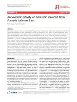
In vitro α-glucosidase inhibitory assay
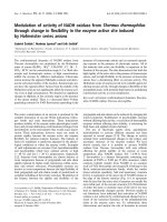
The α-glucosidase inhibitory effects of the isolated
compounds (1–9) were evaluated. The inhibition %
and IC50 values of all compounds are shown in Table 1.
The resulting IC50 values indicated that all the compounds, except 8, showed stronger α-glucosidase
inhibitory activity than acarbose (IC50 127.64 ±
Science & Technology Development Journal, 22(1):106-113
Table 1: In vitroα-glucosidase inhibitory activity of compounds isolated from Lumnitzera littorea
Compound
Concentration
n (μg/mL)
5
25
1
Inhibition (%)
30.22 ±
0.48
36.27
0.43
±
39.38
0.340
±
41.45
0.75
±
53.02
0.62
2
17.81 ±
0.54
46.79
0.68
±
60.25
0.72
±
70.81
0.35
±
>100
38.74 ± 0.63
3
24.02 ±
0.28
53.00
0.43
±
72.67
0.27
±
89.03
0.18
±
>100
28.12 ± 0.37
4
38.51 ±
0.43
55.07
0.53
±
80.95
0.75
±
94.41
0.61
±
>100
18.82 ± 0.59
5
43.16 ±
0.16
50.76
0.37
±
84.98
0.43
±
>100
>100
17.86 ± 0.42
6
31.30 ±
0.27
38.30
0.63
±
63.19
0.21
±
74.81
0.18
±
>100
34.45 ± 0.34
9
28.24 ±
0.17
43.20
0.26
±
62.75
0.34
±
78.32
0.53
±
92.98
0.48
Concentration
n (μg/mL)
10
50
Inhibition (%)
9.92
0.39
±
32.52
0.26
±
49.31
0.17
±
63.19
0.63
±
74.81
0.72
±
114.19
0.61
±
8
2.56
0.39
±
15.63
0.76
±
32.67
0.35
±
45.81
0.49
±
53.67
0.39
±
174.51
0.58
±
Acarbose
(Positive
control)
4.65
0.35
±
10.47
0.21
±
39.54
0.67
±
62.40
0.64
±
79.07
0.51
±
127.64
0.64
±
7
50
75
100
100
150
IC50
(μg/mL)
±
±
200
97.95 ± 0.58
38.18 ± 0.45
IC50
(μg/mL)
Data are presented as mean± SD values of triplicate determinations. A one-way analysis of variance (ANOVA) and positive analysis were done
using Duncan’s multiple test; significance was set at P<0.05.
0.64 mg/mL). Particularly, one ursane-type triterpene (compound 5) and one oleanane-type triterpene
(compound 4) showed outstanding α-glucosidase inhibition activities, with IC50 values of 17.86 ± 0.42
and 18.82 ± 0.59 µg/mL, respectively. Meanwhile,
the other compounds displayed α-glucosidase inhibition activities with IC50 values ranging from of 34.00–
115.00 µg/mL.
DISCUSSION
The 1 H NMR spectrum of compounds 1-5 showed
the presence of several singlet signals in the high
shielded region at d 0.71-1.69, that was characteristic of methyl protons. The 13 C NMR spectrum of
compounds 1-5 revealed 30 carbon signals, including seven methyl carbons, nine methylene carbons,
seven methine carbons, and seven non-hydrogenated
carbons. The result showed characteristic of a pentacyclic triterpenoid. On the other hand, the skeleton
of 1 was recognized to be lupane triterpenoid by the
NMR spectra, with the typical olefinic proton signals
at d 4.56 (s, H-29b) and 4.68 (brs, H-29a) in the 1 H
NMR spectrum and two olefinic carbons of the exocyclic double bond at d 109.5 (C-29) and 151.1 (C-20)
in the 13 C NMR spectrum. Moreover, the assignment
of the hydroxyl group at C-3 was performed by the
presence of one secondary hydroxyl proton signal at
d 3.18 (dd, 11.5, 5.0 Hz, H-3), correlating with a carbon signal at d 79.1 (C-3). Thus, 1 was determined as
lupeol that was consistent with the reported values in
the literature 4 .
The NMR spectra of 2 were similar to those of 1,
including the proton and carbon signals for the terpenoid of lupane skeleton. The 1 H NMR spectrum of
2 differed from that of 1 by having a pair of proton signals at d 3.33 (d, 11.0 Hz, H-28b) and 3.80 (d, 10.5 Hz,
H-28a), instead of a methyl proton signal at d 0.79 (s,
H-28) as in 1. In the 13 C NMR spectrum of 2, besides
an oxygenated methine carbon signal at d 79.1 (C-3),
compound 2 had another oxygenated methylene car-
110
Science & Technology Development Journal, 22(1):106-113
bon signal at d 60.7 (C-28), thus confirming that there
was a second hydroxyl group at C-28 in the structure
of 2. Comparison of the spectroscopic data of 2 with
those in the literature suggested 2 was betulin 4 .
Similar to the NMR spectra of 2, the 1 H NMR and
13
C NMR spectra of 3 also possessed the signals of
a lupane skeleton. However, the 1 H NMR spectrum
of 3 differed from that of 2 in the absence of a pair of
proton signals at d 3.30-3.80 of H-28 position. It corresponded to the presence of a carboxyl carbon signal
at d 180.5, instead of an oxygenated methylene carbon signal at d 60.7 (C-28) as in 2. Thus, compound
3 was betulinic acid whose NMR data were in good
compatibility with those in the literature 5 .
The 1 H NMR spectrum of compound 4 displayed one
olefinic proton signal at d 5.29 (t, 3.5 Hz, H-12), together with a signal at d 2.83 (dd, 13.5, 4.0 Hz, H-18)
which indicated the oleanan-12-ene skeleton. One
methine proton signal at d 3.22 (dd, 11.5, 4.0 Hz, H3) showed that 4 had one hydroxyl group. The 13 C
NMR spectral data exhibited signals at d 122.9 and
143.7, corresponding to the carbons C-12 and C-13,
respectively. The signal at d 177.8 was assigned to the
carboxyl group at C-28. This data allowed the identification of compound 4 as oleanolic acid which is isolated for the first time from Lumnitzera littorea.
The 1 H NMR spectrum of compound 5 showed the
presence of two doublet methyl signals at d 0.82 (d,
6.0 Hz, H-29) and 0.91(d, 7.0 Hz, H-30), which were
characteristics for ursane skeleton. Furthermore, the
olefinic proton signal was observed at d 5.15 (td, 14.5,
3.6 Hz, H-12) along with one methine proton signal
at d 2.11 (d, 11.5 Hz, H-18). Two oxygenated methine
proton signals at d 2.74 (d, 9.5 Hz) and 3.41 (m, overlapped with the solvent signal) were assigned to H3 and H-2, respectively. In the 13 C NMR spectrum
of 5 showed two oxygenated carbons at d 67.2 (C-2)
and 82.3 (C-3), two disubstituted double carbons at d
124.5 (C-12) and 138.3 (C-13), and one carbonyl carbon d 178.4 (C-28). The spectral data were similar to
the ones reported for corosolic acid 6 .
The 1 H NMR spectrum of compound 6 revealed the
presence of six methyl proton signals, including two
methyl singlets at d 0.68 (s, H-19) and 1.01 (s, H-18),
four methyl doublets at d 0.81 (d, 7.0 Hz, H-26), 0.83
(d, 6.5 Hz, H-27), 0.85 (d, 7.5 Hz, H-29), and 0.92 (d,
6.5 Hz, H-21). The olefinic proton signal d 5.35 (d, 5.0
Hz, H-6) appeared to be characteristic of the sterols.
Furthermore, the proton signal connected to the C-3
hydroxyl group appeared as a multiplet at d 3.52 (m,
H-3). The 13 C NMR spectrum exhibited 29 carbon
signals, including two carbon signals at d 121.9 (C-6)
and 140.9 (C-5), characteristic of a double bond and
111
an oxymethine carbon signal at d 72.0 (C-3). Thus,
the structure of 6 was assigned as β-sitosterol and was
consistent with values reported in the literature 7 .
Detailed analysis of NMR spectra of 7 indicated that
it also possessed the proton and carbon signals of a βsitosterol skeleton. Additionally, the 1 H NMR spectrum of 7 confirmed the presence of one β-glucose
unit through a doublet signal at d 4.21 (d, 8.0 Hz, H1’), assigned for anomeric proton, and multiplet signals from d 2.89 to 3.15, assigned for the carbinol protons of the sugar part. In the 13 C NMR spectrum
which displayed an anomeric carbon signal at d 100.9
(C-1’), an oxygenated methylene carbon signal at d
61.2 (C-6’) and four oxymethine carbon signals at d
70.2-77.1 (C-2’-5’) of sugar unit were observed. These
data confirm that compound 7 was β- itosterol 3-Oβ-D-glucopyranoside 8 .
Similar to the NMR spectra of 7, the 1 H NMR and
13
C NMR spectra of 8 indicated that it possessed a
similar structure to that of β-sitosterol glucoside. The
difference was the presence of signals for a palmitoyl moiety in 8. The 1 H NMR spectrum observed
one methylene proton signal adjacent to a carboxyl
group at d 2.35 (t, 7.5 Hz, H-22 ), other methylene
proton signals at d 1.20-1.50 (characteristic for a long
aliphatic chain), and one terminal methyl proton signal at d 0.89 (d, 7.0 Hz, H-162 ). Moreover, the 13 C
NMR spectrum revealed 51 carbon signals, including
29 carbons of a β- itosterol skeleton and 6 carbons of
a glucose unit. The assignment of 1 carbon signal at d
174.9 (C-12 ) was determined by the presence of a carboxyl group, as well as carbon signals at d 29.3-29.7
of the methylene carbons and at d 14.3 of a terminal
methyl group. Besides that, the HMBC correlations of
methylene protons at d 4.26 (dd, 12.0, 2.0 Hz, H-6¢b)
and 4.50 (dd, 12.0, 4.5 Hz, H-6’a) with the carbonyl
carbon at d 174.9 (C-12 ) indicated an attachment of
the palmitoyl moiety at C-6’ of the glucose unit. Comparison of spectroscopic data of 8 with those in the
literature suggested that 8 was stigmast-5-ene-3β-O(6-O-hexadecanoyl-β-D-glucopyranoside) 4 .
The 1 H NMR spectrum of 9 closely resembled that
of 6. In addition, the 1 H NMR spectrum confirmed
the presence of one olefinic proton signal at d 5.72 (s,
H-4) and the absence of a multiplet proton signal at
d 3.10-3.60 of H-3 position. The 13 C NMR spectrum
showed the carbonyl carbon signal at d 199.8 (C-3)
and two olefinic carbon signals at d 171.9 (C-5) and
123.9 (C-4). Based on the spectral data obtained and
comparison with literature data, the structure of 9 was
confirmed as stigmast-4-ene-3-one 9 .
Although 1–5, 8,and 9 are known compounds, this is
the first time their presence in leaves of Lumnitzera
Science & Technology Development Journal, 22(1):106-113
littorea has been reported. α-Glucosidases are a series of enzymes located on the human intestine. The
most important carbohydrates in food are hydrolyzed
to monosaccharide by α-glucosidase, then absorbed
into the blood to increase blood glucose level. This
is the reason for development of diabetes. The αGlucosidase inhibitors may have the potential to delay or prevent the rise of blood glucose level. However, the mechanism of the inhibitions against αglucosidase has not yet clear.
In our experiments, five compounds of triterpenoids
and four compounds of steroids from Lumnitzera littorea showed different activity against α-glucosidase
(Table 1). From the structures of compounds 1–3, we
can infer that the α-glucosidase inhibitory acitvity is
strengthened when the methylene group at C–28 is
altered to an oxygenated methylene or a carboxylic
group. As the result, the IC50 values of lupeol (1),
betulin (2) and betulinic acid (3) were 97.95 ± 0.85,
38.74 ± 0.63 and 28.82 ± 0.37 µg/mL, respectively.
Furthermore, a carboxylic acid group or a CH2 –OH
group at C–17 is important for the action of compounds 2–5.
Comparison of the chemical structures and the αglucosidase inhibitory activity indicates that the presence of a hydroxyl group at C–3 plays an important role in the α-glucosidase inhibitory activity.
Thus, the data from this study also demonstrated
that the IC50 values of compounds 2–6 were lower
than those of compounds 7 and 8–9.Of note, it is interesting that for 9 it is not an –OH group but an
=O group. However, an oxygen is not enough at
7 and 8 have low activity. This could be ascribed
to the more bulky structure of the inhibitor. Thus,
the presence of one β-glucose unit at C–3 of βsitosterol 3-O-β-D-glucopyranoside (7) or the attachment of the palmitoyl moiety at C-6’ of the glucose
unit of stigmast-5-ene-3β-O-(6-O-hexadecanoyl-βD-glucopyranoside) (8) decreased the α-glucosidase
inhibitory activity. This demonstrated that the IC50
values of compounds 6–8 had increased to 34.45 ±
0.34, 114.19 ± 0.61 and 174.51 ± 0.58 µg/mL, respectively.
When the methylene group at C–2 was altered to a
hydroxyl group, the α-glucosidase inhibitory activity increased. This also indicated that IC50 values of
corosolic acid (5), as the most effective compound,
displayed a significantly inhibitory activity against αglucosidase with IC50 values of 17.86 ± 0.42 µg/mL.
CONCLUSIONS
In the investigation of the chemical constituents of
Lumnitzera littorea leaves, nine compounds were iso-
lated. There were five triterpenoids: lupeol (1), betulin (2), betulinic acid (3), oleanolic acid (4), and
corosolic acid (5). As well, there were four steroids: βsitosterol (6), β-sitosterol 3-O-β-D-glucopyranoside
(7), stigmast-5-ene-3β-O-(6-O-hexadecanoyl-β-Dglucopyranoside) (8), and stigmast-4-ene-3-one (9).
Although these compounds were already known in
other species, this is the first time they were reported
in Lumnitzera littorea. All of them were evaluated
for α-glucosidase inhibitory activity and among them,
corosolic acid was the most potent inhibitor with IC50
values of 17.86 ± 0.42 µg/mL, closely followed by
oleanolic acid. Based on our report, one may expect
compound 4 (with a hydroxyl group added at C–3) to
be very active.
ABBREVIATIONS
13
C NMR: Carbon-13 nuclear magnetic resonance
H NMR: Proton nuclear magnetic resonance
CC: column chromatography
CDCl3 : chloroform-d.
DMSO: Dimethyl sulfoxide (CD3 SOCD3 )
HMBC: Heteronuclear multiple bond correlation
TLC: Thin layer chromatography
1
COMPETING INTERESTS
The authors declare that they have no conflicts of interest.
AUTHORS’ CONTRIBUTIONS
Nguyen Thi Le Thuy and Pham Thi Thuy have contributed in conducting experiments, getting hold
of data and writting the manuscript. Poul Erik
Hansen and Nguyen Kim Phi Phung have contributed
significantly explanation of data and revissing the
manuscript.
ACKNOWLEDGMENTS
This research is funded by HoChiMinh City Open
University.
REFERENCES
1. Saad S, Taher M, Susanti D, Qaralleh H, Rahim NA, Abdul R.
Antimicrobial activity of mangrove plant (Lumnitzera littorea).
Asian Pacific Journal of Tropical Medicine. 2011;4:523–5. Available from: Doi:10.1016/s1995-7645(11)60138-7.
2. Thuy NTL, Hung QT, Duc NT, Thuy PT, Hung LV, Hansen PE,
et al. Flavonoids from the leaves of Lumnitzera littorea. Vietnam Journal of Chemistry. 2017;55:606–610.
3. Apostolidis E, Kwon Y, Shetty K. Inhibitory potential of herb,
fruit and fungal-enriched cheese against key enzymes linked
to type 2 diabetes and hypertension. Innovative Food Science
& Emerging Technologies. 2007;8:46–54. Available from: DOI:
10.1016/j.ifset.2006.06.001.
112
Science & Technology Development Journal, 22(1):106-113
4. Tian M, Dai H, Li X, Wang B. Chemical constituents of marine
medicinal mangrove plant Sonneratia caseolaris. Chinese Journal of Oceanology and Limnology. 2009;27:288–96. Available
from: DOI:10.1007/s00343-009-9138-7.
5. Adrielli T, Ariela MB, Catarina CB, Alexandre B, Caio MM, Valdir
C. Chemical composition, antibacterial and antimycoplasma
activities of four Eugenia species growing in Brazil. The Journal
of Medical Research. 2017;11:596–602.
6. Ahmed ME, Azza AK, Soad AB, Hesham MS.
Chemical
constituents of ornamental pomegranate and its antioxidant
and anti-inflammatory activites in comparison with edible
pomegranate. Journal of Pharmacognosy and Phytochemistry.
113
2016;5:88–94.
7. Venkata SP, Indra P. Isolation of stigmasterol and ?-sitosterol
from the dichloromethane extract of Rubus suavissimus. International Current Pharmaceutical Journal. 2012;1:239–42.
8. Tania P, Kar HK. Isolation and Characterization of ?-sitosterol-3O-?-D-glucoside from the extract of the flowers of Viola odorata. British Journal of Pharmaceutical Research. 2017;16:1–8.
Available from: Doi:10.9734/bjpr/2017/33160.
9. Kyun L, Min AK, Seung YL, Jong KH, Jei HL, Kang RL. Phytochemical constituents of Schizonepeta tenuifolia Briquet. Natural Product Sciences. 2008;14:100–6.