Morphological characterization and pathogenicity of oidium Mangiferae on mango
Bạn đang xem bản rút gọn của tài liệu. Xem và tải ngay bản đầy đủ của tài liệu tại đây (184.03 KB, 4 trang )
Int.J.Curr.Microbiol.App.Sci (2019) 8(5): 1297-1300
International Journal of Current Microbiology and Applied Sciences
ISSN: 2319-7706 Volume 8 Number 05 (2019)
Journal homepage:
Original Research Article
/>
Morphological Characterization and Pathogenicity
of Oidium mangiferae on Mango
Lovepreet Kaur1,2*
1
Department of plant pathology, Dr. Y.S. Parmar University of Horticulture and Forestry,
Nauni- 173230, India
2
Chandigarh University, Gharuan, Mohali- 140413, India
*Corresponding author
ABSTRACT
Keywords
Oidium mangiferae,
Mango,
Pathogenicity,
Morphology
Article Info
Accepted:
12 April 2019
Available Online:
10 May 2019
Samples of powdery mildew of mango collected from the different areas of
Himachal Pradesh revealed the absence of cleistothecia. On the basis of
anamorphic characters like presence of abundant, hyaline, barrel- shaped to
ellipsoid and single celled conidia produced singly or in chains of two to
four, germinating conidia produced simple germ tubes, Superficial, hyaline
and septate mycellium. The fungus inciting the disease was identified as
Oidium mangiferae. In the pathogenicity tests during April on one year old
grafted mango cv. Dusheri, symptoms appeared after 8 days and 5 hours of
inoculation on leaves whereas on pedicels symptoms appeared after 10 days
and 20 hours.
Introduction
Mango is universally considered one of the
most important fruit crop. Mango is attacked
by number of diseases. Powdery mildew
caused by Oidium mangiferae is one of the
most serious diseases of mango. The disease
usually manifest during January to March
(flowering time) but at elevation of 600 to
1200 meters a.m.s.l. is known to persist for
longer periods (Palti et al., 1974). Howard et
al., (1994) revealed that powdery mildew
pathogen usually attacks the young tissue of
all parts of the inflorescences, leaves and
fruits. Initially small isolated patches of
powdery white mycelium develops on the
affected organs. These coalesce later to grow
in size and cover both sides of the leaf,
petioles and young stems. The mildew attacks
mango flowers before fertilization and results
in the dropping of unfertilized flowers. Young
fruits may entirely get covered by the mildew.
As the fruit grows, its epidermis in the
infected areas develops cracks and later
formation of corky tissue lead to their
premature dropping at pea size (Kulkarni,
1924). Palti et al., (1974) reported that
penetration of pathogen is restricted to the
1297
Int.J.Curr.Microbiol.App.Sci (2019) 8(5): 1297-1300
epidermal layers of the infected parts. Fungal
development ceases when infected tissues
become necrotic. Crop loss from powdery
mildew of mango mainly results from
blossom infection. Infected flowers fail to
open and drop.
Materials and Methods
Identification and pathogenicity of causal
fungus
Identification
Mango leaves and fruits with typical
symptoms of powdery mildew disease were
collected
during
disease
survey.
Morphological characteristics of mycelium,
conidiophores and conidia of the fungus were
recorded by extracting them from the diseased
samples with the help of needle. The
microscopic observations were taken by
placing them on glass slides under light
microscope (Magnus). The observations on
shape, septation and number of conidia in a
chain as well as size of conidia in terms of
length and breadth were recorded with the
help of computer software (Magnus MIPS:
Micro Image Projection System). These
observations were compared with already
published accounts of Jhooty et al., (1983) to
confirm the identity of the organism.
Microphotographs of the conidia, conidial
chains and germ tubes were also taken.
Pathogenicity test
Koch’s postulates were established to prove
the pathogenicity of causal fungus.
Pathogenicity of fungus was conducted under
laboratory
condition
following
leaf
inoculation method.
Under laboratory conditions
One year old grafted plants of mango cv.
“Dusheri’ were planted in the plastic pots (8 x
12 inches). The newly emerged leaves in the
month of April were surfaced sterilized by
spraying sodium hypochlorite solution (1.0%)
and after 20 minutes of spray leaves were
washed thoroughly with sterilized water
thrice. The conidial suspension of powdery
mildew fungus was prepared in sterilized
distilled water (3.2 x 10⁴ conidia / ml) from
the diseased samples collected from the field.
A sticker Triton was added in the already
prepared conidial suspension @ 0.2 per cent
and sprayed on the leaves with the help of
atomizer. These plants were kept in the
growth chamber at an ideal conditions
(temperature of 25+ 1˚C and 65% RH) to
develop the disease. In another experiment,
the plants were covered with polythene bags
and the leaves were regularly observed for the
development of disease symptoms to calculate
the incubation period. The conidia of the
fungus were re-isolated and applied on the
young fully expanded leaves to prove the
Koch postulates.
Results and Discussion
Identification
The identification of the causal organism of
powdery mildew was done on the basis of
morphological characters as given in Table 1.
The microscopic examination of the fungus
revealed abundant, hyaline, barrel- shaped to
ellipsoid and single celled conidia produced
singly or in chains of two to four (Plate 1).
The size of the conidia ranged from 31.25 –
44.79 x 16.50 - 23.11µm. The mycelium of
the fungus was superficial, septate, hyaline
measuring 3.8 to 7.9 µm.
In the present investigation, the microscopic
examination of the associated fungus with
diseased samples collected during survey of
the different localities indicated that
abundant, hyaline, barrel shaped to ellipsoid
to ovoid, single celled conidia were produced
either singly or in the chains of two, three or
1298
Int.J.Curr.Microbiol.App.Sci (2019) 8(5): 1297-1300
four on hyaline, two to four celled, clavate
conidiophores. Size of conidia and
conidiophores ranged between 31.25 to 44.79
x 16.50 to 23.11 µm and 63.54 to 163.08 µm,
respectively. The conidia germinated mostly
from the terminal end but rarely from the
sides producing a single, hyaline germ tube.
Jhooty et al., (1983) had established similar
dimensions of conidia i.e. 31.57 to 45.92 x
20.09 to 22.96 µm and attempted to assign the
name of the associated fungus as
Microsphaera alphitoides f. sp. erysiphe in
place of Oidium mangiferae based on the
morphology of conidiophores and conidia.
However, Prakash and Srivastava (1987)
reported that conidiophores emerging from
the superficial mycelium were 64 to 163 µm
long and borne unicellular, hyaline, elliptical
conidia at their ends with very variable size
but more frequently measured 33 to 43 µm in
length and 18 to 22 µm in width, preferred to
retain the name Oidium mangiferae Berthet.
Table.1 Morphological characters of powdery mildew causing fungus Oidium mangiferae
Fungal structure
Mycelium
Conidia
Shape
Superficial, hyaline and septate
Size ( µm)
3.8 to 7.9
Barrel shaped to ellipsoid – ovoid, single
celled produced in chains of two to four
31.25 to 44.79 x 16.50 to
23.11
Table.2 Pathogenicity of O. mangiferae causing powdery mildew on mango
Plant parts
Leaves
Pedicel
Incubation periods (hrs)
197 (8 days and 5 hrs)
260 (10 days and 20 hrs)
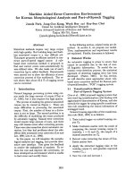
Plate.1
Inoculated Plant
Healthy Plant
Appearance of Symptoms
Plate1. Pathogenicity Of Oidium mangiferae
1299
Int.J.Curr.Microbiol.App.Sci (2019) 8(5): 1297-1300
Since, the morphology and dimensions of
both the conidiophores and conidia, observed
in the present investigations also resembled to
those as documented by earlier workers and
moreover no cleistothecia could be located.
Hence, the name of the pathogen was retained
as Oidium mangiferae Berthet.
Pathogenicity test
The pathogenicity test of powdery mildew
fungus O. mangiferae was conducted on
newly emerged leaves and pedicel of one year
old grafted plants of mango cv. ‘Dusheri’ and
Koch’s postulates were proved. Symptoms on
leaves and pedicels were noticed after 197 hrs
(8 days and 5 hrs) and 260 hrs (10 days and
20 hrs), respectively, of inoculation under the
pot conditions in the laboratory (Table 2) and
(Plate 1).
In the pathogenicity tests, symptoms appeared
after 8 days and 5 hours of inoculation on
leaves whereas on pedicels symptoms
appeared after 10 days and 20 hours.
Adikaram et al., (2002) also calculated the
incubation period to the tune of 10-11 days
for
O.
mangiferae
on
Pedilanthus
tithymaloides var. caculatus. Similarly,
Iliyukhin and Nikitana (1980) reported the
incubation period of 5 to 7 days for powdery
mildew on okra and 12 days in case of
powdery mildew on apple (Kaspers, 1967).
In conclusion, the fungus was of barrel to
ellipsoid – ovoid shape, single celled conidia
produced in chains of two to four and
mycelium was superficial, hyaline and
septate. Newly emerged leaves were found to
be more susceptible to the infection. It clearly
establishes the involvement of O. mangiferae
as the causal agent of this disease in Himachal
Pradesh.
References
Adikaram N K B, Manilewa G and Weerahewa
D. 2002. Changes in pigment composition
and metabolism, etc. in Pedilanthus
tithynaloides leaf following powdery
mildew infection. J. Nati. Sci. Foundation
Sri Lanka. 3(2): 91-111
Howard R J, Garland J A and Seaman W L.
1994. Diseases and pests of vegetables
crops in Canada: an illustrated
compendium.
The
Canadian
Phytopathological
Society,
Ottawa,
Canada. Pp. 1-10.
Iliyukhin G P and Nikitnia M A. 1980. Powdery
mildew of cucumber undercover.
Phytopatho.19:49-51.
Jhooty J S, Kaur J and Munshi G D.
1983.Identify of powdery mildew of
Jujuba and mango. Trop. Pl. Sci. Res. 1:
267-268
Kaspers H. 1967. A contribution to studies on
the biology and control of mildew
(Podosphaera leucotricha) (Ell. And Ev.)
Salm. Pflanzenschuts Naehrichten Bayer.
20: 687–702.
Kulkarni G S. 1924. Report of work done in
Plant Pathology section during the year
1922-23. Annual Report Agriculture. Pp.
23: 167–71.
Palti J, Pinkas Y and Chorin M.1974. Powdery
mildew of mango. Plant Dis.Reptr. 58:
45–49.
Prakash O M and Shrivastava K C. 1987.
Mango diseases. In: Mango diseases and
their management. Today and tomorrow’s
printers and publishers, New Delhi. 121
Pp.
How to cite this article:
Lovepreet Kaur. 2019. Morphological Characterization and Pathogenicity of Oidium mangiferae on
Mango. Int.J.Curr.Microbiol.App.Sci. 8(05): 1297-1300.
doi: />
1300