Haemonchus Contortus infection and associated pathological changes in a goat (Capra hircus)
Bạn đang xem bản rút gọn của tài liệu. Xem và tải ngay bản đầy đủ của tài liệu tại đây (280.44 KB, 4 trang )
Int.J.Curr.Microbiol.App.Sci (2019) 8(3): 2111-2114
International Journal of Current Microbiology and Applied Sciences
ISSN: 2319-7706 Volume 8 Number 03 (2019)
Journal homepage:
Original Research Article
/>
Haemonchus contortus Infection and Associated Pathological
Changes in a Goat (Capra hircus)
Shailesh Kumar Patel1, Jigyasa Rana2*, Poornima Gumasta1, Dhananjay Kumar Jolhe1
Pankaj Kumar Patel3 and Anish Kumar Sonwani1
1
Department of Veterinary Pathology, College of Veterinary Science & A.H., CGKV, Anjora,
Durg-491001 Chhattisgarh, India
2
Department of Veterinary Anatomy and Histology, Nagpur Veterinary College, MAFSU,
Nagpur-440006 Maharashtra, India
3
Division of Medicine, ICAR-Indian Veterinary Research Institute, Izatnagar-243122, India
*Corresponding author
ABSTRACT
Keywords
Goat, Haemonchus
contortus,
Abomasum, Faecal
examination,
McMaster
technique
Article Info
Accepted:
18 February 2019
Available Online:
10 March 2019
An adult female goat was presented for the post-mortem examination to the
Department of Veterinary Pathology, College of Veterinary Science &
A.H., Anjora, Durg, Chhattisgarh. The animal had emaciated body, pale
mucous membranes with the history of anorexia and loss of body weight.
Upon necropsy, the internal organs were found extremely pale, which was
suggestive of anaemia. Liver of the animal was hemorrhagic and enlarged.
Abomasum of the animal was found filled with the blood-sucking parasites
Haemonchus contortus spp. and severe abomasitis was observed. Fecal
sample collected for microscopic examination revealed 1150 egg per gram
indicating severe parasitic infestation.
Introduction
The alimentary canal of vertebrate represents
one of the most favorable habitats for
numerous helminths that cause structural and
functional changes in the digestive tract
(Berrilli et al., 2012). Pathological effects due
to parasitic infection are varied and more
pronounced in sheep and goat compared to
those seen in other species of livestock (Iqbal
et al., 1993). Haemonchus contortus is an
important blood-sucking parasite of sheep and
causes an insidious drain on production
(Hussain et al., 1967). Although the parasite
is prevalent wherever sheep and goats are
raised, but it exerts the greatest economic
losses in temperate and tropical regions
(Blood, 1979). Haemonchus contortus is a
blood-sucking parasite generally found in the
abomasum of the sheep and goat causes
2111
Int.J.Curr.Microbiol.App.Sci (2019) 8(3): 2111-2114
significant loss of blood. Each worm sucks
around 0.05 ml blood per day resulting
production loss, severe anaemia and even
death of the animal (Urquhart et al., 1987).
In the present study severe parasitic infection
of Haemonchus contortus in a goat has been
reported.
Materials and Methods
followed by proper mixing. 1 ml of an above
mixture (faecal suspension) was mixed with
1ml of saturated sugar solution. A drop of the
solution was placed into McMaster slide to
cover the counting chamber and was left for a
few minutes. The eggs were counted using a
microscope. To calculate eggs per gram of
faeces, total count (Chamber 1 + Chamber 2)
was multiplied with 50 (Lyndal-Murphy,
1985).
A female goat of three years of age was
presented for the post-mortem examination to
the Department of Veterinary Pathology,
College of Veterinary Science & A.H.,
Anjora, Durg Chhattisgarh. The detailed
necropsy examination was carried out and all
the gross pathological changes were recorded
carefully. Faecal sample collected from the
abomasum and different parts of the intestine
during necropsy examination subjected to the
qualitative and quantitative examination.
Results and Discussion
A qualitative examination of faecal Sample
has been performed by saturated salt solution,
knowing the fact that parasitic ova, being
lighter, float on the top of fluid and thus can
be concentrated for examination. Briefly, 1 g
faecal sample was taken, mixed with few
milliliters of distilled water and filtered
through a fine sieve. The filtrate was
centrifuged and the supernatant was
discarded. The sediment was mixed with 4-5
ml of saturated salt solution. The material was
transferred into a tube and filled up to the top
with a saturated salt solution. A clean glass
coverslip was placed on the mouth of the tube
and left for 30 minutes. Finally, the coverslip
is removed and examined under a microscope
for the presence of eggs (Chauhan and
Chandra, 2007).
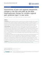
Lungs of the animal were found pneumonic
and liver was slightly enlarged and
hemorrhagic. Abomasum of the goat was
found filled with the adult Haemonchus spp.
most of the parasites were found attached
with the mucous membrane and some were
mixed with the ingesta. Pathological lesions
like the change in colour from pale to pink,
excess secretion of mucous and development
of oedematous folds have been observed in
the abomasum (Fig. 1C). Haemonchus
contortus parasite was identified by observing
bursa of male (Fig. 2B) and vulvar flap of
female (Fig. 2C).
The quantitative examination was performed
by the McMaster technique. In this method, 2
g of faecal sample was collected in a beaker
and 28 ml of distilled water was added
The clinical history taken from the farm
manager revealed that the housed animals
were dull, anorectic, pale and reducing weight
constantly. External body condition of the
animal was rough and emaciated along with
pale conjunctiva (Fig. 1A). Post mortem
examination showed extreme pale viscera
indicating severe anaemia (Fig. 1B). Serous
exudates were found in the abdominal cavity.
Fecal sample subjected to qualitative
examination revealed abundant eggs of
Hemonchus spp. (Fig. 2A). Quantitative
examination of the faecal sample revealed
1150 EPG (egg per gram) showing the severe
category of parasitic infection indicating the
requirement of treatment of the surviving
goats (Machen et al., 1998).
2112
Int.J.Curr.Microbiol.App.Sci (2019) 8(3): 2111-2114
Fig.1 A: Showing pale conjunctiva of dead goat; B: Showing extremely pale viscera of the goat;
C: Showing congested mucous membrane of abomasum along with oedema
Fig.2 A: Eggs of Haemonchus spp. parasite (x400); B: Showing bursa of the male parasite
(x400); C: Showing vulvar flap of the female parasite (x100)
Our results corroborated with many
researchers. Dutta et al., (2017) reported
highly anaemic carcass with pale to papery
white visible mucous membranes. In most
cases, the carcasses were emaciated and
abomasal contents were mixed with blood and
a large number of adult Haemonchus
contortus parasites. Oedematous folds,
petechial haemorrhage and nodule formation
in the infected abomasum may be due to the
piercing activity of the worm (Tehrani, 2012).
Rinaldi et al., (2011) suggested that over
secretion of mucous in the parasitized
abomasum may be due to host reaction
against parasite for their defense, as it has
been reported that mucous layer acts as a
physical barrier for microorganisms, parasites
and their toxins. Faecal eggs of Haemonchus
contortus was also identified by Dutta et al.,
(2017) in his study which was similar to our
case.
In conclusion, the present case indicated that
animal was suffering from severe parasitic
infection
leading
to
anaemia
and
immunosuppression resulting in the death of
the animal. This also provided an idea of
heavy worm infestation of that particular farm
requires antihelminthic drug treatment as soon
as possible.
References
Bailly, V., Zhang, Z., and Meier, W. 2002.
Shedding of kidney injury molecule-1,
a putative adhesion protein involved in
2113
Int.J.Curr.Microbiol.App.Sci (2019) 8(3): 2111-2114
renal
regeneration.
Journal
of
Biological Chemistry. 277: 3973939748.
Berrilli, F., Di Cave, D., Cavallero, S. and
D’Amelio, S. 2012. Interactions
between parasites and microbial
communities in the human gut.
Frontiers
in Cellular and Infection
Microbiology.2: 141.
Blood, D. C., Henderson, J. A., and Radostits,
A. M. 1979. Veterinary Medicine. 5th
Ed., Bailliere Tindall, London, UK.
Chauhan, R.S. and Chandra, D. 2007.
Veterinary Laboratory Diagnosis. 2nd
Ed., International Book Distributing
Co. Lukhnow.
Dutta, B., Konch, P., Rahman, T.,
Upadhyaya, T. N., Pathak, D. C.,
Phangchoo, C. V., and Begum, S. A.
2017. Occurrence and pathology of
Haemonchus contortus infection in
Goats. Journal of Entomology and
Zoology Studies. 5(3): 1284-1287.
Hussain, M. Z., and Akram M. 1967.
Hostparasite relationship Studies on
the productivity of sheep as affected
by haemonchosis. Pakistan Journal of
Medical sciences.5: 247-251.
Iqbal, Z., Akhtar, M., Khan, M. N. and Riaz,
M. 1993. Prevalence and economic
significance of Haemonchosis in
sheep and goats slaughtered at
Faisalabad abattoir. Journal of
Agricultural Science. 30: 51-53.
Lyndal-Murphy.
1985.
The
modified
McMaster method. In Anthelmintic
Resistance in Sheep. Queensland,
Australia: Animal Research Institute,
Queensland Department of Primary
Industries 8.
Machen, R., Craddock, F., Craig, T., and
Fuchs, T. 1998. A
Haemonchus
contortus Management Plan for Sheep
and Goats in Texas. Pamphlet L-5095.
College Station, T.X.: AgriLife
Communications,
Texas
A&M
System.
Rinaldi, M., Dreesen, L., Hoorens, P. R., Li,
R. W., Claerebout, E., Goddeeris, B.,
Vercruysse, J., Broek, W. V. D., and
Geldhof, P. 2011. Infection with the
gastrointestinal nematode Ostertagia
ostertagi in cattle affects mucus
biosynthesis in the abomasum.
Veterinary Research.42: 61.
Tehrani, A., Javanbakht, J., Jani, M., Sasani,
F., Solati, A., Rajabian, M., Khadivar,
F., Akbari, H., and Mohammadian, M.
2012. Histopathological Study of
Haemonchus contortus in Herrik
Sheep
Abomasum.
Journal
of
Bacteriology and Parasitology. 3 (5):
144.
Urquhart, G. M., Armour, J., Duncan, J. L.,
Dunn, D. M., and Jennings, F. W.
1987. Veterinary parasitology. English
Language Book Society, England.
How to cite this article:
Shailesh Kumar Patel, Jigyasa Rana, Poornima Gumasta, Dhananjay Kumar Jolhe Pankaj
Kumar Patel and Anish Kumar Sonwani. 2019. Haemonchus contortus Infection and
Associated Pathological Changes in a Goat (Capra hircus). Int.J.Curr.Microbiol.App.Sci.
8(03): 2111-2114. doi: />
2114