Isolation, pathogenicity and effect of different culture media on growth and sporulation of alternaria brassicae (berk.) Sacc. causing alternaria leaf spot disease in cauliflower
Bạn đang xem bản rút gọn của tài liệu. Xem và tải ngay bản đầy đủ của tài liệu tại đây (540.23 KB, 11 trang )
Int.J.Curr.Microbiol.App.Sci (2019) 8(4): 1900-1910
International Journal of Current Microbiology and Applied Sciences
ISSN: 2319-7706 Volume 8 Number 04 (2019)
Journal homepage:
Original Research Article
/>
Isolation, Pathogenicity and Effect of Different Culture Media on Growth
and Sporulation of Alternaria brassicae (berk.) Sacc. causing Alternaria Leaf
Spot Disease in Cauliflower
H.T. Valvi, J.J. Kadam and V.R. Bangar*
Department of Plant Pathology, College of Agriculture, Dapoli, India
Dr. Balasaheb Sawant Konkan Krishi Vidyapeeth, Dapoli, Dist.,
Ratnagiri- 415 712(M.S.), India
*Corresponding author
ABSTRACT
Keywords
Cauliflower, Isolation,
Pathogenicity,
Alternaria brassicae
(Berk.) Sacc., Culture
media, Sporulation
and mycelial growth
Article Info
Accepted:
15 March 2019
Available Online:
10 April 2019
The leaf spot disease of Cauliflower (Brassica oleracae L. var. Botrytis) caused by A.
brassicae (Berk.) Sacc. was noticed in moderate to severe form on farm of College of
Agriculture Engineering and Technology, Dapoli during 2014-2016. The pathogenic
fungus was isolated on potato dextrose agar medium from infected leaves of cauliflower.
The pathogenicity of the isolated fungus was proved by inoculating healthy seedlings of
cauliflower. On the basis of typical symptoms on foliage, microscopic observations and
cultural characteristics of the fungus, it was identified as Alternaria spp. The Chief
Mycologist, Agharkar Research Institute, Pune identified the pathogenic fungus as
Alternaria brassicae (Berk.) Sacc. Eight culture media were tested among that, the potato
dextrose agar medium was found most suitable and encouraged maximum radial mycelial
growth (90.00 mm) of A. brassicae. The second best culture medium found was host leaf
extract agar medium (87.00 mm). This was followed by Richard’s agar medium (75.33
mm) and oat meal agar medium (71.66 mm). Carrot potato agar medium (55.00 mm),
Asthana and Hawker’s agar medium (51.66 mm) and Czapek’s Dox agar medium (39.00
mm) were moderate in mycelial growth of A. brassicae. Poor mycelial growth was
recorded in V8 juice agar medium (32.66 mm).
Introduction
‘caulis’ meaning
meaning flower.
Cauliflower (Brassica oleracae L. var.
Botrytis) belongs to cruciferae/brassicae
family. It is originated from wild cabbage
known as ‘Cole warts’, through mutation,
human selection and adoption. The name
cauliflower consists of two Latin words,
Cauliflower is one of the most important
winter vegetables of India. India produces
8573.3 MT of cauliflower in the year 2013-14
from 433.9 ha. area with an average
productivity of about 19.8mt/ha. In
1900
cabbage
and
‘floris’,
Int.J.Curr.Microbiol.App.Sci (2019) 8(4): 1900-1910
Maharashtra, the area under cauliflower is
36.0 ha with total production of 813.2mt and
average productivity of 22.6 MT/ha in the
year 2013-14. The major cauliflower
producing states are Bihar, Uttar Pradesh,
Orissa, West Bengal, Assam, Haryana and
Maharashtra (Anonymous, 2013).
Disease sample
Several factors are responsible for low
production of cauliflower crop, among which
diseases also play an important role. The
important diseases of Cauliflower crop are
leaf spot, Downy mildew, Damping off, Club
root, Powdery mildew, White rust, Black rot,
Bacterial soft rot and Cauliflower mosaic.
Among these diseases Alternaria leaf spot is a
serious disease of cauliflower.
Microscopic examination
The disease appears as minute specks on the
leaves, which enlarge over a time and result in
substantial lesions with concentric rings
where spores are produced.
Isolation of causal organism
Defoliation of the outer leaves may occur on
severely infected plants and extensive
trimming may be required to remove infected
leaves from the cabbage head at harvest. In
susceptible varieties, apart from yield,
significant reduction in quality may occur.
Considering importance of the crop and
disease, present study was planned and
conducted with isolation, pathogenicity and
cultural characteristics of pathogen to know
its survival, association with host plant in
Konkan region and in vitro characteristics and
further management studies.
The cauliflower leaves showing typical
symptoms of leaf spot were collected in the
paper bags from the College of Agricultural
Engineering and Technology, Dapoli and
brought to the laboratory for further studies.
These samples were then washed under tap
water to remove extraneous material.
Temporary mounts were prepared from the
diseased specimens in lacto-phenol cotton
blue and examined under compound
microscope for presence of microorganism if
any.
Small bits of desired size of infected samples
were cut by taking care that each bit
contained half infected and half healthy
portion. Such bits were then disinfected with
0.1 per cent mercuric chloride (HgCl2) for 1
minute followed by three washings in distilled
sterile water to remove the traces of mercuric
chloride.
These bits were then placed on sterilized
blotters for drying. Properly dried bits were
transferred aseptically in sterilized Petri plates
containing sterilized, solidified PDA medium.
Examination of diseased samples
The plates were incubated in BOD incubator
at 24 ± 1°C till the fungal mycelium fully
covered the surface of the medium. The
fungal growth obtained was then transferred
to PDA slants and maintained as stock culture
for further studies.
Visual observations
Pathogenicity test of the isolated organism
Visual observations of disease symptoms
were recorded in the field to know the
development of the disease in a plant
population under natural conditions.
Inoculation
Materials and Methods
Three Seeds of cauliflower (variety-super fast
crop) was sown in the earthen pots containing
1901
Int.J.Curr.Microbiol.App.Sci (2019) 8(4): 1900-1910
desired potting mixture. Potting mixture
comprising FYM and soil (1:2) was
autoclaved for three successive days in order
to kill the micro flora present if any.
original culture obtained from naturally
infected leaves under field conditions.
One healthy growing cauliflower seedling per
pot was maintained and watered regularly.
Spore-cum mycelial suspension of the test
pathogen was prepared by pouring the
distilled sterile water in 7-8 days old culture
plates.
The re-isolated, pure fungal culture was
identified in Department of plant pathology,
college of agriculture, Dapoli, Ratnagiri
comparing its morphological and colony
characters with the information available in
the reviewed literature as well as on the
standard websites for fungal identification.
The resultant spore cum-mycelial suspension
was filtered through muslin cloth and filtrate
obtained was suitably diluted with distilled
sterile water to get inoculum concentration of
105 spores/ml (Pattanamahakul and Strange,
1999).
Forty five days old seedlings of cauliflower
already grown in earthen pots were artificially
inoculated by spraying the spore-cummycelial suspension (105 spores/ml) of the
test fungus with an automizer. Seedlings
grown in earthen pots and sprayed only with
sterile water (without inoculum) were
maintained as control.
Identification of the causal organism
Further, culture was sent to chief Mycologist,
Agharkar Research Institute, Pune for
identification of the fungus up to species
level.
Effect of culture media on growth and
sporulation
Five synthetic and three non-synthetic media
were evaluated in the present study. Media
were prepared with given composition. The
initial pH of each medium was adjusted to 6.5
prior to autoclaving. The medium was
prepared with given composition and
dispended in conical flask.
Development of symptom
Pots (both inoculated and non-inoculated)
were incubated in the moist chamber prepared
with a wooden frame covered with a muslin
cloth. Proper humidity (85-90%) was
maintained in the chamber by frequently
spraying sufficient clean water on the muslin
cloth. Seedlings were watered as and when
required till the development of typical
disease symptoms.
The causal organism was re-isolated from the
artificially inoculated leaves showing typical
symptoms of the disease.
The fungal growth obtained on PDA medium
on re-isolation was compared with the
The flasks were plugged with non-absorbent
cotton plugs and sterilized in an autoclave at
15 lbs. psi for 20 minutes. Petri plates were
sterilized in hot air oven at 1600C for 1 hour.
Such sterilized Petri plates were poured with
20 ml of molten medium and allowed to
solidify. Five millimetre diameter disc of the
test fungus was cut with the help of
incinerated cork borer and inoculated at the
centre of Petri plates.
The inoculated plates were then incubated at
room temperature (27 ± 20C) for 7 days. The
compositions of all the media used were
obtained from Ainsworth and Bisby’s
Dictionary of the fungi as mentioned below.
1902
Int.J.Curr.Microbiol.App.Sci (2019) 8(4): 1900-1910
Media used and their composition
1)
A)
Czapek's Dox agar medium
i)
Sucrose (C12H22O11)
ii)
Sodium nitrate (NaNO3)
iii)
Potassium dihydrogen phosphate (KH2PO4)
iv)
Magnesium sulphate (MgSO4.2H2O)
v)
Potassium chloride (KCl)
vi)
Ferric chloride (FeCl3)
vii)
Agar-agar
viii)
Distilled water
Asthana & Hawker's agar medium
i)
Glucose (C6H12O6)
ii)
Potassium Nitrate (KNO3)
iii)
Potassium dihydrogen phosphate (KH2PO4)
iv)
Magnesium sulphate (MgSO4. 2H2O)
v)
Agar-agar
vi)
Distilled water
Richard’s agar medium
i)
Sucrose
ii)
Potassium dihydrogen phosphate (KH2PO4)
iii)
Potassium nitrate (KNO3)
iv)
Magnesium sulphate (MgSO4.2H2O)
v)
Ferric chloride (FeCl3)
vi)
Agar-agar
vii)
Distilled water
B) Non-synthetic media:
1)
Oat meal agar medium
:
:
:
60.00 g
20.00 g
1000 ml
2)
i)
Oat meal
ii)
Agar-agar
iii)
Distilled water
Potato dextrose agar (PDA) medium
:
:
:
:
200.00 g
20.00 g
20.00 g
1000 ml
3)
i) Peeled potato
ii) Dextrose
iii) Agar-agar
iv)
Distilled water
Vegetable 8 (V8) juice agar medium
i)
V8-agar
ii)
Distilled water 1000 ml
Potato carrot agar
i) Potato extract (as made above)
ii) Carrot extract (as made above)
iii)
Agar
iv) Distilled water
Host leaf extract agar medium
:
:
44.3 g
1000 ml
:
:
:
:
250.0 ml
250.0 ml
15.0 g
500.0 ml
i)
ii)
iii)
:
:
:
100 ml
20.00 g
900 ml
2)
3)
4)
5)
Cauliflower leaves extract (10 %)
Agar-agar
Distilled water
1903
:
:
:
:
:
:
:
:
30.00 g
2.00 g
1.00 g
0.50 g
0.50 g
0.01 g
20.00 g
1000 ml
:
:
:
:
:
:
5.00 g
3.50 g
1.75 g
6.75 g
20.00 g
1000 ml
:
:
:
:
:
:
:
50.00 g
5.00 g
10.00 g
2.50 g
0.02 g
20.00 g
1000 ml
Int.J.Curr.Microbiol.App.Sci (2019) 8(4): 1900-1910
For preparing host leaf extract medium,
cauliflower leaves were chopped and ground
with the help of mixer. The extract was
strained through muslin cloth and volume
made to 1 lit.
Twenty millilitre of each medium as listed
above was poured into sterilized Petri plate,
separately. After solidification, 5 mm culture
discs of the test fungus from actively growing
7 days old fungal culture were cut using
sterilized cork borer and a single disc was
placed at the centre of each Petri plate and
incubated at 27 ± 2ºC. Each treatment was
replicated thrice. The measurement of the
colony diameter was taken when the
maximum (90mm) growth was achieved in
any one of the media tested. The cultural
characters such as colony diameter, colony
colour and degree of sporulation were
recorded.
these spot enlarged into gray to black lesions
of 0.5 to 1 cm diameter. As the disease
progressed, the lesions attended target board
pattern due to formation of many concentric
rings with wavy margin. Centres were coated
with sooty black spore masses and later it
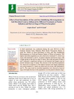
drop out, producing shot holes. (PLATE-I)
Microscopic examination
Temporary mounts were prepared from the
diseased samples in lacto-phenol cotton blue.
Microscopic examination revealed the
presence of fungal structures such as
mycelium and conidia. The conidia were
obclavate, muriform with a long beak with
both transverse (10 to 11) and longitudinal (2
to 3) septa. The conidia were slightly
constricted at transverse septa. Conidia were
light brown to gray coloured and measured
104.0-142.0 × 11.62-16.95 µm. Length of
beak was 43.35-70.57 µm. (PLATE-II)
Statistical analysis
Isolation and proving pathogenicity
The data obtained were statistically analysed
by the methods suggested by Gomez and
Gomez (1986). The standard error and critical
difference were worked out and the results
obtained were compared statistically.
Results and Discussion
Examination of diseased samples
Visual observation
The leaf spot disease of cauliflower (Brassica
oleracae L. var. Botrytis) caused by
Alternaria brassicae (Berk.) Sacc. was
noticed in moderate to severe at the farm of
College of Agriculture Engineering and
Technology, Dapoli during November, 2015.
The disease appeared initially as small,
circular, dark, yellow spots on the lower
leaves. Later on these spots enlarged into
circular areas with concentric rings and
possibly surrounded by yellow halo. Later on
The pathogen was isolated successfully on
potato dextrose agar medium from the
diseased tissue showing well developed
lesions along with healthy portion which were
brought to the laboratory from naturally
infected cauliflower plants. The inoculated
plates were incubated in BOD incubator at 25
± 20C for 5 to 7 days. The culture of the
fungus obtained by isolation from diseased
tissue was transferred to PDA in Petri plates
and multiplied in the laboratory.
Purification of fungal culture
The test fungus produced greenish grey to
black coloured, fluffy, lanose to loose cottony
growth on potato dextrose agar medium after
seven days of incubation. The slants of the
pure culture were sealed with paraffin wax
and maintained in the laboratory in
refrigerator for further use.
1904
Int.J.Curr.Microbiol.App.Sci (2019) 8(4): 1900-1910
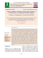
Inoculation of fungal culture
Healthy growing, 25 days old seedlings of
cauliflower (Variety-supper fast crop) were
used for pathogenicity test. Seedlings after
making injuries on leaves by pinning were
inoculated by spraying with the spore
suspension. After 8 - 10 days of incubation,
typical symptoms of blight on foliage of
artificially inoculated plant were observed
(PLATE-III). The lesions on the artificially
inoculated plant also exhibited conidia
formation. However, the plant kept as control,
which was sprayed only with sterilized water
did not produced any kind of symptoms.
The fungus was reisolated on PDA from
artificially inoculated plants showing typical
blight symptoms and was found to be
identical to original isolate. Thus, the
pathogenicity of the isolated fungus was
proved on cauliflower. In present study,
symptoms developed on artificial inoculated
cauliflower plants were similar to those
observed in field. Gaikwad (2013) also
proved pathogenicity of A. brassicae by
inoculating one month old seedlings of
cabbage with the spore-cum-mycelial
suspension (2×10⁵). Similarly Sharma et al.,
(2013) also proved pathogenicity of A.
brassicae on detached leaves of cauliflower
and mustard. The findings are also in
agreement with Giri et al., (2013) and Deep et
al., (2014)
Identification of causal organism
Based on the typical symptoms on foliage,
microscopic observations and cultural
characteristics of the fungus, it was tentatively
identified as Alternaria spp. This proved that
the pathogen responsible for causing leaf spot
disease of cauliflower was Alternaria spp.
Further, the Chief Mycologist, Agharkar
Research Institute, Pune, confirm the
identification of the pathogenic fungus as
Alternaria brassicae (Berk.) Sacc. Thus the
study revealed that leaf spot of cauliflower
under present study was caused by Alternaria
brassicae (Berk.) Sacc. The pathogen was
easily isolated on potato dextrose agar
medium. On PDA, the fungus produced
greenish grey to black fluffy, lanose to loose
cottony growth which resembled to the
colony of Alternaria brassicae. The A.
brassicae was already reported to be isolated
from diseased tissue of cauliflower leaves by
Deep and Sharma (2012), Reshu et al.,
(2012), Gaikwad 2013, Sharma et al., (2013),
Chand and Chandra (2014), Deep et al.,
(2014), Taware et al., (2014) and Koley and
Mahapatra (2015).
Effect of culture media on growth and
sporulation of Alternaria brassicae (Berk.)
Sacc.
Growth and sporulation of A. brassicae were
studied in vitro using eight synthetic and nonsynthetic culture media.
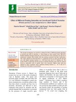
The data from Table 1, PLATE-IV and Figure
1 revealed that of the eight culture media
tested, potato dextrose agar medium was
found most suitable and encouraged
maximum radial mycelial growth (90.00mm)
of A. brassicae. The second best culture
medium found was host leaf extract agar
medium (87.00 mm). This was followed by
Richard’s agar medium (75.33 mm) and oat
meal agar medium (71.66 mm). Carrot potato
agar medium (55.00 mm), Asthana and
Hawker’s agar medium (51.66 mm) and
Czapek’s Dox agar medium (39.00 mm) were
moderate in mycelial growth of A. brassicae.
Poor mycelial growth was recorded in V8
juice agar medium (32.66 mm).
Excellent sporulation of A. brassicae was
observed in potato dextrose agar medium and
host leaf extract agar medium. Good
1905
Int.J.Curr.Microbiol.App.Sci (2019) 8(4): 1900-1910
sporulation was observed in Richards’s agar
medium and oat meal agar medium. Fair
sporulation was observed in carrot potato agar
medium and Asthana and Hawker’s agar
medium and it was poor in Czapek’s Dox
agar medium. The results of present
investigation are in close conformity to Singh
(1980) who reported that oat meal agar media
best for growth and sporulation of A.
brassicae. Similarly, Sharma et al., (2013)
also reported that potato dextrose agar and
cauliflower agar medium were optimum for
fungal growth of A. brassicae. Deep et al.,
(2014) also reported that cauliflower leaf
extract medium and potato dextrose agar were
appeared optimum for growth and sporulation
of the fungus.
Plate.1 Field symptoms of leaf spot of cauliflower caused by Alternaria brassicae
Plate.2 Culture of Alternaria brassicae and its spores
1906
Int.J.Curr.Microbiol.App.Sci (2019) 8(4): 1900-1910
Plate.3 Inoculation of Alternaria brassicae
Uninoculated plant (Healthy)
Inoculated plant (Infected)
Plate.4 Effect of different culture media on mycelial growth and sporulation
1907
Int.J.Curr.Microbiol.App.Sci (2019) 8(4): 1900-1910
Table.1 Effect of different culture media on mycelial growth and sporulation of A. brassicae.
(Berk.) Sacc.
Tr. No
T1
T2
T3
T4
T5
T6
T7
T8
Culture media
V8 juice agar medium
Host leaf extract agar medium
Czapek’s Dox agar medium
Carrot potato agar medium
Oat meal agar medium
Asthana and Hawker’s agar
medium
Richards agar medium
Potato dextrose agar medium
S.Em.±
C.D. at 1%
Average mycelial
growth (mm)
32.66
87.00
39.00
55.00
71.66
51.66
Sporulation
75.33
90.00
0.70
2.94
+++
++++
+
++++
+
++
+++
++
Sporulation
+
++
= No sporulation,
= Poor,
= Fair,
+++
++++
= Good,
= Excellent.
Fig.1 Effect of different culture media on mycelial growth of A. brassicae
1908
Int.J.Curr.Microbiol.App.Sci (2019) 8(4): 1900-1910
In conclusion, on the basis of the results of
present study it can be concluded that
Alternaria leaf spot of cauliflower caused by
Alternaria brassicae (Berk.) Sacc. is an
important disease of cauliflower in Konkan
region. Among the various biotic factors
responsible for low production and
productivity of cauliflower, Alternaria leaf
spot caused by Alternaria brassicae (Berk.)
Sacc. is one of the constraints.
References
Anonymous, 2013. Area, Production and
productivity of cauliflower in India.
Indian Horticulture Database Pp. 141151.
Chand, G. and Chandra, K. K. 2014.
symptomological, cultural and molecular
variability of Alternaria brassicicola leaf
spot in broccoli (Brassica oleracea var.
Ltalica L.). International Journal of
Pharma and Bio Sciences., 5 (2): 680 –
688.
Deep S., Sharma P., Behera N. and
Chowdappa P. 2014. Diversity in Indian
Isolates of Alternaria brassicicola
(Schwein) Wiltshire Causing Black Leaf
Spot Disease in Cauliflower. Plant
Pathology Journal., pp.1-14
Deep Swati and Sharma Pratibha. 2012. Host
age as predisposing factor for incidence
of black leaf spot of cauliflower caused
by Alternaria brassicae and Alternaria
brassicicola. Indian phytopath., 65(1):
71-75.
Gaikawad, P.A. 2013. Studies on Leaf Spot of
Cabbage Caused by Alternaria brassicae
(Berk.) Sacc. M.Sc. (Ag) Thesis,
submitted to V.N.M.K.V., Parbhani,
Maharashtra.
Giri, P., Taj, G., Meena, P.D. and Kumar, A.
2013. Microscopic study of Alternaria
brassicae infection processes in Brassica
juncea cultivars by drop plus agarose
method. Afr. J. Microbial. Res., 7: 4284-
4290.
Gomez, K. A. and Gomez, A.A. 1986.
Statistical Procedures for Agricultural
Research. 2nd ed. John Wiley and Sons,
London, 680.
Koley, S. and Mahapatra, S. S. 2015.
Evaluation of Culture Media for Growth
Characteristics of Alternaria solani,
Causing Early Blight of Tomato. Plant
Pathol Microbiol., 5(5): 312-314.
Pattanamahakul, P. and Strange, R. N. 1999.
Identification and toxicity of Alternaria
brassicicola, the causal agent of dark leaf
spot disease of Brassica species grown in
Thailand. Plant Pathology, 48(6):749755.
Peruch L.A.M., Michereff S., Araujo I. B.
2006. Survey of intensity of Alternaria
black spot and black rot on brassica
species under organic farming systems in
Pernambuco and anta catarina states,
Brazil. Horti.Bras., 24(4).
Reshu and Khan, M. M. 2012. Role of
different Microbial-origin bioactive
antifungal compounds against Alternaria
spp. causing leaf blight of Mustard. Plant
Pathol. J., 11(1): 1-9.
Sharma, P., Deep, S., Sharma, M. and Bhati,
D.S. 2013. Genetic variation of
Alternaria brassicae (Berk) Sacc.
causing dark leaf spot of Cauliflower and
Mustard in India. J. Gen. Plant
Pathology. 79: 41-45.
Singh, D.B 1980. Effect of culture media, pH
and temperature on growth behavior of
Alternaria brassicae and Drechslera
graminae. Proc.Indian natn.Sci.Acad.,
46(3): 393-396.
Sreedhar,
K.N.,
Ashokkumar,
C.T.,
Padamabha, K. and Sudhirkumar, A.S.
(2013). Compatibility of insecticides and
fungicides mixtures against cabbage leaf
spot, Alternaria brassicae (Sacc.) Berk.
The Asian Journal of Horticulture.,
8:659-666.
Surviliene, E. and Dambrauskiene, E. (2006).
1909
Int.J.Curr.Microbiol.App.Sci (2019) 8(4): 1900-1910
Effect of Different active Ingredients of
Fungicides on Alternaria spp. growth in
vitro. Agronomy Research, pp: 403–406.
Thaware, D. S., Fugro, P. A., Jadhav, Y. T.,
Magar, S. V. and Karande, R. A. 2010. In
vitro evaluation of different fungicides,
plant extracts and bio-agents against
Alternaria alternata (Fr.) Keissler
causing leaf blight of cowpea.
International Journal of Plant., 3(2):
356-360.
How to cite this article:
Valvi, H.T, J.J. Kadam and Bangar, V.R. 2019. Isolation, Pathogenicity and Effect of Different
Culture Media on Growth and Sporulation of Alternaria brassicae (berk.) Sacc. causing
Alternaria Leaf Spot Disease in Cauliflower. Int.J.Curr.Microbiol.App.Sci. 8(04): 1900-1910.
doi: />
1910