New records for the macromycota of Turkey from Balikesir province
Bạn đang xem bản rút gọn của tài liệu. Xem và tải ngay bản đầy đủ của tài liệu tại đây (218.63 KB, 4 trang )
Turk J Bot
29 (2005) 333-336
â TĩBTAK
Research Note
New Records for the Macromycota of Turkey from Balkesir
Province
Fadime YILMAZ ERSEL
Fungus Program, Ula Ali Koỗman Vocational High School, MuÔla University, 48640 Ula, MuÔla - TURKEY
Received: 05.08.2004
Accepted: 07.04.2005
Abstract: Macrofungi samples recorded in this study were collected from different localities in Balkesir province from 1998 to
2001. As a result of the field and laboratory studies, 4 taxa belonging to 4 families were recorded for the first time in Turkey. These
are Hebeloma mesophaeum (Pers.) Fr. var. lacteum Vesterh., Lepiota griseovirens Maire, Lactarius violascens (J.Otto) Fr., and
Melanoleuca griseofumosa Secr. ex Singer & Clộmenỗon.
Key Words: Turkish macromycota, Taxonomy, Basidiomycetes
Balkesir linden Tỹrkiye Makromikotas ỗin Yeni Kaytlar
ệzet: Bu ỗalflmada kaydedilen makrofungus ửrnekleri 1998 ile 2001 yllar arasnda Balkesir ilinin farkl lokalitelerinden
toplanmfltr. Arazi ve laboratuar ỗalflmalar sonucunda 4 familyaya ait 4 takson Tỹrkiyede ilk kez kaydedilmifltir. Bunlar Hebeloma
mesophaeum (Pers.) Fr. var. lacteum Vesterh., Lepiota griseovirens Maire, Lactarius violascens (J.Otto) Fr., ve Melanoleuca
griseofumosa Secr. ex Singer & Clộmenỗondir.
Anahtar Sửzcỹkler: Tỹrkiye makromikotas, Taksonomi, Basidiomycetes
Introduction
During field studies from 1998 to 2001, macrofungi
specimens were collected from Balkesir province. Among
these specimens 4 new taxa records for Turkey were
determined.
Many species of macrofungi growing in Balkesir have
already been reported (Ylmaz et al., 1997; Aflkun &
IflloÔlu, 1997; IflloÔlu et al., 2001, 2001a; Ylmaz &
IflloÔlu, 2002; Solak et al., 2002), resulting in a list of
200 taxa. For Turkey, many studies on macrofungi have
been carried out (Mat, 2000), resulting in 300 articles by
many scientists and the number of taxa reaching
approximately 1300. With this study, 4 new taxa have
been added to the macromycota of Turkey.
Materials and Methods
During field studies, the morphological and ecological
characteristics of the macrofungi were recorded and
photographed in their natural habitats and then they
were brought to the laboratory. Their spore prints were
taken and their spores were photographed. Dried
specimens were numbered and placed in sealed bags. In
addition, they were put into a deep freeze for a week to
protect against internal and external parasite attacks.
The specimens were identified with the help of
macroscopic and microscopic features. Keys by Marchand
(1980), Phillips (1981), Moser (1983), Breitenbach &
Krọnzlin (1984), Vesterholt (1989), Zhishu et al. (1993)
and Heilmann-Clausen (2000) were used. All specimens
collected are preserved in the fungarium of MuÔla
University.
Results
The newly recorded taxa from this research area, and
their descriptions, synonyms, localities, photographs,
dates of collection and herbarium numbers are given
below.
Cortinariaceae
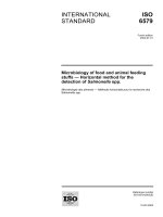
Hebeloma mesophaeum (Pers.) Fr. var. lacteum
Vesterh.
333
New Records for the Macromycota of Turkey from Bal›kesir Province
Cap 30-50 mm, convex to expanded, whitish to
cream, centre light brown, surface smooth, viscid when
moist, dull silky, margin inrolled to incurved. Lamellae
dirty cream, then dark beige, somewhat punctuated
brownish, almost without smell. Stipe cylindric, the same
colours as cap, older stipe browning at base (Figure 1a).
Spores 8-10 x 5-6.5 µ, plum shaped, smooth (Figure 1b).
Balıkesir, De¤irmenbo¤azı picnic area, east of the
watchman’s hut, under cedar trees, 09.11.1998, FY
723.
Balıkesir, De¤irmenbo¤azı picnic area, north of the
children’s playground, in pine forest, 28.11.1998, FY
752.
Cap, 50-120 mm, convex with a slightly depressed
centre, margin at first decurved but gradually expanding,
surface smooth, viscid, ochre-grey-brown to brownish
violet, zonate. Lamellae broadly adnate to decurrent,
crowded, pale cream to ochre, old dark and often rustyspotted, when bruised turning vinaceous purple to slate
purple. Stipe 30-40 x 12-20 mm, cylindric or tapering
downwards, surface smooth, dry, pale cream, then cream
to greyish buff, with yellowish brown spots at the base,
bruised places turning slate purple to dark livid red, flesh
elastic, hollow in the stipe, white, then slowly turning
greyish lilac to dark purple. Milk white (Figure 3a), taste
mild. Spore print pale cream. Spores 8-11 x 6.5-8.5 µ,
subglobose to ellipsoid, ornamented (Figure 3b).
Lepiotaceae
Lepiota griseovirens Maire
Cap 15-25 mm, convex to expanded, markedly
umbonate, surface conspicuously covered with small
grey-green scales, olive black in apex. Lamellae cream to
ochre, darkening slightly at maturity. Stipe 3-5 x 20-40
mm, cylindric, sometimes slightly bulbous, at base same
colour as cap, paler in apex (Figure 2a). Smell unpleasant.
Spores 6-8 x 3-4 µ, slightly angular, hyaline dextrinoid
(Figure 2b).
Russulaceae
Lactarius violascens (J.Otto) Fr.
Syn: Lactarius uvidus var. violascens (J.Otto) Quél.
Figure 1. Hebeloma mesahaeum var. lacteum, a. Fruiting body b. Spores.
334
F. YILMAZ ERSEL
Figure 2. Lepiota griseovirens a. Fruiting body b. Spores.
Figure 3. Lactarius violascens a. Fruiting body b. Spores.
Figure 4. Melanoleuca griseofumosa a. Fruiting body b. Spores.
335
New Records for the Macromycota of Turkey from Balkesir Province
Balkesir, Balkesir-Susurluk road, 25 km, under
bushes, 17.11.1998, FY 778.
Tricholomataceae
Melanoleuca griseofumosa Secr. ex Singer &
Clộmenỗon
Cap 30-40 mm, convex when young, later plain and
sometimes indented in the centre and somewhat obtusely
umbonate, shield shaped, surface smooth, dull, silky,
lighter grey, centre darker, margin incurved and
somewhat frosted. Lamellae broadly adnate, white,
pruinose light grey-cream. Stipe 3-4 x 40-60 mm,
cylindric, slightly enlarged toward the base, grey, surface
longitudinally grey-brown fibrillose, finely pruinose
(Figure 4a). Flesh grey to whitish. Spores print cream.
Spores 7.5-8 x 4.5-5 à, elliptical, smooth (Figure 4b).
Cystidia spindle shaped.
Balkesir, DeÔirmenboÔaz picnic area, east of the
watch-tower, mixed forest, 12.01.2001, FY 1108.
References
Aflkun T & IflloÔlu, M (1997). Macrofungi of Balya (Balkesir) Counry.
Turk J Bot 21: 279-284.
Moser M (1983). Keys to Agarics and Boleti. London: Gustav Fisher
Verlag.
Breitenbach J & Krọnzlin F (1984-1991). Fungi of Switzerland. Vol. 13, Lucerno: Verlag Mycologia.
Phillips R (1981). Mushrooms and Other Fungi of Great Britain and
Europe. London: Pan Books Ltd.
Heilmann-Clausen J, Verbeken A & Vesterholt J (2000). The Genus
Lactarius. Denmark: Jens H. Petersen/ Low Budget Publishing.
Solak MH, Ylmaz Ersel F, Gỹcin F & IflloÔlu M (2002). Macrofungi of
Balkesir Province from Turkey. Bio-Science Research Bulletin
18(2): 137-149.
IflloÔlu M, Ylmaz F & Merdivan M (2001). Concentration of trace
elements in wild edible mushrooms. Food Chemistry 73: 169175.
IflloÔlu M, Merdivan M & Ylmaz F (2001a). Heavy metal contents in
some macrofungi collected in the northwestern part of Turkey.
Archives of Environmental Contamination and Toxicology 41: 17.
Marchand A (1980). Champignons du nord et du midi, Lactaires et
Pholiotes. Tome 6, Perpignan: Sociộtộ Mycologique des Pyrộnộes
Mộditerranộennes,
Mat A (2000). Tỹrkiyede Mantar Zehirlenmeleri ve Zehirli Mantarlar.
stanbul: Nobel Tp Kitabevleri Ltd. fiti.
336
Vesterholt J (1989). A revision of Hebeloma sect. Indusiata in the
Nordic countries. Nordic J Bot 9: 289-319.
Ylmaz F & IflloÔlu M. (2002). Macrofungi of DeÔirmenboÔaz
(Balkesir). Turk J Bot 26: 161-164.
Ylmaz F, ệder N & IflloÔlu M (1997). The macrofungi of Soma
(Manisa) and Sava?tepe (Balkesir) District. Turk J Bot 21: 221220.
Zhishu B, Guoyang Z & Taihui L (1993). The Macrofungus Flora of
Chinas Guangdong Province. Hong Kong: The Chinese University
Press.