Colonial and morphological characteristics of some microfungal species isolated from Agricultural soils in Eskiflehir province (Turkey)
Bạn đang xem bản rút gọn của tài liệu. Xem và tải ngay bản đầy đủ của tài liệu tại đây (1.28 MB, 10 trang )
Turk J Bot
30 (2006) 95-104
â TĩBTAK
Research Article
Colonial and Morphological Characteristics of Some
Microfungal Species Isolated from Agricultural Soils in Eskiflehir
Province (Turkey)
1
2
3
4
5
Semra LHAN , Rasime DEMREL , Ahmet ASAN , Cengiz BAYầU , Engin KINACI
1
Eskiflehir Osmangazi University, Arts and Science Faculty, Biology Department, Eskiflehir - TURKEY
2
Eskiflehir Osmangazi University, Graduate School of Natural and Applies Sciences, Biology Programme, Eskiflehir - TURKEY
3
Trakya University, Arts and Science Faculty, Biology Department, Edirne - TURKEY
4
Eskiflehir Osmangazi University, Medical Faculty, Department of Histology and Embryology, Eskiflehir - TURKEY
5
Eskiflehir Osmangazi University, Agricultural Faculty, Eskiflehir - TURKEY
Received: 04.04.2005
Accepted: 05.12.2005
Abstract: Aspergillus crustosus Raper & Fennell, Eupenicillium egyptiacum (J.F.H.Beyma) Stolk & D.B.Scott, Paecilomyces ramosus
Samson & H.C.Evans, and Penicillium novae-zeelandiae J.F.H.Beyma were examined for their colonial and morphological properties
via visual, light and scanning electron microscopy. These species isolated from soil in different regions of Eskiflehir are recorded for
the first time in Turkey.
Key Words: Soil fungi, Aspergillus, Eupenicillium, Paecilomyces, Penicillium
Eskiflehir Yửresindeki Tarm Topraklarndan zole Edilen Baz Mikrofungus Tỹrlerinin Koloni ve
Morfolojik ệzellikleri
ệzet: Aspergillus crustosus Raper & Fennell, Eupenicillium egyptiacum (J.F.H.Beyma) Stolk & D.B.Scott, Paecilomyces ramosus
Samson & H.C.Evans, ve Penicillium novae-zeelandiae J.F.H.Beyma koloni ve morfolojik ửzellikleri aỗsndan ỗplak gửzle, flk ve
taramal elektron mikroskobu ile incelenmifltir. Eskiflehirde farkl bửlgelerden alnan toprak ửrneklerinden izole edilen bu tỹrlerin
Tỹrkiye iỗin yeni kayt olma olaslÔ yỹksektir.
Anahtar Sửzcỹkler: Toprak funguslar, Aspergillus, Eupenicillium, Paecilomyces, Penicillium
Introduction
Microfungi are important eukaryotic micro-organisms
that affect humans and the majority of living forms in
different ways. Soil microfungi play an important role in
the degradation of organic debris (Barnett & Hunter,
1999). In addition, they are used in industrial and food
fermentation processes, and they exist commonly in
different types of soils, indoor and outdoor air, food and
water. Since microfungi are found almost everywhere,
they are frequently cited in species lists in ecological
studies (Asan, 2004). Aspergillus Link. and Penicillium Fr.
species are commonly found as contaminants in foods
during drying and subsequent storage. Thus, accurate
identification of Aspergillus and Penicillium and related
genera at the species level is essential. Aspergillus and
Penicillium are not easy to identify to the species level. To
further complicate things, the taxonomy of both genera
still needs work, but there appear to be fewer problems
in Aspergillus than in Penicillium. Although molecular,
biochemical and physiological methods are important for
the systematics of these species, morphological properties
are commonly used for identification (Asan, 2004).
The species of Aspergillus, Penicillium and
Paecilomyces Bainer are among the most abundant and
widely distributed microfungi in nature (Pitt, 1979;
Christensen et al., 2000; Klich, 2002; Asan,
95
Colonial and Morphological Characteristics of Some Microfungal Species Isolated from Agricultural Soils in Eskiflehir Province (Turkey)
recorded for the first time in Turkey. Reference strains of
these soil microfungi isolates have been deposited in the
Culture Collections of KUKENS (WDCM101), Centre for
Research and Application of Culture Collections of
Microorganisms. The purpose of this study is to
contribute to the checklist of Aspergillus, Penicillium and
other related species in Turkey, as well as to present
macroscopic and microscopic characteristics of these
species. Descriptions of 4 species which are new records
for the Turkish mycoflora are presented in our study.
2004). A number of species belonging to these genera
have been isolated and identified in studies carried out in
Turkey (Öner, 1970, 1973, 1974; Ekmekçi, 1975;
Haseneko¤lu, 1982, 1985, 1987; Haseneko¤lu & Azaz,
1991; Haseneko¤lu & Sülün, 1990; Asan, 1997; ‹lhan &
Asan, 2001). Morphological studies of microfungi are
rare in Turkey. Eltem et al.’s work in 2004 is an
important investigation about the genus Aspergillus in
Turkey. Since the morphological characteristics of these
genera resemble each other and there are no absolute
criteria for each genus, it can be extremely difficult to
distinguish the species. Pitt & Hocking (1985) discussed
characteristics that could be used to differentiate
Aspergillus and Penicillium from each other, and from the
related genera Raperia Subram & Rajendan,
Paecilomyces, Geosmithia Pitt, Nomuraea Maublanc,
Eladia G.Smith, and Merimbla Pitt.
Materials and Methods
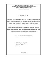
The research areas (Figure 1), Karacahöyük and
Bahçecik, are 25 km and 35 km from the centre of
Eskiflehir (latitude 39° 47', longitude 30° 31') towards
the east, respectively. Osmangazi University experiment
fields I (OGU I) and II (OGU II) are close (approx. 5 km)
to the centre of Eskiflehir. According to the climatologic
data of the past 60 years the annual mean temperature in
this province is 10.8 °C. The mean temperature of the
hottest months (July-August) is 21 °C; the mean
temperature of the coldest months (January- February) is
-0.2 to 1.2 °C. Annual mean precipitation in the region is
25.3 mm and annual relative humidity is 67%. The
climatologic data were obtained from Eskiflehir
Meteorology station.
As a result of the survey, we isolated 110 species
from soil. Identification of the species revealed 13
Aspergillus, 1 Eupenicillium, 4 Paecilomyces and 31
Penicillium species previously reported by our group
(Demirel et al., 2005). According to Asan’s Checklist
(Asan, 2004), Aspergillus crustosus Raper & Fennell,
Eupenicillium egyptiacum (J.F.H.Beyma) Stolk &
D.B.Scott, Paecilomyces ramosus Samson & H.C.Evans,
and Penicillium novae-zeelandiae J.F.H.Beyma are
BOLU
Sar›yar Dam N
Gökçekaya Dam
ANKARA
B‹LEC‹K
Bahçecik
ESK‹fiEH‹R
OGU II
Karacahöyük
OGU I
Sakarya River
Porsuk Stream
KÜTAHYA
ANKARA
ESK‹fiEH‹R
AFYON
10 km
Figure 1. Map of investigation area.
96
KONYA
S. ‹LHAN, R. DEM‹REL, A.ASAN, C. BAYÇU, E. KINACI
The soil plate method (Waksman, 1922) was used to
isolate the soil fungi from 56 composite soil samples from
4 different areas, Karacahöyük, Bahçecik, OGU I, and
OGU II, in Eskiflehir province in 2002 (July and October)
and 2003 (January and April). Peptone dextrose agar
plus Rosebengal-Streptomycine medium containing 10 g
of dextrose, 5 g of peptone, 1 g of KH2PO4, 0.5 g of
MgSO4.7H2O, 10 ml of (1/30,000) Rosebengal (Fluka
Chemika BioChemika, Switzerland), 30 µg of
streptomycin (Deva Inc., Turkey), 15 g of agar and 990
ml of distilled water was employed for the isolation of
fungi.
Isolates were inoculated in Malt Extract Agar (MEA),
Czapek Dox Agar (CZ), and Potato Dextrose Agar (PDA)
media and incubated at 25 ºC for 7 days for identification.
After that colony diameters were measured. Petri dishes
were first examined under a dissecting microscope (a
stereomicroscope) and then under a high resolution light
microscope to determine the colonial features and the
morphological structures of the fungi. During
determination of the morphological structures, a
modified mounting medium, Lacto-Cotton Blue, as
proposed by Sime & Abbott (2002), was used.
Macroscopic and light photomicrographs of fungal species
were obtained using a Nikon CoolPix 5000 digital camera
and an Olympus microscope with a Spot In-IGHT colour
digital camera, respectively.
For scanning electron microscopy (SEM), the cultures
were fixed in 5% (v/v) glutaraldehyde + phosphate buffer
solution for 24 h. The samples were then transferred to
a graded ethanol series (50%, 70%, 90% and 100%)
for 30 min each and finally to amyl acetate solution (Deo
et al., 1983). Critical point dried samples were
(POLARON CPD) coated with gold-palladium using a
Polaron SC7620 Sputter Coater for 90 s. The coated
specimens were examined in a Jeol JSM-5600 LV
scanning electron microscope.
Fungi were identified to genus level according to
Barnett & Hunter (1999). The isolates were identified to
species level according to various mycological references
as below: Penicillium and Eupenicillium species were
grown on 3 different media according to Pitt (1979).
Cultures were inoculated in 3 points onto Czapek Yeast
Extract agar (CYA) and incubated at 3 different
temperatures (5, 25 and 37 ºC) for 7 days in the dark. In
addition, CZ, MEA, and 25% Glycerol Nitrate agar
(G25N) were used for the cultivation of Penicillium
species (at 25 ºC, for 7 days) (Raper & Thom, 1949; Pitt,
1979). Aspergillus species was identified according to
Raper & Fennell (1965) and Klich (2002). Therefore,
MEA, CZ, CYA with 20% sucrose (CY20S), CYA (at 25
and 37 ºC), M40Y, and MY20 medium were prepared
and Aspergillus culture was inoculated into each medium
and incubated at 25 ºC (except CYA37), for 7 days.
Paecilomyces species were inoculated to MEA and PDA
media and incubated at 25 °C for 7 days and then
identified according to Samson (1974). All names of the
identified species and authors were cited according to
Kirk & Ansell (1992). The “Flora of British Fungi Colour
Identification Chart” (CIC) was used for the colour
catalogue (Henderson et al., 1969).
Results
According to results obtained from our previous
studies, A. crustosus was only found in a soil sample
collected from Karacahöyük in winter. E. egyptiacum was
isolated from the Bahçecik area in summer. P. ramosus
was one of the most abundant species and isolated from
4 different areas. P. novae-zeelandiae was found in 2
areas, Karacahöyük and OGU II, in autumn and spring
(Demirel et al., 2005). The Aspergillus, Eupenicillium,
Paecilomyces and Penicillium species are described below.
Aspergillus crustosus Raper & Fennell, The Genus
Aspergillus: 530 (1965).
Colony Characteristics: Colony diameter after 7
days’ incubation on CYA at 25 ºC was 10 mm. Growth
was restrictedly umbonate. Conidia were sparse,
olivaceous buff (CIC: 63) to grey olivaceous (CIC: 61);
mycelium was white and floccose; exudate absent; soluble
pigment light chestnut in colour; reverse bay (CIC: 19).
Colonies on MEA were 13-15 mm in diameter,
centrally umbonate, with floccose white mycelium;
conidia were moderate, lemon yellow (CIC: 54) to grey
olivaceous in colour; exudate and soluble pigment were
absent. Reverse was chestnut (CIC: 23).
Colonies on CY20S were 9-10 mm in diameter,
umbonate; mycelium was floccose; conidia were sparse to
moderate, olivaceous buff in colour; exudate and soluble
pigment were absent; reverse pale, light vinaceous buff
(CIC: 31); margin was low, regular or irregular.
Colonies on CZ were 10-13 mm in diameter,
consisting of a dense basal mycelial felt submerged and
97
Colonial and Morphological Characteristics of Some Microfungal Species Isolated from Agricultural Soils in Eskiflehir Province (Turkey)
nonsporulating in marginal area, 2 to 3 mm wide,
umbonate, with floccose white mycelium; conidia were
sparse, olivaceous buff; exudate and soluble pigment
absent; reverse was clay pink (CIC: 30) at margin while
purplish date (CIC: 22) at centre.
On CYA at 37 °C, no growth (Figure 2). Colonies on
M40Y were 15 mm in diameter, plane, lemon yellow at
near central area, reverse buff. Colonies were 15 to 18
mm on MY20 agar, strongly buckled and wrinkled, in
colour as on M40Y agar. Hulle cells were not produced on
M40Y agar.
Microscopic Characteristics: Stipes were 60-150 x
2.5-4.0 µm, smooth to slightly rough-walled, uncoloured
to pale green or slightly brownish; conidial heads were
columnar to radiate, 18-30 µm. Vesicles pyriform to
spathulate, 6.0-14.0 µm wide, hyaline to pale green.
Aspergilli were biseriate. Metulae were covering only the
upper half of the vesicle, 6.0 x 2.5 µm in size; phialides
were 5.0 x 2.0 µm in size, ampuliform with tapering
collula. Conidia were 2.5-3.5 µm in diameter, globose to
sub-globose, with wall smooth to slightly rough. Hulle
cells were very abundant, globose to sub-globose, 15.0 x
20.0 µm in size, hyaline to light green en masse (Figure
2).
Eupenicillium egyptiacum (C.F.H.Beyma) Stolk &
D.B.Scott, Persoonia 4: 401 (1967).
Anamorph: Penicillium nilense Pitt, The Genus
Penicillium and its teleomorph states Eupenicillium and
Talaromyces (London): 145 (1980) [1979]
Colony Characteristics: Colonies on CYA (25 ºC)
were 22-31 mm in diameter at 7 days, radially sulcate,
convolute, lightly annular, consisting of velutinous or
floccose mycelium, enveloping abundant cleistotesia;
margin was deep, entire or irregular; mycelium was white
or off-white; conidiogenesis was inconspicuous, but after
7th day coloured light grey (CIC: 34). Exudate produced
was clear to clay pink, reverse near brick (CIC: 15) to
salmon, soluble pigment as reverse.
Colonies on MEA (25 ºC) were 21-25 mm in diameter
at 7 days, radially sulcate, plane, slightly centrally
umbonate, consisting of floccose white mycelium;
conidiogenesis was inconspicuous, exudate was clear and
soluble pigment absent; reverse pale or yellow.
On CYA, 5 ºC and 37 ºC, 7 days, no growth. Colonies
on CZ (25 ºC, 7 days) were similar in morphology to
those on CYA25 (Figure 3).
98
Microscopic Characteristics: Cleistothecia were
200-300 µm in diameter, pseudo parenchymatous,
maturing within 3 weeks, asci borne in chains, 6.0-10.0
µm in size. Ascospores were broadly ellipsoidal, 3.0 x 2.5
µm in size, smooth walled and slightly furrowed. Stipes
were 155 x 3.0 µm in size and bearing biverticillate or
occasionally terverticillate penicilli, smooth walled Rami
5.0 x 2.5 µm; metulae 10.0 x 2.5 µm, each metula had 4
phialides; phialides 7.5 x 25 µm in size, ampulliform, with
gradually tapering collula. Conidia were globose, 2.5 µm
in diameter, smooth walled, borne in disordered chains
(Figure 3).
Paecilomyces ramosus Samson & H.C.Evans,
Samson, Stud. Mycol. 6: 44 (1974).
Colony Characteristics: Colonies on MEA (25 ºC, 7
days) were 44-48 mm in diameter, low, plane, with
floccose white mycelium; conidia were sparse, white to
lemon yellow; exudate was clear; soluble pigment was
lemon yellow; reverse was luteus to lemon yellow in
colour.
On PDA colonies were 43-44 mm in diameter, other
properties were similar to those on MEA. Conidia were
moderate to abundant but covered by mycelium; exudate
was clear; soluble pigment was absent or slightly yellow;
reverse was pale to light lemon yellow (Figure 4).
Microscopic Characteristics: Hyphae were hyaline,
septate, smooth-walled. Conidiogenous structures were
synnematous or mononematous. Synnemata with white
powdery heads were cylindrical with many side branches.
Conidiophores were scattered along the synnema, 50110 µm in length and 2.5-4.0 µm in diameter, consisting
of some verticillate branches with whorls of 2 to 4
phialides. Conidiogenous cells were phialidic, consisting of
a cylindrical or swollen basal portion, tapering into a long
distinct neck. Phialides were 8.0-20 x 2.5-3.5 µm in size,
consisting of a cylindrical portion, tapering abruptly into
a long neck of 0.5-2.0 µm. Conidia were hyaline, smoothwalled, 3.5-5.0 x 1.5-3.0 µm in size, in dry, thick-walled,
divergent, basipetal chains, 1 or 2-celled, pyriform,
apiculate (Figure 4).
Penicillium novae-zeelandiae J.F.H.Beyma, Antonie
van Leeuwenhoek 6: 273 (1940).
Colony Characteristics: On CYA, 25 ºC, 7 days,
colonies were 30-36 mm in diameter, radially sulcate,
comprising a surface layer of black sclerotia, often
densely packed and near the margins arranged in radial
S. ‹LHAN, R. DEM‹REL, A.ASAN, C. BAYÇU, E. KINACI
Figure 2. Aspergillus crustosus A) Colonial appearance (7 days); Light microscopic appearance of B) conidial head and C) hulle cells; SEM appearance
of D) conidial heads and E) hulle cell.
99
Colonial and Morphological Characteristics of Some Microfungal Species Isolated from Agricultural Soils in Eskiflehir Province (Turkey)
Figure 3. Eupenicillium egyptiacum A) Colonial appearance (7 days); Light microscopic appearance of B) penicilli C) cleisthotecium and D) ascus; SEM
appearance of E) penicilli and F) cleisthotecium.
100
S. ‹LHAN, R. DEM‹REL, A.ASAN, C. BAYÇU, E. KINACI
Figure 4. Paecilomyces ramosus A) Colonial appearance (7 days); Light microscopic appearance of B) conidiofor and conidia C) synnematous structure; SEM appearance of D) Phialides and tapering collula and E) branching and phialides.
101
Colonial and Morphological Characteristics of Some Microfungal Species Isolated from Agricultural Soils in Eskiflehir Province (Turkey)
lines, consisting of floccose mycelium; margin was low,
irregular; mycelium was white; conidiogenesis was sparse
to moderate; conidia were en masse olivaceous buff (CIC:
64) or grey olivaceous; exudate produced was clear;
soluble pigment absent; reverse dark buff to almost black
especially in areas beneath sclerotia embedded in
medium.
Colonies on MEA (25 ºC, 7 days) were 32-35 mm in
diameter, slightly sulcate, plane, consisting of velutinous
or less floccose mycelium and often with sclerotial
development less extensive; margin was low to deep,
entire; mycelium was white, conidiogenesis was
moderate, in colours similar to those on CYA; exudate and
soluble pigment were absent; reverse buff, usually
blackish, less beneath the sclerotia. On CYA 5 and 37 ºC,
7 days, no growth.
Colonies on CZ were 15-21 mm in diameter, deeply
sulcate, floccose at the margin, velutinous at the central,
with margin deep and irregular; mycelium was white,
conidiogenesis was light to moderate, conidia were en
masse olivaceous buff; exudate produced was clear;
soluble pigment absent; reverse pale. Sclerotia were
borne subsurface, dark brownish green in colour,
becoming black when fully formed (Figure 5).
Microscopic Characteristics: Conidiophores were
borne from surface hyphae, stipes were long, 350 x 3.0
µm with rugose walls, comprising a cluster of 4
appressed metulae, 11.0 x 3.0 µm in size, apically
swollen; phialides were in verticils at least 4-5
ampulliform, 6.0 x 2.0 µm with short tappered collula;
conidia were subglobose to globose, 2.5-3.0 x 2.0 µm in
size, slightly roughened, borne in disordered chains;
sclerotia were irregular in shape and up to 140-150 µm
long (Figure 5).
Discussion
The species belonging to the genera Aspergillus and
Penicillium exist in greater numbers and more frequently
than the other species in soil. In the checklist of mycoflora
in Turkey, Asan (2004) reported that there were 200
Aspergillus species and 116 Penicillium species isolated
from different regions of Turkey. The numbers include P.
novae-zeelandiae, which were isolated in this study. In the
same aforementioned checklist, 11 Eupenicillium and 10
Paecilomyces species were reported for Turkey, including
E. egyptiacum and P. ramosus (Asan, 2004).
102
According to our findings, A. crustosus is quite rare
although Aspergillus species are common. Pitt (1979)
reported that E. egyptiacum is a relatively rare soil
fungus. The low coincidence of the species in soil may be
related to very poor conidiogenesis. P. ramosus is an
enthomopathogen. Although a comparatively rare
species, P. novae-zeelandiae is widely distributed in soils
and decaying vegetation (Pitt, 1979).
The most distinguishing property of A. crustosus is
the presence of globose-subglobose hulle cells as stated
by Raper & Fennell (1965). This feature was distinctly
observed in our investigation. Colonies had an image
consisting of a raised central area and a crusty layer of
intervowen hyphae, hulle cells and conidial heads. Raper
& Fennell reported that the colony of A. crustosus was
crustlike in nature on a variety of common agar media.
The colonies on M40Y agar were plane, were not
crustlike in nature and had no hulle cells.
E. egyptiacum differ from other related species by
some distinguishing features; it forms cleisthotecia which
are pale, and when grown on CYA they sometime produce
a brownish orange pigment in the reverse (Pitt, 1979).
These features were distinctly observed during the
investigation. In addition, the species showed very poor
conidiogenesis on all media used.
The main characteristic of P. ramosus is the typically
branched and erect synnemata, measuring 2.5-5.0 cm in
length in natural habitat (Samson, 1974). In this study
the erect synnemata were not distinguishable on MEA.
However, the synnemata and typically branching were
observed at microscopic investigation. The conidiophores
of P. ramosus strongly resemble those produced in the
genus Penicillium. The species is, however, placed in
Paecilomyces because of its white colour, synnematous
habit, and phialides that terminate into a long thin neck
(Samson, 1974). On the other hand, the shape and size
of Paecilomyces conidia differ from those of Penicillium
conidia. P. ramosus conidia do not have a symmetrical
shape (Figure 4).
The distinguishing feature of P. novae-zeelandiae is its
black partially subsurface sclerotia of irregular shape
(Pitt, 1979). This feature was distinctly observed on the
reverse surface of the colony at the centre. In conclusion,
the descriptions of some soil microfungi are compared in
this paper.
S. ‹LHAN, R. DEM‹REL, A.ASAN, C. BAYÇU, E. KINACI
Figure 5. Penicillium novae-zelandiae A) Colonial appearance (7 days); Light microscopic appearance of B) entire sclerotia in solid medium, C) one
sclerotium and D) polygonal cells of sclerotium, E) penicilli; SEM appearance of F) penicilli and G) phialides and conidia.
103
Colonial and Morphological Characteristics of Some Microfungal Species Isolated from Agricultural Soils in Eskiflehir Province (Turkey)
Acknowledgements
We would like to thank Osmangazi University
Scientific Research Projects Commitee for its financial
support (Project No: 2003 19 003) and Arzu ‹fiCAN for
the SEM preparation.
References
Asan A (1997). Trakya Bölgesi m›s›r tarlalar› mikrofungus floras› I.
Turk J Biol 21: 89-101.
Asan A & Ekmekçi S (2002). Contribution to the colonial and
morphological characteristics of some Aspergillus species isolated
from soil. J Fac Sci Ege Univ 25: 121-139.
Asan A (2004). Aspergillus, Penicillium and related species reported
from Turkey. Mycotaxon 89: 155-157. Link: http://www.
mycotaxon.com/resources/checklists/Checklist001.pdf
Barnett HL & Hunter BB (1999). Illustrated Genera of Imperfect Fungi.
The Amer. Phytopathol. Soc. St. Paul, Minnesota (USA): Aps Pres.
Christensen M, Frisvad JC & Tuthill DE (2000). Penicillium species
diversity in soil and taxonomic and ecological notes. In: Samson
RA, Pitt JI (Eds) Integration of Modern Taxonomic Methods for
Penicillium and Aspergillus Classification, pp. 309-320.
Singapore: Harwood Academic Publishers.
Demirel R, Ilhan S, Asan A, Kinaci E & Öner S (2005). Microfungi in
cultivated fields in Eskiflehir province (Turkey). J Basic Microbiol
45: 279-293.
Deo YM, Costerson JW & Gaucher GM (1983). Examination of
immobilized fungal cells by phase-contrast and scanning electron
microscopy. Can J Microbiol 29: 1642-1649.
Ekmekçi S (1975). Güney yar› Ege Bölgesi topraklar›ndan izole edilen
Penicillium ve Aspergillus türleri. Bitki 2: 19-29.
Eltem R, Aflkun T, Sar›gül N, Özkale E & Efendiler H. (2004). Colonial
and Morphological characteristics of some Aspergillus Fr.:Fr.
species isolated from vineyards in Manisa and ‹zmir provinces
(Turkey). Turk J Bot 28: 287-298.
Haseneko¤lu ‹ (1982). Erzurum et kombinas› civar›ndaki kirlenmifl
topraklar›n mikrofungus populasyonu. Atatürk Univ Fen Fak Derg
1: 409-416.
Haseneko¤lu ‹ (1985). Sar›kam›fl civar› orman, çay›r ve tarla
topraklar›n›n mikrofungus floras›. Kükem Dergisi 8: 40-46.
Haseneko¤lu ‹ (1987). Do¤u Igd›r ovas› çorak topraklar›n›n
mikrofungus populasyonu üzerine bir ön araflt›rma. Kükem Derg
10: 53-59.
Haseneko¤lu ‹ & Azaz AD (1991). Sar›kam›fl civar› trafllanm›fl orman
alanlar› topraklar›n›n mikrofungus floras› ve bunun normal orman
topraklar› floras› ile karfl›laflt›r›lmas› üzerine bir çal›flma. Turk J
Bot 15: 214-226.
104
Haseneko¤lu ‹ & Sülün Y (1990). Erzurum Aflkale çimento fabrikas›n›n
kirletti¤i topraklar›n mikrofungus floras› üzerine bir araflt›rma.
Turk J Bot 15: 20-27.
Hendersen DM, Orton PD & Watling R (1969). Flora of British Fungi
Colour Identification Chart, Edinburgh (UK): HMSO.
Ilhan S & Asan A (2001). Soilborne fungi in wheat fields of K›rka
Vicinity (Eskiflehir-Turkey). Biologia 56: 363-371
Kirk PM & Ansell AE (1992). Authors of Fungal Names. Index of Fungi
Supplement. International Mycological Institute. UK: Latimer
Trend & Co. Ltd. Link: www.indexfungorum.org
Klich MA (2002). Identification of Common Aspergillus Species. 122 pp
Utrecht, The Netherlands: Centraalbureau voor Schimmelcultures.
Öner M (1970). Soil microfungi of Turkey. Mycopathol Mycol Appl 42:
81-87.
Öner M (1973). Atatürk Üniversitesi Erzurum çiftli¤i E¤erli da¤› kuzey
yamac› ve Trabzon-Hopa sahil fleridi mikrofungus floras› ile ilgili
bir araflt›rma. Erzurum: No 21: 17: Atatürk Üniv. Yay.
Öner M (1974). Seasonal distribution of some Fungi Imperfecti in the
soils of Western part of Anatolia. Mycopathol Mycol Appl 52:
267-288.
Pitt JI (1979). The Genus Penicillium and Teleomorphic States
Eupenicillium and Talaromyces. London: Academic Press Inc.
Pitt JI & Hocking AD (1985). Interfaces among genera related to
Aspergillus and Penicillium. Mycologia 77: 810-824.
Raper KB & Fennell DI (1965). The Genus Aspergillus. Baltimore (USA):
The Williams and Wilkins Company.
Raper KB & Thom C (1949). A Manual of the Penicillia. 875 pp
Baltimore (USA): The Williams Wilkins Company.
Samson RA (1974). Paecilomyces and some allied Hypomycetes. Stud in
Mycol. />Sime AD & Abbott SP (2002). Mounting medium for use in indoor air
quality spore trap analyses. Mycologia 94: 1087-1088.
Waksman SA (1922). A method of counting the number of fungi in the
soil. J Bacteriol 7: 339-341.