Zinc phosphide toxicity with a trial of tranexamic acid in its management
Bạn đang xem bản rút gọn của tài liệu. Xem và tải ngay bản đầy đủ của tài liệu tại đây (362.98 KB, 8 trang )
Journal of Advanced Research (2011) 2, 149–156
Cairo University
Journal of Advanced Research
ORIGINAL ARTICLE
Zinc phosphide toxicity with a trial of tranexamic acid
in its management
Abdel Rahman M. El Naggar, Nashwa M. El Mahdy
*
National Egyptian Center of Clinical and Environmental Toxicological Research (NECTR), Faculty of Medicine,
Cairo University, Cairo, Egypt
Received 25 May 2010; revised 21 September 2010; accepted 18 November 2010
Available online 10 February 2011
KEYWORDS
Zinc phosphide;
Suicidal attempts;
Sodium bicarbonate;
Tranexamic acid;
Psychosocial counseling
Abstract Zinc phosphide is a highly effective rodenticide used widely to protect grain in stores and
domestically to kill rodents. Acute poisoning may be direct by ingestion or indirect through accidental inhalation of phosphine gas generated during its use. This study aims to identify the patterns
of intoxication with zinc phosphide among Egyptian patients admitted to the National Egyptian
Center of Clinical and Environmental Toxicological Research (NECTR); to study the role of antifibrinolytics in management of zinc phosphide toxicity; and to publish the results of the study,
which include recommendations for action towards planning prevention and education programs.
The study provides descriptive data and analysis of 188 cases admitted to the NECTR with acute
zinc phosphide poisoning over a period of 22 months. Results show that poisoning is more common
among females (60.6% of cases) than males (39.4%); the mean age is nearly 21 years old. The most
common cause of poisoning is suicidal attempts (83.6%) followed by accidental exposure (16.4%).
The most common causative factors that lead to self-poisoning are marital disharmony, economic
hardship, social problems and scolding from other family members. Signs and symptoms of toxicity
include gastrointestinal disturbances, respiratory compromise and changes in mental status. Other
features include disseminated intravascular coagulation, hepatic and renal impairment. Metabolic
disturbances had been reported. Death can result immediately due to pulmonary edema or delayed
due to cardiotoxicity. Patients must be admitted to hospital and observed for at least 3 days.
Symptomatic and supportive care is the mainstay of therapy. Zinc phosphide poisoning requires
gastric lavage with excessive sodium bicarbonate solution. Tranexamic acid – an antifibrinolytic
* Corresponding author. Tel.: +20 2 25254011; fax: +20 2 23643129.
E-mail address: (N.M. El Mahdy).
2090-1232 ª 2011 Cairo University. Production and hosting by
Elsevier B.V. All rights reserved.
Peer review under responsibility of Cairo University.
doi:10.1016/j.jare.2011.01.001
Production and hosting by Elsevier
150
A.M. El Naggar and N.M. El Mahdy
agent – was found to be of help in some cases. Psychosocial counseling in cases of intentional poisoning is an important aspect of overall management of the problem.
ª 2011 Cairo University. Production and hosting by Elsevier B.V. All rights reserved.
Introduction
It has been estimated that some form of poison directly or indirectly is responsible for more than one million illnesses worldwide annually. This figure could be just the tip of the iceberg
since most cases of poisoning actually go unreported, particularly in Third World countries [1].
The problem of poisoning, both unintentional and intentional, is getting worse with time as newer drugs and chemicals
are developing in huge numbers. Poisoning cases are increasing
day-by-day due to changes in lifestyle and social behavior.
Deliberate self-poisoning has reached epidemic proportions
in parts of the developing world [2]. Moreover, the problem
is not confined to developing countries; it frequently occurs
in developed countries as well.
Pesticide poisoning from occupational, accidental and
intentional exposure is a major developing world public health
problem [3]. Millions of people are exposed to danger through
hazardous occupational practices and unsafe storage of pesticides. Based on extrapolations from limited data, it was estimated that three million cases of pesticide poisoning occur
world-wide annually with 220,000 deaths, the majority intentional [4].
In Egypt in 1996 Abdelmegid and Salem surveyed 5913 patients admitted to the Alexandria Poison Center (APC) during
the previous year and recorded that pesticide poisoning accounted for 14.3% of admitted cases [5]. In 2006, at Ain Shams
University, insecticide poisoning represented 51.0% of admitted cases (a total of 21,805 cases); of this number, organophosphorus insecticides accounted for 75.0%, zinc phosphide
20.0% and carbamates 5.0% [6].
Zinc phosphide is a highly effective rodenticide. It is a crystalline, dark grey powder mixed into food as rodent bait. It is
also used widely to protect grain in stores and during
transportation.
Acute poisoning with this compound may be direct due to
ingestion of the salt or indirect from accidental inhalation of
phosphine gas (PH3) generated during its use [7].
Phosphides are normally found as powders or pellets, usually in the form of zinc or aluminum phosphide (Zn3P2 and
AlP, respectively); calcium and magnesium phosphides are also
available [8]. Due to its low price and easy availability, zinc
phosphide is emerging as a common self-poisoning agent in
Egypt.
Zinc phosphide ingested orally reacts with water and acid in
the stomach and produces phosphine gas, which may account
in large part for observed toxicity. Phosphine is an extremely
toxic gas, being highly irritating to the respiratory tract and
also producing severe systemic toxicity [9].
Phosphine acts by disrupting mitochondrial function
through blocking cytochrome C oxidase. In addition to producing energy failure in cells, free radical generation increases,
resulting in lipid peroxidation [10,11]. Phosphine also inhibits
cholinesterases in rats [12].
Phosphides produce toxicity rapidly, generally within
30 min of ingestion; and death may follow in less than 6 h
[13]. The ingestion of fresh unopened tablets consistently results in death [14]. Phosphide ingestions over 500 mg are often
fatal [15].
Phosphides are potent gastric irritants; profuse vomiting
and abdominal pain are often the first symptoms [14]. Respiratory signs and symptoms include tachypnea, hyperpnea, dyspnea, cough and chest tightness that may progress to acute lung
injury over days [16–19]. Also, delayed onset non-cardiogenic
pulmonary edema can develop and once incurred it should
be managed aggressively using endotracheal intubation and
positive end-expiratory pressure (PEEP) ventilation [20].
Tachycardia, hypotension and dysrhythmias may develop.
Phosphine-induced dysrhythmias include atrial fibrillation,
flutter, heart block and ventricular tachycardia and fibrillation
[21]. Other effects include central nervous system toxicity manifested as coma, seizures, tonic-clonic convulsions and delirium
[13,18].
Hepatomegaly, raised transaminases, hepatic failure, severe
hypoglycemia or severe metabolic acidosis with acute distal renal tubular acidosis, have been associated with zinc phosphide
ingestion [22,23]. Also acute pancreatitis has been reported
[7,24].
The aim of this study is to identify the patterns of intoxication with zinc phosphide among Egyptian patients admitted to
the National Egyptian Center of Clinical and Environmental
Toxicological Research (NECTR); to study the role of antifibrinolytics in management of zinc phosphide toxicity; and to
publish the results of the study, which includes recommendations for action towards planning prevention and education
programs.
Subjects and methodology
This retrospective study provides descriptive data and analysis
of 188 cases with a history of acute zinc phosphide poisoning
admitted to the NECTR over a period of 22 months (from
January 2007 to October 2008). Data of poisoning cases were
collected and analyzed with respect to age, sex, geographic distribution, causes of poisoning, clinical picture, management
procedures, outcome and length of hospital stay.
Results
There were 188 cases with a history of acute poisoning with
zinc phosphide, of which 159 (84.5%) were zinc phosphide
alone; 6 cases (3%) involved food contaminated with zinc
phosphide; 19 cases (10%) involved zinc phosphide combined
with organophosphorus poisoning; and 4 cases (2%) involved
zinc phosphide combined with drugs. The last 23 cases were
analyzed separately to illustrate the clinical picture of combined effects.
The amount of intentional ingested powder varies from 1/2
to two sachets (a sachet is 2–3 g).
Table 1 shows that the studied group comprised 100
females (60.6%) and 65 males (39.4%) and that age ranged
Zinc phosphide toxicity among Egyptian patients
Table 1
Descriptive analysis of the studied group.
Sex
Number
Males
Females
65
100
Total
165
%
151
Age incidence Mean (in years) SD
(in years)
Min
Max
39.4 1
60.6 1.6
55
45
21.9
19.8
±8.8
±6.6
100
Min = Minimum.
Max = Maximum.
Graph 1
Geographic distribution.
Table 2 Age group distribution among a total number of 188
patients admitted to NECTR.
Age groups (in years)
Number of patients
0–9
10–19
20–29
30–39
>40
9
85
79
9
6
Table 4
4.8
45.2
42
4.8
3.2
Initial presentation
Asymptomatic
Minor symptoms
Moderate symptoms
Major symptoms
Total
from 1 to 55 years with a mean of 21.9 years ± 8.8 in males
and from 1.6 to 45 years old in females with a mean of
19.8 years ± 6.6.
Table 2 shows that 45.2% of patients were 10–19 years old,
42% were 20–29 years old, 4.8% were less than 9 years old,
4.8% were 30–39 years old and 3.2% were more than 40 years
old.
Table 3 shows reasons for exposure, which were intentional
suicidal attempts in 83.6% of cases and accidental exposure in
16.4% of cases.
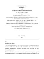
The geographic distribution shows that most cases were
from the Giza district (58.8%), then the Cairo district
(35.2%), then the other governorates including El-Fayoum,
El-Dakahleya, Assuit, Benisweif, El-Menia, El-Kalyoubeya
and El Behera at lower percentages up to 6% (Graph 1).
Table 4 shows the different clinical presentations of poisoned patients who were classified according to an internal
protocol applied in our centre as either asymptomatic or with
minor, moderate or major symptoms. Accordingly, 24.2% of
the patients were asymptomatic on arrival at the emergency
department, while 36.4%, 29.1% and 10.3%, presented with
minor, moderate and major symptoms, respectively.
Minor symptoms were in the form of GIT manifestations
(epigastric pain, nausea or vomiting); moderate symptoms included respiratory manifestations in addition (dyspnea and
excessive secretions); and major symptoms involved signs of ef-
Table 3
Sex
Causes of poisoning.
Cause of poisoning
No. of suicide
attempts
Males
Females
Total
54
84
138
Analysis of recorded symptoms.
%
%
No. of accidental
poisoning
%
83
84
11
16
17
16
83.6
27
16.4
No. of
females
No. of
males
Total
number
30
38
20
12
10
22
28
5
40
60
48
17
100
65
165
%
24.2
36.4
29.1
10.3
100
Asymptomatic = without any symptom or only dizziness.
Minor symptoms = in the form of GIT manifestations (epigastric
pain, nausea/or vomiting).
Moderate symptoms = included respiratory manifestations in
addition (dyspnea and excessive secretions).
Major symptoms = involved signs of other system affection
(tachycardia and hypotension, agitation, hallucinations and
depression).
fects on other systems (tachycardia and hypotension, agitation, hallucinations and depression).
Table 5 shows that 33.3% of cases were metabolically stable
on admission; 11.5% of cases presented with metabolic acidosis, of which 9.2% were in a compensated state; 10.3% presented with respiratory acidosis, of which 5.7% were in a
compensated state; 18.4% presented with respiratory alkalosis,
of which 10.3% were in a compensated state; 26.5% of cases
presented with mixed metabolic acidosis and respiratory alkalosis, of which 19.6% were in a compensated state.
Table 6 shows that 30% of patients had elevated ALT levels; 45% had elevated AST levels. Kidney function tests were
nearly within normal range; only 2.4% had an elevated urea
level. There were some electrolyte disturbances; 50% of patients had low sodium levels; 25% had low potassium levels.
Synthetic liver functions were performed in 1/3 of patients so
as to assess the condition of the liver and the need for treatment with antifibrinolytic or anticoagulation therapy. Results
revealed that 33% of them had prolonged PT, 10% had low
prothrombin concentration while INR and platelet count were
within normal range on admission.
The majority of cases (50%) presented to the emergency
department within 2 h of exposure; meanwhile 20% of cases
presented within 1 h, 10% after 4 h and the remaining 20%
within 6–12 h of exposure.
As regards management procedures, Table 7 shows that patients who presented within 2 h of exposure underwent gastric
lavage with sodium bicarbonate solution (61%); 32.7%
152
Table 5
A.M. El Naggar and N.M. El Mahdy
Metabolic status of patients.
Metabolic status
Table 6
Number %
Normal
58
Metabolic acidosis (uncompensated)
4
Compensated metabolic acidosis
16
Respiratory acidosis (uncompensated)
8
Compensated respiratory acidosis
10
Respiratory alkalosis (uncompensated)
14
Compensated respiratory alkalosis
18
Mixed metabolic acidosis and respiratory alkalosis 12
Compensated mixed
34
Total
174
33.3
2.3
9.2
4.6
5.7
8.1
10.3
6.9
19.6
100
Differences recorded in the total number of cases were due to some
data missed in history taking.
Normal ABG values:
pH = 7.35–7.45.
PO2 = 95–100 mm Hg.
PCO2 = 35–45 mm Hg.
HCO3 = 24–28 mmol/L.
Diagnosis is based on the following criterion according to the
Henderson–Hasselbach parameters:
If the pH is within normal range but associated with respiratory or
metabolic disturbance, the patient is in a compensated state; if not
the case is uncompensated.
A respiratory disturbance alters the arterial PCO2, while metabolic
disturbance alters the HCO3:
Metabolic acidosis = pH value, PCO2 and HCO3 are below
normal ranges.
Metabolic alkalosis = pH and PCO2 and HCO3 are above
normal ranges.
Respiratory acidosis = pH is below normal range, HCO3 and
PCO2 are above normal ranges.
Respiratory alkalosis = pH is above normal range, HCO3 and
PCO2 are below normal ranges.
Mixed types = is diagnosed by Winter’s formula Expected
PCO2 = (1.5 · HCO3) + 8 ± 2.
This means that if a metabolic acidosis is present, we use Winter’s
formula to determine the respiratory response; in cases of simple
metabolic acidosis, the measured PCO2 will fall within the range
determined by the equation; if a respiratory disturbance is
coexisting, then PCO2 varies outside the range; if below the range,
a respiratory alkalosis is also occurring; if above the range, there is
respiratory acidosis.
N.B. The diagnosis did not depend upon measuring the anion gap,
as the studied cases were of ZP poisoning, so there was no need to
measure the anion gap as it would be essential only to differentiate
between the causes of metabolic acidosis.
received oral sodium bicarbonate solution; activated charcoal
was given to 74% of patients; other symptomatic treatment
used according to the condition of the patient included fluid
administration, antispasmodics, antiemetics, sedatives, antibiotics and cathartics. Tranexamic acid as an antifibrinolytic
agent was found to be of help in 9.7% of patients.
Table 8 shows the medical outcome of the studied cases:
complete cure was recorded in 35.7% of cases; 7.2% were discharged with some residual effects such as mild abdominal
pain; 56.3% were discharged before 48 h on personal consent
for social or financial reasons but most of these were completely cured as revealed on follow up in the outpatient clinic,
a percentage which should be added to the completely cured
figure; death was recorded only in 0.6% of cases due to delay
Analysis of different laboratory investigations.
Other investigations Patient values Normal values Number (%)
ALT
AST
Urea
Creatinine
Sodium
Potassium
Prothrombin time
Prothrombin
concentration
INR
Platelet count
>40 U/L
>38 U/L
>50 mg/dL
>1.5 mg/dL
<135 mmol/L
<3.5 mmol/L
>12.3 s
Out of range
Up to 40
Up to 38
Up to 50
Up to 1.5
136–145
3.5–5
Control 12.3
80–100%
30 (28.5)
45 (42.8)
3 (2.4)
0 (0)
72 (50)
36 (25)
55 (33)
10 (6)
<1
<150
1–1.2
150–450
0
0
Aspartate aminotransferase (AST) = up to 38 U/L.
Alanine aminotransferase (ALT) = up to 40 U/L.
Urea = 15–50 mg/dl.
Creatinine = 0.5–1.5 mg/dl.
Sodium (Na) = 136–145 mM/L.
Potassium (K) = 3.5–5.0 mM/L.
Prothrombin time (PT) = 12.3 s.
Prothrombin concentration (PC) = 80–100%.
International normalized ratio (INR) = 1.0–1.2.
Table 7
Analysis of management procedures.
Procedures
Number of patients
%
Gastric lavage with sodium
bicarbonate
Oral sodium bicarbonate
Activated charcoal
Others (fluids, antispasmodic,
antiemetics, sedatives,
antibiotics, cathartics, etc.)
Tranexamic acid
100
61
54
122
138
32.7
74
83.6
16
9.7
Table 8
Analysis of medical outcome.
Medical outcome
Completely cured
Discharged with residual effects
Discharged on consent
Death
Total
Number of patients
59
12
93
1
165
%
35.7
7.2
56.3
0.6
100
in reaching the hospital, deterioration of the patient’s general
condition and development of pulmonary complications.
Depression was the commonest residual outcome in most cases
of intentional poisoning and these needed psychological assessment and follow up with a specialist.
The length of hospital stay varied from 2 h to up to 2 days
with a mean of 13.36 h according to clinical condition and
other factors related to agony, social factors or economic
causes.
Patients who accidently ingested food contaminated with
zinc phosphide (3%) showed milder manifestations compared
with other cases; this could be explained by ingestion of a small
amount of poison, early presentation to the emergency department and the performance of gastric lavage; there was one
Zinc phosphide toxicity among Egyptian patients
exception, a case that came late and showed the full picture of
toxicity.
The patients who presented with suicidal ingestion of zinc
phosphide combined with carbamazepine overdose (1%) (20–
30 tablets), an enzyme inducer [25], exhibited a rapid appearance of the toxic effects of poisoning with zinc phosphide in
the form of severe vomiting and epigastric pain; one of them
presented to the emergency unit within 1 h; the other presented
after 12 h with deterioration of his general condition in the
form of drowsiness, hypotension, hypokalemia, hallucinations
from the effects of carbamazepine overdose with metabolic acidosis and pneumonic patches revealed on X-ray, and so
needed prolonged hospital stay and close monitoring.
A patient with suicidal ingestion of zinc phosphide combined with alcohol, an enzyme inducing agent, presented with
severe metabolic acidosis, increased levels of liver enzymes,
hypokalemia and bradycardia, a picture that necessitates
aggressive therapy.
Patients who presented with suicidal ingestion of zinc phosphide combined with theophylline (1%) presented with their
combined effects on the GIT in the form of severe and repeated vomiting, which was not relieved by the usual antiemetic drugs and so needed aggressive management; also,
metabolic acidosis was reported with tachycardia, hypotension
and hypokalemia, which necessitated intravenous fluid and
electrolyte replacement and prolonged hospital stay compared
with other cases.
Ten percent of patients presented with ingestion of zinc
phosphide combined with organophosphorus compounds;
90% of them were suicide attempts and 10% were accidental
exposure. Most cases (80%) presented with severe vomiting
and abdominal pain in addition to muscle twitches and diarrhea, which indicated the combined effects of toxicity that required antidotal therapy (pralidoxime); 10% of cases needed
endotracheal intubation and artificial ventilation due to late
arrival, development of metabolic acidosis and deterioration
of the respiratory functions.
Discussion
Acute poisoning with pesticides is a global public health problem. The easy availability of highly toxic pesticides in the
homes of farming communities has made pesticides the easiest
means of suicide with an extremely high case fatality. Similarly, the extensive use of pesticides exposes the community
to both long-term and acute occupational health problems
[26].
In Egypt most farmers outside Cairo use phosphide tablets
extensively in grain stores, allowing the phosphine to be released once the storage sites are sealed. Phosphine exposure
and toxicity from phosphide salts occurs during grain fumigation in both transport and storage areas, causing inhalation of
the gas and reported cases of sudden death [13]; an important
event occurred in June 2009, when death was reported in nine
workers who were engaged in grain transportation in the Kalyoubeya governorate; the condition was not discovered at the
time and caused a lot of controversy until diagnosed. Similarly,
an agricultural health study (AHS) cohort study performed in
Iowa and North Carolina (a cohort that included 52,629 private applicators) revealed that 16% of workers who apply pesticides in agriculture experienced a serious pesticide exposure
153
event. Fifty major type of pesticides were included, falling into
four main classes; herbicides, insecticides, fungicides and fumigants. The most frequent were alachlor, trifluralin, atrazine
and phorate [27].
Our study was limited to admissions to the NECTR that
represent only a small percentage of hospital admissions of
cases of zinc phosphide poisoning in Egypt; other cases would
present to other poison control centers such as the Ain Shams
hospital or general hospitals; in addition there are calls received at the information unit and missed cases due to misdiagnosis or to finding difficulty in reaching any centre due to
scarcity of specialized centers in other governorates.
Unfortunately, it is not currently possible to link all the databases to provide a more comprehensive overview of poisoning
with zinc phosphide in our country; we think it is important
therefore to highlight our views on this type of poisoning
and to call for the publishing of statistical records of all cases
of poisoning in specialized and other general hospitals.
A total of 10,638 cases attended the NECTR during the
period of our study: 11.8% were pesticide poisoning and zinc
phosphide poisoning represented 15% of this percentage.
These results had not been previously published elsewhere
since our reference was the information unit of the NECTR.
The diagnosis is usually made from the history since phosphine tissue concentrations are not routinely available.
The results of this study show that oral ingestion was the
main route of poisoning, which is in agreement with other publications [28,29]. Most cases were teenagers and younger
adults, as in the majority of centers across the world, which
also record that the highest incidence of poisoning is in younger age groups [30–32] (Tables 1 and 2).
The differences recorded in geographical distribution might
be attributable to the easier access to the centre; the culture;
and to the magnitude of the problems encountered in overcrowded rural areas, where easy access to toxic pesticides turns
many impulsive acts of self-poisoning into suicide.
The incidence of attempted suicide in females was greater
than in males (a ratio of 1.5:1) (Table 3), due to marital disharmony, economic hardship, social problems and scolding from
other family members, which is in agreement with other studies
[30,33–35].
Due to their easy availability, pesticides have become the
most commonly used agent for suicide in the developing world
[36,37]. Since intentional self-poisoning is often impulsive and
simplified by easy availability of the poison, a proportion of
self-poisoning by pesticides can be prevented by reducing its
access. So regulating availability of pesticides and improving
medical management may reduce fatality from pesticide poisoning and also reduce the number of suicide attempts in
younger age groups [38].
Most patients in the study presented with abdominal pain
and vomiting (75.8%), followed by respiratory manifestations
(29.1%); systemic affection, which was evident in those who
did not seek medical help immediately or to their ingestion
of large amounts, presented to a lesser extent.
Initially asymptomatic patients (24.2%) attended the centre
because they feared death even though no symptoms had manifested. Their actions were the result of a transient period of
distress, they did not have the actual desire to kill themselves;
they were only giving a cry for help to gain sympathy from
their relatives and help with their problems whether social or
economic.
154
Nearly 33% of cases were metabolically stable on admission so they were put under observation in the intermediate
care unit (ICU). The rest (67%) required admission to the
intensive care unit (ICU) for adjustment and careful monitoring; 11.5% of them presented with metabolic acidosis, which is
in agreement with other studies [23,39–41]; 26.5% presented
with mixed metabolic acidosis and respiratory alkalosis in
accordance with other studies [39,42] (Table 5); patients with
severe respiratory compromise (20%) required intubation
and artificial ventilation.
With regard to systemic toxicity encountered with phosphine, it has been proved that the major targets of PH3 poisoning in the human body are the lungs, heart, brain,
gastrointestinal tract, kidney and liver [43,44], in our study,
30% of patients showed elevated ALT levels, 45% had elevated AST levels without history of previous liver affection,
which reflected the hepatotoxicity [7,42]. Saleki et al. in 2007
[45] stated that PH3 can cause liver dysfunction, especially
after the first day of poisoning, and that the main histopathologic changes found were fine cytoplasmic vacuolization of
hepatocytes and sinusoidal congestion. Similar condition was
recorded in the case presentation of Khurana et al. in 2009
[46] who found elevated transaminases levels; also Karanth
and Nayyar in 2003 [47] recorded severe hepatic dysfunction
in their cases. Congestion, oedema and centrizonal necrosis
of the liver were found on histopathological examination in
the study of Musshoff et al. in 2008 [48].
No kidney affection was recorded in our study; kidney
function tests were nearly within normal range; only 2.4%
had an elevated urea level.
There were some electrolytes disturbances; 50% of patients
had hyponatremia, similar to the case study of Khurana et al.
in 2009 [46]; it is caused most probably by excessive vomiting
and diarrhea or may be related to adrenal insufficiency.
Twenty-five percent suffered hypokalemia, which may be related to repeated vomiting; Proudfoot [39] also attributed its
occurrence to catecholamine release.
Zinc phosphide poisoned patients were carefully evaluated
with complete history and physical examination. Symptomatic
and supportive care was the mainstay of therapy. Some toxicology references suggest induction of emesis within 30 min
post ingestion [41]. Gastric lavage with water or 3–5% sodium
bicarbonate (to reduce gastric acid and production of phosphine) [39], or 1:5000 potassium permanganate (to oxidize
phosphine to less absorbable phosphide) has been advised;
however, other researchers stated that its effectiveness is unproven [49]. Each of these decontamination techniques carries
its own risks and physicians must be aware of these dangers before undertaking any decontamination measures, taking into
consideration the importance of airway protection.
Gastrointestinal decontamination with administration of
sodium bicarbonate solution was performed in nearly 61%
of our patients who presented within 4 h of poisoning; they
showed better outcome than cases who presented later and
who did not receive gastric lavage (Table 6).
Those who came late (32.7%) received oral sodium bicarbonate solution in a trial to oxidize any remnants in the gastrointestinal tract; activated charcoal as adsorbent was given in
74% of patients; other symptomatic treatments were used
according to the condition of the patient as follows: fluid
administration, antispasmodics, antiemetics, sedatives, antibiotics and cathartics (Table 7).
A.M. El Naggar and N.M. El Mahdy
During the observation period, patients were closely monitored for cardiac affection by ECG monitoring, with assessment of hepatic and renal functions as well as fluid and
electrolyte status.
Death was recorded in one case only who presented after
24 h of poisoning with bad general condition; he received basic
therapy but unexpectedly developed sudden repeated hemoptysis suggesting the development of a hemorrhagic diathesis;
intubation and artificial ventilation was performed but the patient’s condition deteriorated rapidly and before receiving any
corrective therapy he developed respiratory failure and died.
This case raised our attention to the possibility of the development of disseminated intravascular coagulopathy (DIC) as
recorded in previous studies [39,42,46,50–52]. Proudfoot in
2009 [39] attributed its occurrence to the highly toxic phosphine gas not to the metal phosphides. Sood et al. in 1997
[50] stated that metabolic acidosis accompanying aluminium
phosphide poisoning could have been responsible for the acute
hemolytic episode recorded in their patient; a picture similar to
that observed in our patient. Srinivas et al. in 2007 [51] described a patient with ALP poisoning presenting with intravascular hemolysis secondary to G6PD deficiency. However,
Aggarwal et al. in 1999 [42] had proved the occurrence of
intravascular hemolysis with aluminium phosphide poisoning
in a patient with normal G-6-PD levels. Khurana et al. in
2009 [46] proved in addition the occurrence of microangiopathic hemolytic anemia (MAHA) in a patient who ingested
aluminum phosphide. They stated that aluminum phosphide,
being a redox substance, can theoretically cause hemolytic anemia. Aluminum phosphide could be associated with DIC,
which is a well-recognized cause of MAHA. From this research, we can justify the possibility of the occurrence of
DIC in phosphide poisoning.
Some patients presented with the usual picture of zinc phosphide poisoning (profuse vomiting, abdominal pain); then the
condition deteriorated rapidly, from drowsiness, pallor, restlessness, irritability, tachycardia, hypotension, bleeding from
gums or at the site of injection, to a disturbed conscious level,
and shock and respiratory distress in some patients. Close clinical monitoring was done with full investigations that revealed
the development of metabolic disturbances, elevated transaminases, low hemoglobin levels, prolonged prothrombin times
and thrombocytopenia in most of them. Although not routinely used in management, but owing to the development of
DIC, we began our trial using tranexamic acid, an antifibrinolytic agent, which competitively inhibits the activation of plasminogen to plasmin, a molecule responsible for the
degradation of fibrin, which is the basic framework for the formation of a blood clot in hemostasis [53]. Tranexamic acid was
started by a loading dose of 10 mg/kg intravenously followed
by a maintenance dose of 10 mg/kg three times daily for 2–
8 days according to the condition; two cases needed blood
transfusion in addition due to low hemoglobin level; 2 days later, with continuous supportive care, patients showed a gradual improvement in their general condition with progressive
improvement in liver function tests, coagulation profile and
platelet count. Complete cure occurred within 1–2 weeks in
all cases.
The use of steroids in the management of such patients was
also greatly recommended for several reasons including stressful situations and hypotension [54,55], to minimize the possibility of pulmonary injury [54,56], as well as of
Zinc phosphide toxicity among Egyptian patients
adrenocortical insufficiency, which might develop due to hemorrhage in the adrenal cortex as recorded in the study of
Chugh et al. in 1989 [57] who detected severe adreno-cortical
involvement in some patients; histopathological examination
revealed severe changes in the adrenal cortex (complete lipid
depletion, hemorrhage, necrosis, etc.) and all these patients
died. They attributed these changes to shock or to the cellular
toxic effect of phosphine, a finding also recorded by Arora
et al. in 1995 [58] where histopathological changes were observed in various body organs such as lungs, liver, kidneys,
heart, brain, stomach and adrenals, including varying degrees
of congestion, edema and leucocytic infiltration changes suggestive of cellular hypoxia. Even adrenal gland damage can occur, as recorded by Proudfoot in 2009 [39].
Recommendations
Our recommendations focus on the importance of providing
awareness to medical and paramedical staff of the manifestations of zinc phosphide poisoning especially in rural areas, to
improve case management and to raise public awareness so
as to limit the unregulated sale of this toxic compound.
Collaboration between different poison control centers for
data-gathering about cases of zinc phosphide and other pesticide poisonings will help to create a solid database and a full
picture of the actual condition in our country and will subsequently assist us to assess the magnitude of the problem and
to perform the required actions.
Further research including studies on a larger scale should
be considered concerning the use of antifibrinolytics and steroids in the management of poisoned patients, to improve their
prognosis.
Another important issue is psychosocial counseling, which
is an important aspect of the overall management of the problem of attempted suicide.
155
[10]
[11]
[12]
[13]
[14]
[15]
[16]
[17]
[18]
[19]
[20]
[21]
[22]
References
[1] Singh B, Unnikrishnan B. A profile of acute poisoning at
Mangalore
(South
India).
J
Clin
Forensic
Med
2006;13(3):112–6.
[2] Eddleston M. Patterns and problems of deliberate self-poisoning
in the developing world. QJM 2000;93(11):715–31.
[3] Jeyaratnam J. Acute pesticide poisoning: a major global health
problem. World Health Stat Q 1990;43(3):139–44.
[4] Konradsen F, van der Hoek W, Cole DC, Hutchinson G,
Daisley H, Singh S, et al. Reducing acute poisoning in
developing countries––options for restricting the availability of
pesticides. Toxicology 2003;192(2–3):249–61.
[5] Abdelmegid LAM, Salem EM. Trends in the pattern of acute
poisoning in Alexandria Poison Center in 1994. In: Proceedings
of the 3rd congress on toxicology in developing countries, vol. 1,
November 19–23, Cairo, Egypt; 1996. p. 237–50.
[6] Mansour SA. Environmental impact of pesticides in Egypt. Rev
Environ Contam Toxicol 2008;196:1–51.
[7] Chomchai S. Phosphine and phosphides. In: Olson KR, editor.
Poisoning and drug overdose. New York: Mc Graw Hill; 2004.
p. 306–7.
[8] Burkhart KK. Methyl bromide and other fumigants. In:
Flomenbaum NE, Goldfrank LR, Hoffman RS, Howland
MA, Lewin NA, Nelson LS, editors. Goldfrank’s toxicologic
emergencies. New York: McGraw-Hill; 2006. p. 1556–63.
[9] Perry HE. Rodenticides. In: Shannon MW, Borron SW, Burns
MJ, editors. Haddad and Winchester’s clinical management of
[23]
[24]
[25]
[26]
[27]
[28]
[29]
[30]
[31]
poisoning and drug overdose. Philadelphia, PA: Saunders –
Elsevier; 2007. p. 1225–6.
Chugh SN, Arora V, Sharma A, Chugh K. Free radical
scavengers and lipid peroxidation in acute aluminium
phosphide poisoning. Indian J Med Res 1996;104:190–3.
Rodenberg HD, Chang CC, Watson WA. Zinc phosphide
ingestion: a case report and review. Vet Hum Toxicol
1989;31(6):559–62.
Mittra S, Peshin SS, Lall SB. Cholinesterase inhibition by
aluminium phosphide poisoning in rats and effects of atropine
and pralidoxime chloride. Acta Pharmacol Sin 2001;22(1):37–9.
Ahmad SH, Fakhir S, Gupta S, Singh RK. Celphos poisoning.
Indian Pediatr 1991;28(3):300–1.
Chopra JS, Kalra OP, Malik VS, Sharma R, Chandna A.
Aluminium phosphide poisoning: a prospective study of 16 cases
in one year. Postgrad Med J 1986;62(734):1113–5.
Banjaj R, Wasir HS. Epidemic aluminium phosphide poisoning
in Northern India. Lancet 1988;1(8589):820–1.
Andersen TS, Holm JW, Andersen TS. Forgiftning med
muldvarpegasningsmidlet aluminiumfosfid [Poisoning with
aluminum phospholipide used as a poison against moles].
Ugeskr Laeger 1996;158(38):5308–9.
Duenas A, Perez Castrillon JL, Cobos MA, Herreros V.
Treatment of the cardiovascular manifestations of phosphine
poisoning with trimetazidine, a new antiischemic drug. Am J
Emerg Med 1999;17(2):219–20.
Misra UK, Tripathi AK, Pandey R, Bhargwa B. Acute
phosphine poisoning following ingestion of aluminium
phosphide. Hum Toxicol 1988;7(4):343–5.
Singh RB, Singh RG, Singh U. Hypermagnesemia following
aluminum phosphide poisoning. Int J Clin Pharmacol Ther
Toxicol 1991;29(2):82–5.
Lohani Shyam P, Rajendra Kumar BC, Bidur O. An
epidemiological study on acute zinc phosphide poisoning in
Nepal. J Nepal Health Res Council 2002;1(1):13–6.
Siwach SB, Singh H, Jagdish Katyal VK, Bhardwaj G. Cardiac
arrhythmias in aluminium phosphide poisoning studied by on
continuous holter and cardioscopic monitoring. J Assoc
Physicians India 1998;46(7):598–601.
Frangides CY, Pneumatikos IA. Persistent severe hypoglycemia
in acute zinc phosphide poisoning. Intensive Care Med
2002;28(2):223.
Orak M, Ustundag M, Sayhan MB. Severe metabolic acidosis
secondary to zinc phosphide poisoning. J Pak Med Assoc
2008;58(5):289–90.
Sarma PS, Narula J. Acute pancreatitis due to zinc phosphide
ingestion. Postgrad Med J 1996;72(846):237–8.
Calvey N. Enzyme inducers and inhibitors: addition, subtraction
and synergism. Anaesth Intensive Care Med 2005;6(4):139–40.
Konradsen F. Acute pesticide poisoning – a global public health
problem. Danish Med Bull 2007;54(1):58–9.
Keim SA, Alavanja MC. Pesticide use by persons who reported
a high pesticide exposure event in the agricultural health study.
Environ Res 2001;85(3):256–9.
Chan TY, Critchley JA. Hospital admissions due to acute
poisoning in the new territories, Hong Kong. Southeast Asian J
Trop Med Public Health 1994;25(3):579–82.
Ekeberg O, Ellingsen O, Jacobsen D. Suicide and other causes of
death in a five-year follow-up of patients treated for selfpoisoning in Oslo. Acta Psychiatr Scand 1991;83(6):432–7.
Schmidtke A, Bille Brahe U, DeLeo D, Kerkhof A, Bjerke T,
Crepet P, et al. Attempted suicide in Europe: rates, trends and
sociodemographic characteristics of suicide attempters during
the period 1989–1992. Results of the WHO/EURO Multicentre
Study on Parasuicide. Acta Psychiatr Scand 1996;93(5):327–38.
Hawton K, Houston K, Shepperd R. Suicide in young people.
Study of 174 cases, aged under 25 years, based on coroners’ and
medical records. Br J Psychiatry 1999;175:271–6.
156
[32] Hulten A, Wasserman D, Hawton K, Jiang GX, Salander
Renberg E, Schmidtke A, et al. Recommended care for young
people (15–19 years) after suicide attempts in certain European
countries. Eur Child Adolesc Psychiatry 2000;9(2):100–8.
[33] Beautrais AL, Joyce PR, Mulder RT. Youth suicide attempts: a
social and demographic profile. Aust NZ J Psychiatry
1998;32(3):349–57.
[34] Dudley MJ, Kelk NJ, Florio TM, Howard JP, Waters BG.
Suicide among young Australians, 1964–1993: an interstate
comparison of metropolitan and rural trends. Med J Aust
1998;169(2):77–80.
[35] Lecomte D, Fornes P. Suicide among youth and young adults,
15 through 24 years of age. A report of 392 cases from Paris,
1989–1996. J Forensic Sci 1998;43(5):964–8.
[36] Gunnell D, Eddleston M. Suicide by intentional ingestion of
pesticides: a continuing tragedy in developing countries. Int J
Epidemiol 2003;32(6):902–9.
[37] Eddleston M, Phillips MR. Self poisoning with pesticides. BMJ
2004;328(7430):42–4.
[38] Eddleston M, Karunaratne A, Weerakoon M, Kumarasinghe S,
Rajapakshe M, Sheriff MH, et al. Choice of poison for
intentional self-poisoning in rural Sri Lanka. Clin Toxicol
(Phila) 2006;44(3):283–6.
[39] Proudfoot AT. Aluminium and zinc phosphide poisoning. Clin
Toxicol 2009;47(2):89–100.
[40] Amr MM, El Batanouni MM, El Samra GM, El Dosouky S,
Awadallah R. Metabolic changes due to zinc phosphide
exposure. Egypt J Ind Med 1984;8(2):211–21.
[41] Chugh SN, Aggarwal HK, Mahajan SK. Zinc phosphide
intoxication symptoms: analysis of 20 cases. Int J Clin
Pharmacol Ther 1998;36(7):406–7.
[42] Aggarwal P, Handa R, Wig N, Biswas A, Saxena R, Wali JP.
Intravascular hemolysis in aluminium phosphide poisoning. Am
J Emerg Med 1999;17(5):488–9.
[43] Sudakin DL. Occupational exposure to aluminium phosphide
and phosphine gas? A suspected case report and review of the
literature. Hum Exp Toxicol 2005;24(1):27–33.
[44] Hsu C, Han B, Liu M, Yeh C, Casida JE. Phosphine-induced
oxidative damage in rats: attenuation by melatonin. Free Radic
Biol Med 2000;28(4):636–42.
[45] Saleki S, Ardalan FA, Javidan Nejad A. Liver histopathology of
fatal phosphine poisoning. Forensic Sci Int 2007;166(2–
3):190–3.
A.M. El Naggar and N.M. El Mahdy
[46] Khurana V, Gambhir I, Kishore D. Microangiopathic hemolytic
anemia following disseminated intravascular coagulation in
aluminum phosphide poisoning. Indian J Med Sci
2009;63(6):257–9.
[47] Karanth S, Nayyar V. Rodenticide-induced hepatotoxicity. J
Assoc Physicians India 2003;51:816–7.
[48] Musshoff F, Preuss J, Lignitz E, Madea B. A gas
chromatographic analysis of phosphine in biological material
in a case of suicide. Forensic Sci Int 2008;177(2-3):e35–8.
[49] Gupta S, Ahlawat SK. Aluminum phosphide poisoning––a
review. J Toxicol Clin Toxicol 1995;33(1):19–24.
[50] Sood AK, Mahajan A, Dua A. Intravascular haemolysis after
aluminium phosphide ingestion. J R Soc Med 1997;90(1):47–8.
[51] Srinivas R, Agarwal R, Jairam A, Sakhuja V. Intravascular
haemolysis due to glucose-6-phosphate dehydrogenase
deficiency in a patient with aluminium phosphide poisoning.
Emerg Med J 2007;24(1):67–8.
[52] Bogle RG, Theron P, Brooks P, Dargan PI, Redhead J.
Aluminium phosphide poisoning. Emerg Med J 2006;23(1):e3.
[53] Wikipedi. Tranexamic acid. < />wiki/Tranexamicacid>.
[54] World Health Organization (WHO). International programme
on chemical safety: phosphine and metal phosphides. Health
and Safety Guide no. 28. Geneva: World Health Organization
(WHO); 1989.
[55] Finer NN, Powers RJ, Ou CH, Durand D, Wirtschafter D,
Gould JB. Prospective evaluation of postnatal steroid
administration: a 1-year experience from the California
Perinatal
Quality
Care
Collaborative.
Pediatrics
2006;117(3):704–13.
[56] Nin N, Penuelas O, de Paula M, Lorente JA, Fernandez
Segoviano P, Esteban A. Ventilation-induced lung injury in rats
is associated with organ injury and systemic inflammation that is
attenuated
by
dexamethasone.
Crit
Care
Med
2006;34(4):1093–8.
[57] Chugh SN, Ram S, Sharma A, Arora BB, Saini AS, Malhotra
KC. Adrenocortical involvement in aluminium phosphide
poisoning. Indian J Med Res 1989;90:289–94.
[58] Arora B, Punia RS, Kalra R, Chugh SN, Arora DR.
Histopathological changes in aluminium phosphide poisoning.
J Indian Med Assoc 1995;93(10):380–1.