Effect of P2O5 and MnO2 on crystallization of magnetic glass ceramics
Bạn đang xem bản rút gọn của tài liệu. Xem và tải ngay bản đầy đủ của tài liệu tại đây (1.08 MB, 8 trang )
Journal of Advanced Research (2014) 5, 543–550
Cairo University
Journal of Advanced Research
ORIGINAL ARTICLE
Effect of P2O5 and MnO2 on crystallization
of magnetic glass ceramics
Salwa A.M. Abdel-Hameed
a
b
a,*
, Mohamed A. Marzouk a, Mohamed M. Farag
b
Glass Department, National Research Center, Dokki, Cairo, Egypt
Biomaterial Department, National Research Center, Dokki, Cairo, Egypt
A R T I C L E
I N F O
Article history:
Received 8 May 2013
Received in revised form 4 July 2013
Available online 17 July 2013
Keywords:
X-ray method
Magnetic properties
Glass ceramics
Magnetite
A B S T R A C T
This work pointed out the effect of adding P2O5 and/or MnO2 on the crystallization behavior of
magnetic glass ceramic in the system Fe2O3ÆZnOÆCaOÆSiO2ÆB2O3. The differential thermal analysis of the quenched samples revealed decrease in the thermal effects by adding P2O5 and/or
MnO2 to the base sample. The X-ray diffraction patterns show the development of nanometric
magnetite crystals in a glassy matrix. Heat treatment at 800 °C for 2 h, under reducing atmosphere, caused an increase in the amount of the crystallized magnetite with the appearance of
minor hematite and Ca2SiO4. The transmission electron microscope revealed a crystallite size
in the range 10–30 nm. Magnetic hysteresis cycles were analyzed with a maximum applied field
of 25 kOe at room temperature. The prepared magnetic glass ceramics are expected to be useful
for localized treatment of cancer.
ª 2013 Production and hosting by Elsevier B.V. on behalf of Cairo University.
Introduction
Hyperthermia destroys cancer cells by raising the tumor temperature to a ‘‘high fever’’ range, similar to the way that the
body naturally uses to combat other forms of disease [1]. Generally, tumors are more easily heated than the surrounding
normal tissues, since blood vessels and nervous systems are
poorly developed in the tumor, and cancer cells are easily
killed by heat treatment, since oxygen supply via the blood vessels is not sufficient in the tumor. Hence hyperthermia is expected to be a most useful treatment for cancer with no side
* Corresponding author. Tel.: +20 33371312; fax: +20 33387803.
E-mail address: (S.A.M. Abdel-Hameed).
Peer review under responsibility of Cairo University.
Production and hosting by Elsevier
effects [2]. On the contrary, these temperatures are safe for surrounding healthy tissues with normal and efficient blood cooling systems [2].
Bioactive ferromagnetic glass–ceramics are expected to be
useful as thermoseeds for hyperthermia treatment of cancer,
especially deep-seated cancers such as bone tumors. When ferromagnetic glass–ceramics are implanted around tumors in
granular form, they are bonded to each other so as not to be
moved by forming biologically active bone-like apatite on
them [3], and stably fixed around the tumors if they are located
near bones. Moreover, when they are placed under an alternating magnetic field, they generally heat effectively cancer cells to
be necrotized by magnetic hysteresis loss. After heating, they
can also reinforced weakened timorous bone by bonding to
bone.
Several materials that generate heat by hysteresis loss have
been developed [4–9]. Among them, bioactive ferro and
ferrimagnetic glass–ceramics have been investigated [10–13].
2090-1232 ª 2013 Production and hosting by Elsevier B.V. on behalf of Cairo University.
/>
544
Preparation of magnetite-containing glass ceramics has been
reported by several workers [2,14,15]. It is known that heat
generation depends mainly on the magnetic properties of the
implant, the magnetic field parameters and the characteristics
of the tissue [16].
Bretcanu et al. [16] prepared ferrimagnetic bioglass–
ceramics containing 45 wt% of magnetite revealing a saturation magnetization of 34 emu/g and a coercive force of
85 Oe. The estimated heat generation of this glass–ceramic
using a magnetic field of 40 kA/m and a frequency of
440 kHz was 25 W/g. The previous material showed a bioactive behavior after 2 weeks of soaking in a simulated body
fluid. Ebisawa et al., in 1997 [2] prepared glass ceramic contains 36% magnetite, in a matrix of CaOÆSiO2 based glass,
and b-wollastonite which showed ferrimagnetisms and no
bioactivity [17].
Kuwashita et al. [18] prepare zinc-iron ferrite (ZnxFe3ÀxO4)
in a CaO–SiO2 glassy matrix by heat-treatment under 95
CO2 + 5H2 atmosphere. The prepared material showed a
large amount of heat generation of 12.4 W gÀ1 under conditions of 300 Oe and 100 kHz.
Wu et al. [19] found that, Zn ions play an important role in
the human body, as reported to be involved in bone metabolism and can stimulate bone formation and increase bone protein, calcium content, and alkaline phosphates activity in
humans and animals. They crystallize hardystonite (Ca2ZnSi2O7) which might be biocompatible and used as biomaterials
[19].
From all the above mentioned materials manganese zinc
ferrite (Mn–ZnFe2O4) have special importance due to its high
initial permeability, saturation magnetization and relatively
lower eddy current loss compared to alloy cores [20], moreover
Mn–Zn ferrites are very important in biomedicine as magnetic
carriers, such as in bioseparation, enzyme and protein immobilization [21].
This work aimed at preparation and characterization of
magnetic glass ceramic in the Fe2O3ÆZnOÆCaOÆSiO2ÆB2O3 system containing P2O5 and MnO2. The influence of adding different addition from P2O5 and/or MnO2 on sequence of
crystallization, amount and crystal size of the developed ferrite
and microstructure were studied.
P2O5 was added to study its effect as nucleating agents on
the crystallization of magnetite, while MnO2 were added to
study the effect of replacing Fe2+ by Mn2+, in the magnetite
crystals, on the crystallization process.
Experimental
Theoretical considerations on designing glass ceramic
composition
In previous work [22], the authors succeed to precipitate
$60% nanoparticles magnetite in two different systems,
Fe2O3ÆCaOÆZnOÆSiO2ÆB2O3 and Fe2O3ÆCaOÆSiO2ÆB2O3. The
results showed that, crystallization of large amount of nanoparticles of magnetite in the presence of Zn ions; consequently
the saturation magnetization was increased to reach
52.13 emu/g. In order to improve the amount and nano-crystallite size of magnetite, we got before in the Zn-containing
sample, different oxides such as TiO2, Na2O and P2O5 were
added to this composition. The results showed that, addition
S.A.M. Abdel-Hameed et al.
of the P2O5 was greatly enhancing the amount and nano-crystallite size of magnetite.
Preparation of glasses
The chemical compositions of the examined glasses are shown
in Table 1. About 100 g powder mixtures of our compositions
were prepared from reagent grades of CaO as Ca2CO3, SiO2,
Fe2O3, ZnO and B2O3 as H3BO3. Different amounts of
MnO2 (0.5–40 gm) as MnCO3 and/or P2O5 (3–10 gm) as
NH4H2PO4 were added over 100% batch composition.
Our target was to obtain a glass–ceramic, not a ceramic
material, so a melting step was necessary. Based on their compositions, the batches were melted in a platinum crucible at
1350–1400 °C for 2 h in an electric furnace, with occasional
swirling every 30 min to ensure homogenization. As the
amount of P2O5 and/or MnO2 increased the melting temperatures decreased. The glass melted at 1400 °C was poured onto
a stainless steel plate at room temperature and pressed into a
plate 1–2 mm thick by another cold steel plate. An additional
sample was poured at 1450 °C to study the effect of increasing
temperature on the crystallization of magnetite.
Crystallization of glasses
The samples were thermally examined using Differential Thermal Analysis (DTA). According to the DTA results the obtained samples were covered with active carbon powders, to
apply a reducing atmosphere preventing ferrous ions from oxidation, and heated up to various temperatures at a rate of
10 °C min in a SiC electric furnace for crystallization. It was
noticed that the synthesis parameters (such as temperature,
time, heating rate, and atmosphere) play a fundamental role
for magnetite crystallization.
Characterization
The quenched samples were subjected to powder X-ray diffraction using Ni-filled Cu Ka radiation for determining the types
and contents of the precipitated crystalline phases. The average
crystallite size of magnetite in the heat treated and untreated
samples for the most intense peaks (220, 311, 400, 511 and
440) was determined from the XRD using Debye–Scherrer formula: D = kk/B cos H, where D is particle size, k is constant, k
for Cu is 1.54 A˚, B is full half wide and 2h = 4°. The microstructures of prepared samples were studied using TEM. The
sample was crushed and sonically suspended in ethanol and
few drops of the suspended solution were placed on an amorphous carbon film held by copper microgrid mesh and then observed under transmission electron microscope.
Results and discussion
Characterization
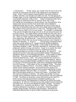
Fig. 1 shows thermal behavior of samples under investigation.
All curves show a glass transformation temperature (Tg) typical of an amorphous phase in the range of 634–675 °C and
exothermic peak in the range of 700–892 °C. The transformation temperature is accompanied by absorption of the heat
Effect of P2O5 and MnO2 on crystallization of magnetic glass ceramics
Table 1
545
Chemical composition of the studied glasses in wt%.
Sample
Fe2O3
CaO
ZnO
SiO2
B2O3
P2O5*
MnO2*
FHP
FHP1
FHP2
FHP3
FHPMn0.5
FHPMn10
FHPMn20
FHPMn 40
58.26
58.26
58.26
58.26
58.26
58.26
58.26
58.26
13.88
13.88
13.88
13.88
13.88
13.88
13.88
13.88
10.07
10.07
10.07
10.07
10.07
10.07
10.07
10.07
14.88
14.88
14.88
14.88
14.88
14.88
14.88
14.88
2.91
2.91
2.91
2.91
2.91
2.91
2.91
2.91
3
5
7
10
10
10
10
10
–
–
–
–
0.5
10
20
40
*
P2O5 and MnO2 were added above 100%.
The transformation temperature is known to be a good
indicator for the relative amount of amorphous phase present
in the samples. In the present study, an increase of both exothermic and endothermic peaks was noticed on samples contain MnO2, than base composition (FHP), thus indicate the
mineralizing role of MnO2 in enhancing the magnetite crystallization at the melting temperature. Consequently, the amount
of the amorphous phase was decreased; leading to thermal
transformation processes occur at higher temperatures. The increase in the area under the exothermic peak and consequently
the enthalpy in FHPMn40 sample certify the above result.
Fig. 1 Differential
investigation.
thermal
analysis
of
samples
under
required for rearrangement of different atoms as a pre-crystallization step. The presence of the glass transition temperature
confirms the presence of reasonable amounts of residual amorphous phases for the glass–ceramic samples [23]. The glass
transition temperatures of quenched samples are similar for
glasses containing iron ions [24] especially for FHPMn40 sample (639 °C). Tg was followed by an exothermic peak(s) corresponding to the crystallization process with an energy release.
In general, a significant decrease in the thermal effects was observed by adding P2O5 and/or MnO2.
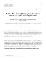
Fig. 2 XRD of different samples after cooling from melting
temperature.
546
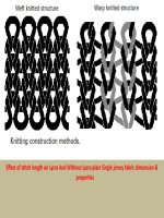
Fig. 3 Effect of P2O5 and MnO2 amount on the lattice
parameters of crystallized ferrites after cooling from melting
temperature.
From XRD results, the only crystallized phase after cooling
from the melting temperature was magnetite. It can be clearly
seen that, sharp peaks of all treated samples reflect high degree
of crystallinity. Economically, although the samples melted at
1450 °C showed slight higher magnetite peak intensities, only
samples melted at 1400 °C will be considered.
The X-ray diffraction patterns of glass–ceramics after cooling from melting temperature (1400 °C) are shown in Fig. 2.
The results present patterns corresponding to the common
structure of magnetite (Fe3O4) in a pure phase without any
other phase’s interference.
These XRD diagrams coincide with those reported in the
previous work for the parent sample (Fe3O4) [25]. They are
dominated by a strong Bragg peak located at ca. 2h = 35°
and peaks with medium intensity at 30°, 57° and 63°. Considering the intensity and position of the peaks, it is well known
that patterns of the Cubic unit-cell (Fd3m space group) can
be identified as magnetite phase.
Generally, the cumulative results of all samples were verified by cell volume calculation which revealed that, the cell volume increased by adding MnO2 and slightly decreased by
adding P2O5. The explanation of the cell volume enlargement
Fig. 4 Effect of P2O5 and MnO2 on the average crystallite size of
crystallized ferrites after cooling from melting temperature.
S.A.M. Abdel-Hameed et al.
could be explained as follows: the diffraction lines of the crystallized magnetite with an increasing amount of MnO2 were
shifted to a higher d-spacing with an increase in the (a) lattice
parameter than that recorded in the reference data card of
JCPDS (Joint Committee on Powder Diffraction Standards)
which show a value of a0 = 8.393–8.399 A˚. This shift may be
attributed to the incorporation of Mn2+ with its high ionic radii in the developed crystals of the magnetite solid solution (ss).
Contrary, the P2O5 addition caused slight lowering in the lattice parameter values of magnetite.
In order to illustrate how the unit cell and cell volume
changes as a function of P2O5 and MnO2 content, the lattice
parameters a, and cell volume were determined as a function
of P2O5 and MnO2 content by the least-squares method,
Fig. 3. The cubic parameters calculated via a least-squares
refinement method using 14 well-defined XRD lines are
a = 8.406 (2) and a = 8.466 (9) A˚ for FHP3 and FHPMn
40, respectively. For these materials the spinal structure is well
preserved upon considerable P2O5 and MnO2 addition. As
shown in Fig. 3, adding P2O5 results in a slightly decrease of
the cubic parameter; the a-axis shrinks can be explained as follow: P2O5 provides an example of a network-former which
exhibits the characteristics of nucleating agent. The phosphorus ion, P5+, assumes tetrahedral co-ordination and therefore
provides an example of phase separation due to a charge difference between the principal network-former, Si4+, and the
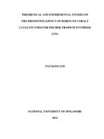
Fig. 5 XRD patterns of different samples after heat treatment at
800 °C for 2 h.
Effect of P2O5 and MnO2 on crystallization of magnetic glass ceramics
Fig. 6
547
TEM of different samples after cooling from melting temperature.
‘‘foreign’’ network-former ions, P5+ [26]. It is interesting to remark that the lattice parameter variations are very similar for
the different dopant concentrations. On the contrary, adding
Mn2+ cations caused progressive increase in lattice parameter,
where Mn2+ which have large ionic radius (93 pm) replaced
the lower ionic radius Fe2+ ions (65 pm).
Several factors can contribute to the broadening of peaks in
X-ray diffraction [25,27]. For example, the instrumental factors related to the resolution and the incident X-ray wavelength, as well as the sample factors such as crystallite size
and non-uniform microstrain. In the case of an instrumental
broadening, the line width will vary smoothly with 2h or d
spacing. On the other hand, the line broadening originating
from the sample characteristics will have a different relationship. Combining the Scherrer’s equation for crystallite size
and the Bragg’s law for diffraction, crystallite size and microstrain components are estimated by using the following
equation:
B2 cos2 h ¼ 16he2 i sin2 h þ
K2 k2
;
L2
ð1Þ
where B is the full-width at half-maximum (FWHM) after correction of the instrumental broadening for finely powdered silicon powder, h is the diffraction angle, Æe2æ denotes local
548
strains (defined as Dd/d being the interplanar spacing), L is the
crystallite size and K is a near-unity constant related to crystallite shape.
Crystallite size obtained from XRD, Fig. 4 shows a common crystallization of magnetite nano particles (<100 nm).
Increasing amount of MnO2 added leads to significant increase
in the crystallite size which may attributed to the replacement
of Fe ions (smaller ionic radius) by Mn ions (larger ionic
radius).
The broadness of magnetite peaks shown in XRD data are
in the order of FHPMn40 > FHPMn20 > FHPMn10 >
FHPMn0.5 and consequently the crystallite size are increased
in the same order.
Heat treatment of different samples at 800 °C for 2 h under
reducing atmosphere, Fig. 5 revealed increase in the magnetite
phase and crystallization of minor hematite and calcium silicate (Ca2SiO4). Two different structures of magnetite were
developed. As the amount of magnetite increased there are
chance to crystallize to different structure forms from
magnetite.
Comparing the XRD patterns of glass–ceramics obtained
by cooling of molten glass Fig. 1 and after the additional thermal treatment, Fig. 5, it can be concluded that, the difference
in the relative amount of crystallized magnetite in the samples
(in spite of containing the same amount of iron oxide) could be
attributed to the effects of adding MnO2 on lowering the viscosity of the melt and formation of solid solution with magnetite, consequently, some iron oxides remain entrapped in the
matrix.
The lattice strain has an opposite direction to lattice constant. Increasing the lattice strain leads to an increase in the
internal forces/stresses which may oppose the crystal growth
of magnetite. Thus, the crystallite size of magnetite, in case
of FHP sample, will be slightly lowered than that in
FHPMn0.5, FHPMn10, FHPMn20, FHPMn40 respectively;
which is confirmed by TEM.
TEM of some selected samples are shown in Fig. 6. The
crystallization of one or different phases is evidenced in
TEM micrographs. TEM revealed precipitation of nano size
rounded crystals of only magnetite phase dispersed in amorphous glassy phase in the quenched FHPMn0.5 and
FHPMn20 samples. The crystallite size was increased by adding MnO2 as seen before from XRD analysis. Diffraction of
FHPMn20 revealed crystallization of single phase, which mean
the incorporation of Mn ions in magnetite.
S.A.M. Abdel-Hameed et al.
40 gm Mn led to significant decrease in Ms to 23.94 emu/g,
which can be attributed to incorporation of the lower magnetic
element (Mn) in the higher magnetic one (Fe3O4).
The remanence is the amount of magnetic materials which
could be magnetized, even in the absence of external magnetic
field. The remanance magnetization values were much lower
than the saturation magnetization values. This could be due
to structural features of glass–ceramic [16]. The coercive field
Magnetic properties
Fig. 7 and Table 2 illustrate the variation of hysteresis curves
and magnetic values with heat treatment temperatures under
a magnetic field of 25 kOe. It could be observed that all the
samples exhibited a similar magnetic behavior, which is characteristic for soft magnetic materials, with a thin hysteresis cycle and low coercive field. Magnetite is the only crystallized
phase regarded as ferromagnetic phase, accordingly, increasing
or decreasing in Ms will be related to the amount of crystallized magnetite. FHP sample have the highest Ms value reaching 58.99 emu/g, this value was decreased to 38.59 emu/g by
increasing the amount of P2O5 added. These results reflect
the effect of adding high quantity of P2O5 on decreasing the
degree of crystallinity in quenched samples. Addition of
Fig. 7 Magnetic hysteresis of quenched samples under a
maximum magnetic field of 25 kOe.
Effect of P2O5 and MnO2 on crystallization of magnetic glass ceramics
Table 2
549
Magnetic properties of quenched samples under a maximum magnetic field of 25 kOe.
Sample no.
FHP
FHP3
FHPMn40
Magnetic properties
Ms (emu/g)
Mr (emu/g)
Hc (Oe)
58.99
38.59
23.941
4.634
3.32
0.99
24.76
76.494
27.358
depends on the microstructure. In general, as the particle size
increase the coercive field decrease.
Role of P2O5
It has been reported that, P2O5 enhance the nucleation density
and thus restricts the crystal growth and induces amorphous
phase separation. P2O5 can induce phase separation to promote heterogeneous nucleation and then produce a finegrained interlocking morphology. The heterogeneous nucleation was favorable in reducing nucleation energy.
The addition of P2O5, greatly affects phase formation and
morphology. It led to a slight increase in the peak crystallization temperature observed in DTA curves due to the reduction
in the number of remaining sites available for magnetite crystallization, where most of the magnetite was crystallized from
melt during cooling to room temperature.
Yinnon and Uhlmann [28] suggested that the nucleating
agents could be selectively enriched in one separated phase
thus providing the cites for nucleus. Ryerson and Hess [29]
proposed that when a modifier cation is added in a silica melt,
it is surrounded by both Ob and Onb. This oxygen isolates the
network-modifier cations from each other by providing screens
that masks the positive charge. However, modifier cations that
are partly or wholly coordinated by bridging oxygen are
poorly shielded from each. Consequently, substantial columbic
repulsions occur between network-modifier cation with give
rise to the enthalpy of unmixing and consequently lead to
phase separation. Markhasev and Sedletskii [30] found that
immiscibility fields expand with a decrease of the ionic radius
of the modifier cation in binary silicate melt.
Role of MnO2
MnO2 have a significant effect on lowering viscosity of the
melt, so increasing amount of MnO2 added leads to decreasing
the melt viscosity and facilitating the mobility of different ions
causing a relatively larger crystallite size as imprinted from
XRD and TEM results. On the other hand, Mn ions can replace Fe ions and make it possible for higher amounts of magnetite to be crystallized.
Conclusions
A series of magnetic glass ceramics contains different additions
of P2O5 and/or MnO2 in the system Fe2O3ÆZnOÆCaOÆSiO2ÆB2O3were prepared. A significant decrease in the thermal effects was observed by adding P2O5 and/or MnO2. XRD of
quenched samples show crystallization of magnetite with particle size $10–30 nm. Addition of MnO2 enhanced crystallization of more magnetite during cooling from melting
temperature. Heat treatment at 800 °C for 2 h, under reducing
atmosphere, caused an increase in the amount of the crystallized magnetite with the appearance of minor hematite and
Ca2SiO4. Ms value reaching 58.99 emu/g. The prepared magnetic glass ceramics are expected to be useful for localized
treatment of cancer.
Conflict of interest
The authors have declared no conflict of interest.
Compliance with Ethics Requirements
This article does not contain any studies with human or animal
subjects.
Acknowledgment
This project was supported financially by the Science and
Technology Development Fund (STDF), Egypt, Grant No.
240.
References
[1] Bretcanu O, Verne´ E, Coisson M, Tiberto P, Allia P.
Temperature effect on the magnetic properties of the co
precipitation derived ferrimagnetic glass–ceramics. J Magn
Magn Mater 2006;300:412–7.
[2] Ebisawa Y, Miyaji F, Kokubo T, Ohura K, Nakamura T.
Bioactivity of ferrimagnetic glass–ceramics in the system FeO–
Fe2O3–CaO–SiO2. Biomaterials 1997;18:1277–84.
[3] Kokubo T, Hayashi T, Sakka S, Kitsugi T, Yamamuro T.
Bonding between bioactive glasses, glass ceramics or ceramics in
simulated body fluid. Yogyo-Kyokai-Shi 1987;95:785–91.
[4] Cetas TC, Gross EJ, Contractor Y. A ferrite core/metallic
sheath thermo seed for interstitial thermal therapies. IEEE
Trans Biomed Eng 1998;45:68–77.
[5] Jordan A, Scholz R, Wust P, Fa¨hling H, Felix R. Magnetic fluid
hyperthermia (MFH): cancer treatment with AC magnetic field
induced excitation of biocompatible superparamagnetic
nanoparticles. J Magn Magn Mater 1999;201:413–9.
[6] Go´mez-Lopera SA, Plaza RC, Delgado AV. Synthesis and
characterization of spherical magnetite/biodegradable polymer
composite particles. J Colloid Interf Sci 2001;240:40–7.
[7] Lee YK, Kim DH, Lee YJ, Kim KN, Shim IB. Ceramics, cells
and tissues. In: Eighth annual seminar and meeting, Faenza;
2003.
[8] Takegami K, Sano T, Wakabayashi H, Sonoda J, Yamazaki T,
Morita S, Shibuya T, Uchida A. New ferromagnetic bone
cement for local hyperthermia. J Biomed Mater Res 1998;43:
210–4.
[9] Borrelli et al. Radio frequency induced hyperthermia for tumor
therapy, US Patent 4323056; 1982.
550
[10] Kokubo T, Yamamuro T, Ebisawa Y, Ohura K. European
patent 361797; 1990.
[11] Oh SH, Choi SY, Lee YK, Kim KN. Research on annihilation
of cancer cells by glass–ceramics for cancer treatment with
external magnetic field. I. Preparation and cytotoxicity. J
Biomed Mater Res 2001;54:360–5.
[12] Ebisawa Y, Miyaji F, Kokubo T, Ohura K, Nakamura T.
Surface reaction of bioactive and ferrimagnetic glass–ceramics in
the system FeO–Fe2O3–CaO–SiO2. J Ceram Soc Jpn 1997;105:
947–51.
[13] Arcos D, del Real RP, Vallet-Regi M. A novel bioactive and
magnetic biphasic material. Biomaterials 2002;23:2151–8.
[14] O’Horo M, Steinitz R. Characterization of devitrification of an
iron-containing glass by electrical and magnetic properties.
Mater Res Bull 1968;3:117–25.
[15] Auric P, Dang NV, Bandyopadhyay AK, Zarzycki J.
Superparamagnetism and ferrimagnetism of the small particles
of magnetite in a silicate matrix. J Non-Cryst Solids 1982;50:
97–106.
[16] Bretcanu O, Spriano S, Verne´ E, Coisson M, Tiberto P, Allia P.
The influence of crystallized Fe3O4 on the magnetic properties of
co precipitation-derived ferrimagnetic glass–ceramics. Acta
Biomat 2005;1:421–9.
[17] Ebisawa Y, Sugimoto Y, Hayashi T, Kokubo T, Ohura K,
Yamamurao T. Crystallization of (FeO, Fe2O3)–CaO–SiO2
glasses and magnetic properties of their crystallized products.
Seram Ronbun 1991;1:7–13.
[18] Kuwashita M, Iwahashi Y, Kokubo T, Yao T, Hamada S,
Shinjo T. Preparation of glass–ceramics containing
ferrimagnetic zinc-iron ferrite for the hyperthermal treatment
of cancer. J Cerm Soc Jpn 2004;112:373–9.
[19] Wu C, Chang J, Zhai W. A novel hardystonite bioceramic:
preparation and characteristics. Ceram Int 2005;31:27–31.
S.A.M. Abdel-Hameed et al.
[20] Hessien MM, Rashad MM, ElBarawy K, Ibrahim IA. Influence
of manganese substitution and annealing temperature on the
formation, microstructure and magnetic properties of Mn–Zn
ferrites. J Magn Magn Mater 2008;320:1615–21.
[21] Papazoglou P, Eleftheriou F, Zaspalis VT. Low sintering
temperature MnZn-ferrites for power applications in the
frequency region of 400 kHz. J Magn Magn Mater
2006;296:25–31.
[22] Abdel-Hameed SAM, Hessien MM, Azooz MA. Preparation
and characterization of some ferromagnetic glass–ceramics
contains high quantity of magnetite. Ceram Int 2009;35:
1539–44.
[23] Arvind A, Sarkar A, Shrikhande VK, Tyagi AK, Kothiyal GP.
The effect of TiO2 addition on the crystallization and phase
formation in lithium aluminum silicate (LAS) glasses nucleated
by P2O5. J Phys Chem Solids 2008;69(11):2622–7.
[24] Karamanov A, Pelino M. Crystallization phenomena in ironrich glasses. J Non-Cryst Solids 2001;281:139–51.
[25] Klug HP, Alexander LE. X-ray diffraction procedures for
polycrystalline and amorphous materials. New York: Wiley;
1974.
[26] McMillan PW. Glass ceramics. 2nd ed. London: Academic
Press; 1979.
[27] Hammond C.
The
basic
of
crystallography
and
diffraction. New York: Oxford University Press; 1997.
[28] Yinnon H, Uhlmann DR. A kinetic treatment of glass formation
V: surface and bulk heterogeneous nucleation. J Non-Cryst
Solids 1981;44:37–55.
[29] Ryerson FJ, Hess PC. The role of P2O5 in silicate melts.
Geochim Cosmochim Acta 1980;44:611–24.
[30] Markhasev BI, Sedletskii ID. Dokl Akad Nauk SSSR (Engl
Transl) Chem Sect 1963;148:112.