Effect of surface roughness and adhesive system on repair potential of silorane-based resin composite
Bạn đang xem bản rút gọn của tài liệu. Xem và tải ngay bản đầy đủ của tài liệu tại đây (1.13 MB, 8 trang )
Journal of Advanced Research (2012) 3, 279–286
Cairo University
Journal of Advanced Research
ORIGINAL ARTICLE
Effect of surface roughness and adhesive system on repair
potential of silorane-based resin composite
Enas H. Mobarak
*
Restorative Dentistry Department, Faculty of Oral and Dental Medicine, Cairo University, Cairo, Egypt
Received 4 July 2011; revised 18 September 2011; accepted 23 September 2011
Available online 6 November 2011
KEYWORDS
Bond strength;
Methacrylate-based resin
composite;
Repair;
Silorane-based resin composite;
Surface roughness
Abstract This study was performed to evaluate the influence of surface roughness and adhesive
system on the repair strength of silorane-based resin composite. Twenty-four substrate discs from
silorane-based Filtek P90 were made and stored for 24 h. Half of the discs were roughened against
320 grit SiC paper while the other half was polished against 4000 grit SiC paper. All discs were
etched with phosphoric acid. Repair resin composite, Filtek P90 or Filtek Z250, was bonded to
the treated surfaces using their corresponding adhesive; P90 System Adhesive (SA) or Adper
Scotchbond Multipurpose (SBMP) ending up with four repair groups. The groups were as follows:
G1: Smooth + SA + Filtek P90; G2: Roughened + SA + Filtek P90; G3: Smooth +
SBMP + Filtek Z250; G4: Roughened + SBMP + Filtek Z250. Additional six unrepaired discs
from each resin composite (G5 and G6) were prepared to test the cohesive strength. After 24 h, discs
(n = 6/group) were serially sectioned to obtain sticks (n = 30/group) for microtensile bond strength
(lTBS) testing. Scanning electron microscopic (SEM) evaluation of substrates that received different treatments as well as representative substrate-repair sticks from each group were performed.
Modes of failure were also determined. Two-way ANOVA with Repeated-Measures revealed that
surface treatment and repair material had no significant effect on repair bond strength of siloranebased composite material. Paired t-test showed that all repair strength values were significantly
lower than the cohesive strength of Filtek P90. Adhesive failure was the predominant mode of failure which was confirmed by SEM. Surface treated Filtek P90 composite showed different textures
under SEM whereas phosphoric acid did not produce clear changes. An interaction layer between
* Tel.: +20 1 01641166; fax: +20 2 33385 775.
E-mail address:
2090-1232 ª 2011 Cairo University. Production and hosting by
Elsevier B.V. All rights reserved.
Peer review under responsibility of Cairo University.
doi:10.1016/j.jare.2011.09.003
Production and hosting by Elsevier
280
E.H. Mobarak
SBMP adhesive and Filtek Z250 repairing composite was detected. Repair of the silorane composite
was successful irrespective of the surface roughness and chemistry of the repair material used. However, it did not reach the cohesive strength of the material.
ª 2011 Cairo University. Production and hosting by Elsevier B.V. All rights reserved.
Introduction
Polymerization shrinkage is one of the clinicians’ main problems when placing direct resin-based composite restorations
[1]. Therefore, several attempts were done to reduce the shrinkage by changing the nature of the resin. Currently, a new cationic ring opening silorane-based monomer system, with the
target profile of a low shrinking and biocompatible composite
that withstands the aggressive oral environment, became available in the market [2,3]. However, fractures and failures, such
as discoloration, worn areas and poor anatomic form of these
restorations can still occur [4].
Successful resin repair requires development of an adequate
interfacial bond between the old and new resins. Composite repair studies have addressed several ways to improve the composite–composite bond including mechanical and/or chemical
surface treatments [5–7]. Surface treatments include roughening with diamond burs [5], silicon papers, carborundom stones
[8], finishing discs [9] sandblasting [8,10], air abrasion with
AlO3 or silica [11] as well as chemical conditioning using
hydrofluoric acid or phosphoric acid [6]. Silane coupling agent
may or may not be added [9,12]. The use of an intermediate
adhesive agent was also found to play an important role in
the repair bond [12,13]. While surface roughness promotes
mechanical interlocking, the adhesive agent may enhance surface wetting and chemical bonding with the new composite
[12]. Self-etching adhesive systems were developed to simplify
adhesion procedures [14]. Self-etching systems can be used to
condition both the surrounding tooth and the composite to
be repaired in one procedure which is more practical [7]. The
effectiveness of those systems in repairing composites that were
six years old was confirmed [7].
Repair of light-cured dimethacrylate resin composites has
been extensively investigated and reported [5–7,12,15–20].
Clinically, immediate repair by primary bonding between resin
composite layers could be achieved due to the presence of an
oxygen inhibited layer (OIL) of unpolymerized resin as well
as due to direct cross-linking with unreacted active radicals
[21,22]. On the other side, delayed repairing is fraught with difficulties including the exposure of the restoration to an oral
environment that enhances the decaying of unreactive methacrylate groups with time [23]. Finishing and polishing of composites accelerate the reduction of reactive groups and
expose the inorganic filler particles to the surface that may
not present further bonding ability [22]. It has been stated that
the greatest monomer functional groups’ radical activity can
be found on the composite surface during the first 24 h after
polymerization [24]. Meanwhile, there are some clinical situations that may require the repair of a restoration after 24 h
from its placement. This emphasizes the fact that repair
strength is strongly dependant on the time frame between
restorative procedure and repair. Also, in many clinical situations, when the clinician decides to repair a composite restoration, he or she may not have complete information about the
nature of the restorations to be repaired including whether
they are dimethacrylate-based or silorane-based resin composite. As silorane-based resin composite represents a new version
of the resin composite, there is no enough information to reach
consensus about its proper repairing method. Immediate
bonding to silorane was first addressed by Tezvergil-Mutluay
et al. [25] and Shawkat et al. [26]. Shawkat et al. [26] confirmed
the possibility of immediate bonding to silorane resin
composite.
Therefore, the aim of this study was to evaluate the influence of surface roughness and adhesive systems on the repair
bond strength of 24 h aged silorane-based composite. The null
hypothesis tested was that the difference in surface roughness
and repair material would not influence the 24 h composite-repair microtensile bond strength of silorane-based resin
composite.
Material and methods
Specimens preparation and grouping
The resin composite restorative materials and adhesive systems
used in the present study are listed in Table 1. Twenty-four cylinder-shaped substrate composite discs (5 mm diameter and
4 mm height) were made. This was done by insertion of Filtek P90 (3M ESPE, St. Paul, MN, USA) in a split Teflon mold
(5 mm in diameter and 4 mm height) placed on top of a Mylar
strip and a glass slab. Caution was taken during the insertion
in order to avoid entrapment of air voids. The top of the increment was also covered with a Mylar strip and compressed with
a glass slide in order to obtain a flat surface of the specimen
after light curing. The top and bottom surfaces of the resin
composite were cured from both sides for 40 s each using
LED light curing unit (Blue phase C5, Ivoclar Vivadent,
Schaan, Liechtenstein) with an output light intensity
of P500 mW/cm2 that was periodically checked using a LED
radiometer (Kerr Corp., Orange, CA, USA).
After curing, the disc was removed from the mold and
cured from two vertical sides that were previously in contact
with the internal surfaces of the mold for an extra 20 s each.
The composite discs (substrate) were stored in water at 37 °C
for 24 h. Half of the discs were wet-roughened against 320 grit
SiC paper for 15 s corresponding to the roughness obtained by
diamond bur grinding [27] (roughened group). The mean surface roughness value (Ra) was determined as 0.42 ± 0.06 lm
with the Surface Roughness Tester (Model TR1, Time Group
Inc., Shangdi, China). The other half were wet-polished using
2500 then 4000 grit SiC paper for the same period (smooth
group). All discs received acid etching with 37% phosphoric
acid (3M ESPE) for 15 s followed by rinsing with water for another 15 s and then were air-dried for 15 s from a distance of
1 cm. Discs of each group (roughened or smooth groups) were
further equally divided (n = 6), to receive either P90 System
Adhesive (SA) or Adper Scotchbond Multipurpose (SBMP).
Adhesive systems were applied as presented in Table 1. Each
Repair potential of silorane-based resin composite
Table 1
281
Materials used in the present study.
Material (manufacturer)
Composition (Lot number)
Application steps
P90 System Adhesive
(3M ESPE, Seefeld, Germany)
Primer: phosphoric acid-methacryloxy-hexylesters mixture,
1,6-hexanediol dimethacrylate, copolymer of acrylic and
itaconic acid, phosphine oxide, (dimethylamino) ethyl
methacrylate, Bis-GMA and HEMA, water and ethanol,
camphorquinone, silane treated silica filler with a primary
particle size of about 7 nm
Filler loading = 8–12 wt% (8AY)
Bond: Substituted dimethacrylate, TEGDMA,
Phosphoric acid methacryloxyhexylesters,
1,6-hexanediol dimethacrylate,
camphorquinone, silane-treated silica fillers.
Filler loading = 5–10 wt% (8AY)
Primer was applied with a microbrush
on the substrate surface and rubbed
for 15 s, gently air dried and cured for 10 s
Adper Scotch Bond Multipurpose
(3M ESPE, St. Paul, MN, USA)
Conditioner: 35% H3PO4, silica thickened (7KL)
Primer: HEMA, polyalkenoic acid copolymer,
water, ethanol (7BL)
Adhesive: Bis-GMA, HEMA (7PY)
Primer was applied on the substrate
surface with a microbrush and left
for 30 s; then gently air dried for 5 s
Adhesive resin that was applied
with a microbrush and cured for 10 s
Filtek P90 (3M ESPE, St. Paul,
MN, USA) shades (A3 and B2)
Resin: ECHCPMS,
bis-3,4-epoxycyclohexylethyl-phenyl-methylsilane,
camphorquinone.
Fillers: Silanized quartz/ yttrium fluoride 0.1–2.0 lm
Filler loading = 76 wt% (53 vol%) (9ET and 9BH)
Applied in two increments
Filtek Z250 Shade (B2)
(3MESPE, St. Paul, MN, USA)
Resin: Bis-GMA,UDMA, Bis-EMA
Fillers: Zirconia/silica 0.01–3.5 lm
Filler loading = 84 wt% (60 vol%) (9AL)
Applied in two increments
The bottle of the bond was agitated first,
then the bond was applied with a
microbrush, exposed to a gentle air
stream for 10 s and cured for 10 s
Bis-GMA: bisphenol A glycol dimethacrylate; HEMA: 2-hydroxyethyl methacrylate;TEGDMA: triethylene glycol dimethacrylate; ECHCPMS:
3,4-epoxycyclohexylcyclopolymethylsiloxane; UDMA: urethane dimethacrylate; Bis-EMA: bisphenol A ethoxylated dimethacrylate.
disc was then reinserted while the treated surface was directed
upwards in another specially constructed repair mold (5.1 mm
in diameter and 7.5 mm in height). Such height was obtained
by assembling three split Teflon molds over each other; the
first one has a height of 3.5 mm, the second one with a height
of 2 mm and the last one with a height of 2 mm. Resin composite discs that received P90 System Adhesive were repaired using
Filtek P90 resin composite (shade B2) and those treated with
SBMP were repaired using Filtek Z250 resin composite (shade
B2). A different shade was chosen for the repairing composite
in order to enable visual identification and orientation of the
repair interface during lTBS testing and failure mode observation. The repairing composite was packed against the treated
side of the Filtek P90 substrate discs incrementally (1.5 mm
thick followed by 2 mm thick). Each increment was cured for
40 s ending up with 7.5 mm in height. Six additional discs of
5 mm diameter and 7.5 mm height were prepared from each
resin composite (Filtek P90 and Filtek Z250) to test their cohesive strength. All specimens were stored in water at 37 °C for
24 h before sectioning.
For microtensile testing, each stick was fixed to the testing jig
attached to the universal testing machine (Lloyd LRX; Lloyd
Instruments Ltd., Fareham Hants, UK) using cyanoacrylate
adhesive (Rocket, Dental Venture of America, Inc., Corona,
CA, USA). The sticks were stressed in tension at a crosshead
speed of 0.5 mm/min. The load at failure was recorded in N
and the bond strength was calculated as MPa by dividing the
load by the cross sectional area at the bonded interface.
Data were analyzed using the SPSS program for windows
(Statistical package for Social Sciences, release 15 for MS Windows, 2006, SPSS Inc., Chicago, IL, USA). Two-way ANOVA
with Repeated-Measures was used to test for surface roughness and adhesive system effects and their interaction. Paired
t-test was used to compare between the cohesive strength values of Filtek P90 and Filtek Z250. Student’s t test was, also,
used to compare between the mean values of the repair groups
and the cohesive strength values of Filtek P90. p < 0.05 was
considered statistically significant.
Scanning electron microscope examination of treated surfaces,
repair interfaces and failed sticks
Microtensile bond strength testing
Specimens were fixed to the cutting machine (Isomet, low speed
saw, Lake Bluff, Ill) and serially sectioned to obtain multiple
beam-shaped sticks. From each disc within each group, five
sticks were tested, resulting in 30 specimens. The cross-sectional
area (0.9 ± 0.01 mm2) was confirmed with a digital caliper
(Mitutoyo digital caliper, Mitutoyo Corp., Kawasaki, Japan).
Two additional Filtek P90 substrate composite specimens from
each different treatment (smooth, smooth with acid etching,
roughened, roughened with acid etching) were prepared to be
examined. Also, five representative substrate-to-repair sticks
from each group were processed for scanning electron microscope (SEM) observation, in order to examine the surface texture and to characterize the repair interfaces in longitudinal
282
E.H. Mobarak
section. Each substrate-repair stick was wet polished using SiC
paper of increasing grit size (1000, 1200, 2500, 4000), rinsed
with water for 30 s, and then left to air-dry in a desiccator.
The two parts of each failed stick were removed from the fixture
with a scalpel blade to be examined to determine the mode of
failure as cohesive in resin composite, adhesive at the interface
(substrate side or repairing material side) or mixed (adhesive at
the interface and cohesive in the resin composite). Treated composite substrates, repaired sticks, and failed parts were then
mounted on an aluminum stubs, sputter coated with gold,
and observed using SEM (SEM 515; Philips, Eindhoven, Netherlands) at magnifications of 80·, 150·, and 500·.
Results
The results of the present study are summarized in Table 2.
Two-way ANOVA with Repeated-Measures indicated that
there were no statistically significant effects for surface roughness (p = 0.88) and repair material (p = 0.59). Also, there was
no significant interaction (p = 0.57) between the variables
(surface roughness and repair material). The cohesive strength
of the Filtek P90 and Filtek Z250 resin composites were
52.1 ± 20.2 MPa and 60.0 ± 22.5 MPa, respectively which
were not statistically significant (p > 0.05). Paired t-test revealed a significantly lower repair strengths’ mean value for
G1 (p = 0.002), G2 (p = 0.007), G3 (p = 0.007), and G4
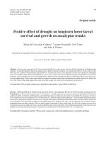
(p = 0.009) when compared with the cohesive strength of Filtek P90 resin composite. Fig. 1 shows the distribution of failure
modes among the experimental groups. The predominant failure mode was adhesive. Mixed mode of failure was recognized
only for the roughened groups.
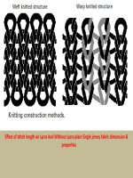
SEM evaluation of surface-treated Filtek P90 composite
substrates revealed different textures, whereas roughening with
320 grit SiC paper (roughened group) produced superficial
scratches (Fig. 2 and 3). Chemical treatment with 37% phosphoric acid did not produce clear changes in the superficial texture of the composite similar to the untreated one (Fig. 2).
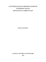
SEM evaluation of substrate-to-repair slabs showed different
interfacial features (Fig. 3). It was observed that there was
an interactive layer (about 13–20 lm) between SBMP adhesive
and Filtek Z250 repairing composite material (Fig. 3d). Such
an interactive layer was not present between P90 System Adhesive and Filtek P90 resin composite (Fig. 3a and b). SEM confirmed the predominance of adhesive failure in all tested
groups especially at the substrate side (Figs. 4 and 5) while
some mixed modes were detected for roughened groups
(Fig. 5).
Table 2
G1
G2
G3
G4
G5
G6
Discussion
The aim of the present study was to evaluate the repair bond
strength of 24 h aged silorane resin composite using, as much
as possible, a practical protocol. The protocol encountered
either surface roughness of silorane resin composite or not, followed by the use of phosphoric acid etching after which an
intermediate adhesive system of either P90 System Adhesive
or Adper Scotchbond Multipurpose was applied. This protocol was chosen for several reasons; one of them is that many
practitioners do not have additional tools in their dental practice such as chair-side air abrasion or silica coating devices.
The usage of diamond finishing burs and acid-etching with
phosphoric acid as surface treatment in repair procedure are
the most common repair approach taught by European [28]
and North American dental schools [29]. Another reason is
that often the repair process includes both enamel and dentin
together with old composite, thus, etching with phosphoric
acid followed by application of an adhesive system is clinically
mandatory. In the present study, two different adhesive systems with their corresponding resin composites were used as
it is not always possible for the dentist to determine the composite brand required to be repaired.
Based on the results of the present study, the null hypothesis has to be accepted since there was an insignificant difference
between the delayed repair bond strength of the tested groups.
SEM observations revealed that chemical treatment with 37%
phosphoric acid for smooth and roughened surfaces did not
produce obvious changes in the superficial texture of the substrate composite compared to that of untreated surfaces. Consequently, acid etching seems to exert only a cleaning effect,
without contributing to composite/composite micromechanical adhesion, as mentioned in previous studies [5,12,15].
The present study revealed an insignificant difference between the bond strength of roughened and smooth groups.
This finding is supported with Cavalcanti et al., findings [10].
On the contrary, others have reported significant differences
between roughened and smooth groups [12,15,20]. Previous researches have explained such inconsistent results in different
ways. Some authors referred this variability to the difference
in coarseness of the diamond burs used among the studies
and consequently the obtained surface roughness [6,15].
However, others attributed such diversity to the resulting
surface debris. Surface debris might interfere with proper
penetration of primers and/or monomers into the underlying
layer [30]. However, those who found such differences between
roughened and smooth specimens have used the shear test
lTBS values (MPa) of the repairing groups of FiltekP90.
Repairing method
lTBS Mean and standard
deviation (SD)
Smooth/acid etch/SA/Filtek P90
Roughened/acid etch/SA/Filtek P90
Smooth/acid etch/SBMP/Filtek Z250
Roughened/acid etch/SBMP/Filtek Z250
Control Filtek P90 resin composite
Control Filtek Z250 resin composite
29.9
32.8
32.7
33.4
52.1
60.0
(9.7)a
(8.7)a
(13.4)a
(10.3)a
(20.2)b
(22.5)b
% of repair strength
in relation to FiltekP90
cohesive strength
58
63
63
64
100
–
Same letter means no significant difference (paired t-test, p > 0.05). n = 30/each group. SA: P90 System Adhesive; SBMP: Adper Scotchbond
Multipurpose.
Repair potential of silorane-based resin composite
Fig. 1 Percentage distribution of failure modes in all tested
groups.
[12,15,20] which differs from the lTBS testing method applied
in the present study. Consequently, such dissimilarity in the
findings could be explained based on the difference in testing
methods, particularly the direction of load application, among
the studies. The present study SEM micrographs supports this
explanation by showing that the resin filled depressions were in
283
the same direction that coincide with the direction of pulling
tensile forces. Meanwhile, in shear testing such depressions
were perpendicular to the applied forces increasing the chance
to reveal high bond values and more cohesive failure modes for
roughened groups. This suggestion may also explain why
Luhrs et al. [31] found that sand blasting which creates multidirectional depressions revealed better lTBS than wet-polishing with 600-grit abrasive paper. Many researchers [12,15,20]
have used shear bond strength (SBS) for testing the repair
bond strength; however, such method has been criticized for
the test arrangement that produces high stress concentration
at the point of contact [32].
The findings of the present study may highlight the importance of the intermediate adhesive system (IAS) application.
During composite repair, there are three possible mechanisms
for bonding; chemical bond to the matrix, chemical bond to
the exposed filler particles as well as micromechanical retention
caused by penetration of the monomer components to the
micro-irregularities in the matrix [8]. For the fillers, in case
of no surface treatment, the quartz in Filtek P90 is surface treated with an oxirane functionalized silane. Therefore, there is
expected chemical affinity between the treated fillers and the
P90 System Adhesive. However, in the present study, mechanical (with finishing or polishing) and chemical (after acid etching) treatments were applied to the surface of Filtek P90, which
in turn, removed the functional silane from the exposed fillers
rendering them with no affinity to both adhesives tested. Based
on this, the micromechanical and/or the chemical coupling to
the resin matrix is expected to be the cause of the obtained repair bond strength of Filtek P90 and its adhesive. According to
Tezvergil-Mutluay et al. [25], immediate chemical bonding is
expected to occur between the phosphate group with oxirane
and the acrylate group with dimethacrylate. The present
study’s new finding may point out that some reactive unreacted monomers may still present after 24 h in Filtek P90.
For the SBMP, there is no chemical affinity between its components and Filtek P90; thus, micromechanical retention may
Fig. 2 SEM micrographs of Filtek P90 after different surface treatments 150·: (A) Smooth; (B) Smooth/acid etched; (C) Roughened; (D)
Roughened/acid etched.
284
E.H. Mobarak
Fig. 3 SEM micrographs of Filtek P90 substrate-to-repair resin composite (FS) at 500·: (a) Smooth substrate repaired with P90 System
Adhesive and Filtek P90 resin composite; (b) Roughened substrate repaired with P90 System Adhesive and Filtek P90 resin composite; (c)
Smooth substrate repaired with SBMP and Filtek Z250 resin composite; (d) Roughened substrate repaired with SBMP and Filtek Z250
resin composite. A = adhesive layer; ID = interdiffusion zone.
Fig. 4 SEM micrographs for representative two resulting surfaces of a stick after failure. S-FS = substrate of Filtek P90; R-FS = repair
Filtek P90; IAA = intermediate adhesive agent. Left: Shows failure at substrate surface of Filtek P90; Right: Shows IAA attached to the
repair Filtek P90.
Fig. 5 SEM micrographs for representative stick after failure. S-FS = substrate of Filtek P90; R-FS = repair Filtek P90; IAA = intermediate bonding agent. Left: Shows adhesive failure at the roughened Filtek P90 substrate; Right: Shows mixed failure.
Repair potential of silorane-based resin composite
contribute to the repair mechanism [13,16,21]. The ability of
monomers and solvent systems to penetrate into the composite
surface depends on the chemical affinity of materials and the
degree of hydration of the composites [7,33]. Most composites
are hydrophobic in nature but contain some absorbed water
that might improve surface penetration by hydrophilic bonding systems. The effectiveness of the studied adhesive systems
may be improved by their low viscosity and hydrophilicity,
which produces a small contact angle and good wetting properties [7,34,35].
An interesting point was that the difference between the
intermediate adhesive systems in composition and in filler content (whether filled or not) did not influence the repairing bond
strength outcome. However, no sufficient data is available
regarding this issue. Previous researchers have claimed that
the repair bond strength was much improved with filled adhesive resins than with unfilled adhesives [7]. This was based on
the fact that the addition of fillers increases the cohesive
strength. Regarding this point, further research is necessary.
Some investigators have reported that interfacial bond
strength to fresh composite was not different from the cohesive
strength of the resin composite itself [13]. On the other side, others reported that delayed repairing of resin composite revealed
widely variable repair bond strengths, which are in the range
of 25–82% of the cohesive strength of the substrate material
[7,10,13,15,30]. In the present study, bond strength values of
the repaired specimens were between 58% and 64% of the cohesive values of the Filtek P90 resin composite. This corresponds
with others’ findings although the test materials and methodologies are different [10,12,15]. Also, the obtained bond strength
values could be considered within the acceptable limits according to Teixeira et al. [7]. However, such results should be interpreted with caution when applied to clinical situation because
whether or not such values will survive in the oral environment
is not yet validated. Therefore, long-term clinical performance
of the repaired materials is the ultimate test. Further investigation regarding the clinical durability of the repair bond strength
of silorane-based composite is still required.
Regarding the mode of failure, the present study showed
the predominance of the adhesive failure mode which denotes
that the repair interface is still the weakest part especially at
the substrate side. The occurrence of failure mainly at the
substrate side may indicate that there is higher bonding affinity
between the IAS and the fresh repair composite more than that
obtained between the IAS and the substrate composite. The
detected inter-diffusion zone between SBMP and Filtek Z250
resin composite may support such speculation.
The previous findings emphasized that the evaluation of
the quality of the bond should not be assessed on the basis
of bond strength data alone. SEM observation of surface
texture and observation of the mode of failure could provide
important information that could potentially help in assessment of repair.
Conclusion
Repair of the silorane composite was successful irrespective of
the surface roughness and chemistry of the repair material
used, However, it did not reach the cohesive strength of the
material.
285
Acknowledgement
Author would like to thank Mr. Mohamed El Shahat for the
molds fabrication.
References
[1] Weinmann W, Thalacker C, Guggenberger R. Siloranes in
dental composites. Dent Mater 2005;21(1):68–74.
[2] Ilie N, Jelen E, Clementino-Luedemann T, Hickel R. Lowshrinkage composite for dental application. Dent Mater J
2007;26(2):149–55.
[3] Palin WM, Fleming GJ, Burke FJ, Marquis PM, Randall RC.
The influence of short and medium-term water immersion on the
hydrolytic stability of novel low-shrink dental composites. Dent
Mater 2005;21(9):852–63.
[4] Gordan VV, Mjor IA, Blum IR, Wilson N. Teaching students
the repair of resin-based composite restorations: a survey of
North American dental schools. J Am Dent Assoc
2003;134(3):317–23.
[5] Papacchini F, Dall’Oca S, Chieffi N, Goracci C, Sadek FT,
Suh BI, et al. Composite-to-composite microtensile bond
strength in the repair of a microfilled hybrid resin: effect of
surface treatment and oxygen inhibition. J Adhes Dent
2007;9(1):25–31.
[6] Rodrigues Jr SA, Ferracane JL, Della Bona A. Influence of
surface treatments on the bond strength of repaired resin
composite
restorative
materials.
Dent
Mater
2009;25(4):442–51.
[7] Teixeira EC, Bayne SC, Thompson JY, Ritter AV, Swift EJ.
Shear bond strength of self-etching bonding systems in
combination with various composites used for repairing aged
composites. J Adhes Dent 2005;7(2):159–64.
[8] Brosh T, Pilo R, Bichacho N, Blutstein R. Effect of
combinations of surface treatments and bonding agents on the
bond strength of repaired composites. J Prosthet Dent
1997;77(2):122–6.
[9] Furuse AY, da Cunha LF, Benetti AR, Mondelli J. Bond
strength of resin–resin interfaces contaminated with saliva and
submitted to different surface treatments. J Appl Oral Sci
2007;15(6):501–5.
[10] Cavalcanti AN, De Lima AF, Peris AR, Mitsui FH, Marchi
GM. Effect of surface treatments and bonding agents on the
bond strength of repaired composites. J Esthet Restor Dent
2007;19(2):90–8.
[11] Bouschlicher MR, Reinhardt JW, Vargas MA. Surface
treatment techniques for resin composite repair. Am J Dent
1997;10(6):279–83.
[12] Shahdad SA, Kennedy JG. Bond strength of repaired anterior
composite resins: an in vitro study. J Dent 1998;26(8):685–94.
[13] Turner CW, Meiers JC. Repair of an aged, contaminated
indirect composite resin with a direct, visible-light-cured
composite resin. Oper Dent 1993;18(5):187–94.
[14] Molla K, Park HJ, Haller B. Bond strength of adhesive/
composite combinations to dentin involving total- and self-etch
adhesives. J Adhes Dent 2002;4(3):171–80.
[15] Bonstein T, Garlapo D, Donarummo Jr J, Bush PJ. Evaluation
of varied repair protocols applied to aged composite resin. J
Adhes Dent 2005;7(1):41–9.
[16] Gordan VV, Shen C, Riley 3rd J, Mjor IA. Two-year clinical
evaluation of repair versus replacement of composite
restorations. J Esthet Restor Dent 2006;18(3):144–53.
[17] Hannig C, Laubach S, Hahn P, Attin T. Shear bond strength of
repaired adhesive filling materials using different repair
procedures. J Adhes Dent 2006;8(1):35–40.
286
[18] Kallio TT, Lastumaki TM, Vallittu PK. Effect of resin
application time on bond strength of polymer substrate
repaired with particulate filler composite. J Mater Sci Mater
Med 2003;14(11):999–1004.
[19] Shen C, Mondragon E, Gordan VV, Mjor IA. The effect of
mechanical undercuts on the strength of composite repair. J Am
Dent Assoc 2004;135(10):1406–12.
[20] Tezvergil A, Lassila LV, Vallittu PK. Composite–composite
repair bond strength: effect of different adhesion primers. J Dent
2003;31(8):521–5.
[21] Sau CW, Oh GS, Koh H, Chee CS, Lim CC. Shear bond
strength of repaired composite resins using a hybrid composite
resin. Oper Dent 1999;24(3):156–61.
[22] Vankerckhoven H, Lambrechts P, van Beylen M, Davidson CL,
Vanherle G. Unreacted methacrylate groups on the surfaces of
composite resins. J Dent Res 1982;61(6):791–5.
[23] Swift Jr EJ, LeValley BD, Boyer DB. Evaluation of new
methods for composite repair. Dent Mater 1992;8(6):362–5.
[24] Lewis G, Johnson W, Martin W, Canerdy A, Claburn C, Collier
M. Shear bond strength of immediately repaired light-cured
composite resin restorations. Oper Dent 1998;23(3):121–7.
[25] Tezvergil-Mutluay A, Lassila LV, Vallittu PK. Incremental
layers bonding of silorane composite: the initial bonding
properties. J Dent 2008;36(7):560–3.
[26] Shawkat ES, Shortall AC, Addison O, Palin WM. Oxygen
inhibition and incremental layer bond strengths of resin
composites. Dent Mater 2009;25(11):1338–46.
E.H. Mobarak
[27] Anusavice KJ. Philips’ science of dental materials. 10th
ed. Pennsylvania: W.B. Saunders Company; 1996, p. 298–99.
[28] Gordan VV. In vitro evaluation of margins of replaced resinbased composite restorations. J Esthet Dent 2000;12(4):209–15.
[29] Blum IR, Schriever A, Heidemann D, Mjor IA, Wilson NH. The
repair of direct composite restorations: an international survey
of the teaching of operative techniques and materials. Eur J
Dent Educ 2003;7(1):41–8.
[30] Gregory WA, Pounder B, Bakus E. Bond strengths of
chemically dissimilar repaired composite resins. J Prosthet
Dent 1990;64(6):664–8.
[31] Luhrs AK, Gormann B, Jacker-Guhr S, Geurtsen W.
Repairability of dental siloranes in vitro. Dent Mater
2011;27(2):144–9.
[32] Versluis A, Tantbirojn D, Douglas WH. Why do shear bond
tests pull out dentin? J Dent Res 1997;76(6):1298–307.
[33] Lastumaki TM, Kallio TT, Vallittu PK. The bond strength of
light-curing composite resin to finally polymerized and aged
glass fiber-reinforced composite substrate. Biomaterials
2002;23(23):4533–9.
[34] Rosales-Leal JI, Osorio R, Holgado-Terriza JA, CabrerizoVilchez MA, Toledano M. Dentin wetting by four adhesive
systems. Dent Mater 2001;17(6):526–32.
[35] Toledano M, Osorio R, de Leonardi G, Rosales-Leal JI,
Ceballos L, Cabrerizo-Vilchez MA. Influence of self-etching
primer on the resin adhesion to enamel and dentin. Am J Dent
2001;14(4):205–10.