Using NMR, X-ray, and CD analysis in the study on natural products obtained from Vietnamese plant and fungi in terms of pharmaceutical product development
Bạn đang xem bản rút gọn của tài liệu. Xem và tải ngay bản đầy đủ của tài liệu tại đây (1.35 MB, 7 trang )
Physical Sciences | Chemistry
Using NMR, X-ray, and CD analysis in the study
on natural products obtained from Vietnamese plant
and fungi in terms of pharmaceutical product development
Dinh Thang Tran1*, Cong Dung Vo1, Ngoc Tuan Nguyen1, Manh Dung Doan2, Yang-Chang Wu3, Tian-Shung Wu4
1
Faculty of Chemistry, Vinh University, Vietnam
Faculty of Chemistry, Hue University of sciences - Hue University, Vietnam
3
School of Pharmacy, College of Pharmacy, China Medical University, Taiwan
4
School of Pharmacy, National Cheng Kung University, Taiwan
2
Received 8 June 2017; accepted 7 November 2017
Abstract:
NMR, X-ray analysis, and CD methods are powerful techniques for the study
of absolute configuration of bioactive compounds from natural resources. This
study presents the results of a joint-study between Vietnam and Taiwan on the
bioactive compounds obtained from Vietnamese plants and fungi. Among the
tested compounds, hexatenuin A displayed the most significant inhibition of
superoxide anion generation and elastase release. These triterpenoids may be
used as potential anti-inflammatory agents.
Keywords: absolute configuration, circular dichroism, NMR, X-ray analysis.
Classification number: 2.2
Introduction
Natural products are an important
source for drug discovery. The
determination of absolute configuration
is one of the most challenging tasks in
the structure elucidation of chiral natural
products, especially those with complex
structures. The available methods
include
NMR
spectroscopy/chiral
derivatization, analytical chemistry,
X-ray crystallography for crystalline
compounds, chemical synthesis, and
chiroptical approaches [1]. Among
these, X-ray crystallography probably
remains the most powerful and effective
approach. However, the complete
structure elucidation of new compound
may require considerable effort and
involve many different spectroscopic
and,
sometimes,
computational
techniques.
The purpose of this review is to use
several examples, representing different
classes of natural products, to illustrate
the applicability of these approaches in
determining the absolute configuration
of natural products obtained from
Vietnamese plants and fungi. Moreover,
the purified constituents were examined
for their anti-inflammatory activity.
Among
the
tested
compounds,
hexatenuin A displayed the most
significant inhibition of superoxide
anion generation and elastase release.
These triterpenoids may have potential
to be used as anti-inflammatory agents.
Experimental
General experimental procedures
The optical rotations were measured
with a JASCO P-2000 digital polarimeter
in a 0.5 dm cell. The UV spectra were
obtained with a Hitachi UV-3210
spectrophotometer while the IR spectra
were measured with a Shimadzu FTIR
Prestige-21 spectrometer. The ECD
spectra were recorded on a JASCO J-720
spectrometer. The 1H- and 13C-NMR
*Corresponding author: Email:
14
Vietnam Journal of Science,
Technology and Engineering
December 2017 • Vol.59 Number 4
spectra were measured using Bruker
AMX-400 and AV500 spectrometers
with TMS as the internal reference, while
the chemical shifts were expressed in δ
(ppm). The ESIMS and HRESIMS were
collected on a Bruker Daltonics APEX II
30e spectrometer. HPLC was performed
on a Shimadzu LC-10ATVP (Japan)
system, equipped with a Shimadzu
SPD-M20A diode array detector at 250
nm, a Purospher STAR RP-8e c (5 μm,
250×4.6 mm), Cosmosil 5C18 ARII
(250×4.6 mm i.d. Nacalai Tesque Inc.),
and Astec Cellulose DMP (150×4.6
mm i.d. 5 μm) columns. The X-ray
diffraction experiments were performed
on a Bruker D8 Venture with a Photon
100 CMOS detector system equipped
with a Cu Incoatec IμS microfocus
source (λ = 1.54178 Å).
Preparation of human neutrophils
Neutrophils were isolated by a
standard
method
of
dextran
sedimentation,
prior
to
their
centrifugation in a Ficoll Hypaque
gradient and hypotonic lysis of
erythrocytes. Blood was drawn from
healthy human donors (20-30 years
old) by venipuncture into heparincoated Vacutainer tubes, using a
protocol approved by the institutional
review board at Chang Gung Memorial
Hospital [2]. The blood samples were
mixed gently with an equal volume
of 3% dextran solution. After the
sedimentation of the red cells for 30 min
at room temperature, the leukocyte-rich
plasma was collected,. The leukocyte-
Physical sciences | Chemistry
rich plasma was transferred on top of a
20 ml Ficoll solution (1.077 g/ml) and
spun down at 400 g for 40 min at 20°C.
The granulocyte/erythrocyte pellets
were resuspended in ice-cold 0.2%
NaCl to lyse the erythrocytes. After
30 s, the same volume of 1.6% NaCl
solution was added to reconstitute the
isotonic condition. Purified neutrophils
were pelleted and then resuspended in
a calcium (Ca2+)- free Hank’s balanced
salt solution (HBSS) buffer at pH 7.4
and maintained at 4°C before use [2].
Measurement of superoxide anion
generation
The assay of the superoxide anion
generation was based on the SODinhibitable reduction of ferricytochrome
c [2]. Briefly, after supplementation
with 0.5 mg/ml ferricytochrome c and
1 mM Ca2+, the neutrophils (6×10⁵
cells/ml) were equilibrated at 37°C
for 2 min and incubated with drugs
or an equal volume of vehicle (0.1%
DMSO, negative control) for 5 min.
The cells were activated with 100 nM
FMLP during the preincubation of 1
μg/ml cytochalasin B (FMLP/CB) for
3 min. Changes in the absorbance, with
a reduction in ferricytochrome c at
550 nm, were continuously monitored
in a double-beam, six-cell positioner
spectrophotometer
with
constant
stirring (Hitachi U-3010, Tokyo, Japan).
Then calculations were based on the
differences in the reactions with and
without SOD (100 U/ml), divided by the
extinction coefficient for the reduction
of ferricytochrome c (ε = 21.1/mM/10
mm) [2].
activated by 100 nM FMLP and 0.5 μg/
ml cytochalasin B while the changes in
absorbance at 405 nm were continuously
monitored to assay the elastase release.
The results were expressed as the
percentage of elastase release in the
FMLP/CB-activated, drug-free control
system [2].
Hexagonin A (16): white powder
(CHCl3); mp 184-185°C; [α]25D +57
(c 0.6, MeOH); UV (MeOH) λ max (log
ε) 262 (2.65) nm; IR (neat) nmax 2946,
1759, 1693, 1455, 1376, 1256, 1219,
1156 cm-1; 1H-NMR (500 MHz, CDCl3)
(d ppm): 4.71 (1H, br s, H-3), 4.32 (1H,
ddd, J = 11.5, 11.5, 5.0 Hz, H-16), 3.72
(3H, s, CH3-4’), 3.40 (2H, s, CH3-2’),
2.27 (1H, dd, J = 14.0, 11.5 Hz, H-15),
2.18 (1H, m, H-20), 2.05 (2H, m, H-6,
-11), 1.89 (1H, m, H-2), 1.84 (1H, m,
H-12), 1.71 (1H, m, H-2), 1.60 (3H, m,
H-7, -12, -22), 1.49 (3H, m, H-1, -7, -22),
1.41 (3H, m, H-1, -5, -17), 1.20 (1H, dd,
J = 14.0, 5.0 Hz, H-15), 1.94 (3H, d, J
= 0.5 Hz, CH3-31), 1.81 (3H, d, J = 0.5
Hz, CH3-27), 1.08 (3H, s, CH3-30), 1.00
(3H, s, CH3-19), 0.93 (3H, s, CH3-29),
0.95 (3H, d, J = 6.5 Hz, CH3-21), 0.88
(3H, s, CH3-28), 0.68 (3H, s, CH3-18);
13
C-NMR (125 MHz, CDCl3) (d ppm):
172.2 (C-26), 165.9 (C-1’), 167.2 (C-3’),
157.4 (C-24), 135.1 (C-9), 133.8 (C-8),
125.2 (C-25), 108.2 (C-23), 79.8 (C-16),
79.6 (C-3), 54.6 (C-17), 52.3 (C-4’), 48.6
(C-14), 45.3 (C-5), 43.5 (C-13), 41.8 (C2’), 41.1 (C-22), 37.1 (C-10), 36.8 (C4), 35.4 (C-15), 30.7 (C-20), 30.5 (C-1),
30.1 (C-12), 28.0 (C-30), 27.6 (C-28),
26.5 (C-6), 23.1 (C-2), 21.7(C-29), 20.2
(C-11), 19.4 (C-21), 18.8 (C-19), 17.9
(C-7), 16.5 (C-18), 10.8 (C-31), 8.5 (C27); ESIMS m/z 621 ([M+K]+, 60), 605
([M+Na]+, 26), 521 (33), 505 (100), 483
(48); HRESIMS m/z 605.3451 ([M +
Na]+, calcd for C35H50O7Na, 605.3454).
Results and discussions
A joint-study between Vietnam and
Taiwan on bioactive compounds from
the Vietnamese plant, Clausena lansium
Skeels (Rutaceae), was conducted.
The methanol extract from the dried
leaves of C. lansium was partitioned
between H2O and CHCl3. The
purification of the CHCl3 fraction by a
combination of column chromatographic
methods afforded eight new lactams,
including γ-lactams (1-3), δ-lactams
(4-7), and amide (8), along with seven
known lactams (9-15), which were
characterized from the leaves of C.
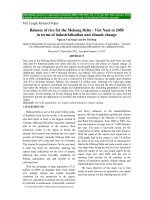
lansium (Fig. 1). Their structures were
elucidated using spectroscopic methods
[3] and the absolute configurations were
determined using electronic circular
dichroism (ECD) and single-crystal
X-ray diffraction analyses with Cu Kα
radiation.
Measurement of elastase release
The degranulation of azurophilic
granules was determined by the
elastase release, as described previously
[2]. Experiments were performed
using
MeO-Suc-Ala-Ala-Pro-Val-pnitroanilide as the elastase substrate.
Briefly, after supplementation with MeOSuc-Ala-Ala-Pro-Val-p-nitroanilide (100
μM), the neutrophils (6×10⁵ cells/ml)
were equilibrated at 37°C for 2 min and
incubated with drugs or an equal volume
of vehicle (0.1% DMSO, negative
control) for 5 min. The cells were
Fig. 1. The lactam compounds 1-15.
December 2017 • Vol.59 Number 4
Vietnam Journal of Science,
Technology and Engineering
15
Physical Sciences | Chemistry
The ECD sign and red shift of
the Cotton effect were shown to
experimentally determine the C-3
configuration as well as the sign and the
magnitude of the n → π* Cotton effect,
which are sensitive to the nature of the
C-3 substituent [4]. Therefore, the C-3
configuration of compound 1 with a
hydroxyl functionality was determined
as S, because it displayed a positive
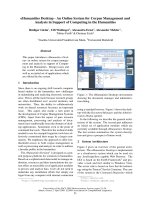
Cotton effect near 230 nm. The absolute
configuration of compound 1 was
unambiguously defined, by a singlecrystal X-ray diffraction analysis with
Cu Kα radiation, as 3S, 4R, 5S, and 6R
(Fig. 2). Consequently, the structure of
the 6-O-methylneoclausenamide (1)
was characterized, as shown in Fig.
1. The 2D structure of compound 2
was similar to compound 1, while the
relative configuration of the lactam
ring was assigned as being similar
to compound 1, through the analysis
of their NOESY spectra (Fig. 3). In
addition, the absolute configurations
at C-4, C-5, and C-6 were determined
by the single-crystal X-ray diffraction
pattern using the anomalous scattering
of Cu Kα radiation (Fig. 2). Therefore,
the absolute configuration was
determined as 3S, 4R, 5S, and 6S. In
effect, the structure of 6-O-methyl-epineoclausenamide (2) was assigned as
shown. The 2D structure of compound
3 was assigned to be identical to those
of compounds 1 and 2 by a comparison
of their UV, IR, MS, and NMR data
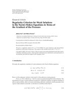
[2]. The ECD spectrum of compound
3 showed a low-amplitude positive
Cotton effect near 236 nm. The ECD
spectrum of compound 12 showed a
high-amplitude positive Cotton effect
at 230 nm. Thus, the low-amplitude
positive Cotton effect at 238 nm in
the ECD spectrum of compound 3
(Fig. 4) suggested 3S and 4S absolute
configurations [5]. By comparing
the specific rotation and absolute
configuration of compound 3 with the
16 stereoisomers of clausenamide,
the 3S, 4S, 5R, 6S and 3S, 4S, 5R,
6R configurations could be further
considered [3]. Therefore, the absolute
configuration
of
6-O-methyl-epi-
16
Vietnam Journal of Science,
Technology and Engineering
Fig. 2. ORTEP drawings of compounds 1, 2, 5, 7, 8, and 10.
cisneoclausenamide (3) was established
as 3S, 4S, 5R, and 6R. The absolute
configuration of C-3 in compound 4 was
deduced by the ECD spectrum. In this
case, the ECD spectrum of compound
4 (Fig. 4) showed a positive Cotton
effect at 231 nm, which evidenced a 3S
absolute configuration. Consequently,
the absolute configuration of compound
December 2017 • Vol.59 Number 4
4 was deduced as 3S, 4S, 5R, and 6R,
the structure of which was illustrated
as shown. To determine the absolute
configuration, compound 5 was
subjected to a single-crystal X-ray
diffraction analysis with Cu Kα radiation
(Fig. 2) which confirmed the structure
unambiguously. Therefore, the absolute
configuration was established as 3S, 4S,
Fig. 2. ORTEP drawings of compounds 1, 2, 5, 7, 8, and 10.
1
O
HO
Physical sciences | Chemistry
2
3
4
6
8
9
N
OCH3
5
Fig. 3. Selected NOESY (↔) correlations for compounds 1-6, 8, and 9.
5S, and
6S (Fig.
2). Hence, compound
(c 0.8,
MeOH)
andspectra
-71.8 (c 1.8,
MeOH)].
of the ECD
spectra of compounds 3 and
Some
relationships
between
the
ECD
and
the absolute
configurations
Compounds 14 and 15 were reported as 9 showed that the absolute configuration
5 wascould
characterized
as
lansamide-6.
be found from the above results. In the ECD spectra, δ-lactams 4, 14,
A positive Cotton effect at 223 nm in racemates in a previous study [5], but at C-5 may influence the wavelength of
andspectrum
15, with
4S, and 5R
absolute
configurations,
negative
the negative
specific
rotation [-107.8 (cexhibited
the ECD
(Fig.3S,
4) suggested
the Cotton
effect.
a 3S absolute configuration. The 1.4, MeOH) and -117.1 (c 0.7, MeOH)]
In the other joint-study, air-dried and
absolute configuration was established and a high-amplitude Cotton effect
powdered fruiting bodies of H. apiaria
as 3S, 4S, and 5S, while the structure (Fig. 4) confirmed that they were pure
were extracted with methanol and the
of lansamide-7 (6) was characterized enantiomers. Their structures were
combined extracts were concentrated
as shown. Based on these results and confirmed by the positive Cotton effects
under reduced pressure to produce a
the single-crystal X-ray diffraction in their ECD spectra [at 230 and 231
deep brown syrup. The crude extract
analyses using Cu Kα radiation (Fig. nm] (Fig. 4) and single-crystal, X-ray
was suspended in water and partitioned
2), the structure of lansamide-8 (7) diffraction analyses (Fig. 2).
with ethyl acetate to afford ethyl acetate
was identified as shown. The crystals
Some relationships between the ECD and water-soluble fractions. Purification
of compound 7 were orthorhombic and spectra and the absolute configurations
of the ethyl acetate fraction by a
belonged to the space group, Pbca. As could be found from the above results.
conventional combination of column
shown in the ORTEP drawing (Fig. 2), the In the ECD spectra, δ-lactams 4, 14,
chromatographies yielded four new
X-ray analysis revealed that compound and 15, with 3S, 4S, and 5R absolute
triterpenoids (16-19) and hexatenuin A
7 was a racemic mixture presumably configurations, exhibited negative and
[6].
originating from the reaction between positive Cotton effects near 210 and
Compound 16 was obtained as an
pyridine-2,3,6-trione
and
acetone. 230 nm, respectively. Compound 5 and
From the spectroscopic analysis and 6, possessing 3S, 4S, and 5S absolute optically active white powder, with
the single-crystal X-ray diffraction data configurations, displayed ECD spectra [α]25D +57 (c 0.6, MeOH). The positive(Fig. 2), the absolute configuration was with a positive Cotton effect at 220 nm. mode HRESIMS of compound 16
confirmed by the Flack parameter 0.0(2) For the γ-lactam group, compounds 1, 12, showed a pseudo-molecular ion peak
and defined as 3S, 4S, 5R, and 6S. The and 13, with 3S and 4R stereochemistry, at m/z 605.3451 ([M+Na]+, calcd for
structures of compounds 9 and 10 were exhibited similar ECD spectra. However, C35H50O7Na, 605.3454), corresponding
confirmed by the HRESIMS data and the absolute configurations of compound to the molecular formula of C35H50O7
single-crystal X-ray diffraction analysis 12 at C-5 and C-6 were different from with 11 indices of hydrogen deficiency
(Fig. 2). These structures have been those of compounds 1 and 13. This (IHD). The UV spectrum of compound
reported as synthetic products, but they implied that the absolute configuration 16 exhibited an absorption maxima
were isolated from their natural sources of C-5 and C-6 had little contribution to at 262 nm, compatible with an α,βfor the first time. Compounds 12 and the ECD spectra. In contrast, compounds unsaturated carbonyl chromophore [7].
13 were identified as (-)-clausenamide 3 and 9 possessed 3S and 4S absolute The IR absorption bands at 2946, 1759,
and (-)-neoclausenamide through the 1H configurations and showed different and 1693 cm-1 suggested the presence
and 13C NMR [1], the positive Cotton ECD spectra, as compared to those of of aliphatic C-H, lactonic carbonyl,
effect in the ECD spectrum [at 230 compounds 1, 12, and 13. This indicated and carbon-carbon double bond
and 229 nm] (Fig. 4), single-crystal that the C-4 phenyl group may have a functionalities. The 1H NMR spectrum
X-ray diffraction analysis (Fig. 2), and significant influence on the Cotton effect of compound 16 displayed five methyl
its negative specific rotation [-148.5 near 230 nm. Furthermore, a comparison singlets at δ 0.68 (3H, CH3-18), 0.88
December 2017 • Vol.59 Number 4
Vietnam Journal of Science,
Technology and Engineering
17
Physical Sciences | Chemistry
Fig. 4. ECD spectra of compounds 1-6 and 8-15.
(3H, CH3-28), 0.93 (3H, CH3-29), 1.00
(3H, CH3-19), and 1.08 (3H, CH3-30),
respectively. In addition, one doublet
methyl group at δ 0.95 (3H, J = 6.5 Hz,
CH3-21) suggested the presence of the
lanostane skeleton. Two vinyl methyl
signals at δ 1.81 (3H, d, J = 0.5 Hz,
CH3-27) and 1.94 (3H, d, J = 0.5 Hz,
CH3-31), along with the 13C NMR signals
at δ 8.5 (C-27), 10.8 (C-31), 108.2
(C-23), 125.2 (C-25), 157.4 (C-24), and
172.2 (C-26), indicated a γ-lactone ring
cyclized between C-23 and C-26. This
was verified by the HMBC correlations
from CH3-31 to C-23, -24, and -25
as well as from CH3-27 to C-24, -25,
and -26, respectively. In the downfield
region of the 13C NMR spectrum, there
were two oxygenated methines at δ 79.6
(C-3) and 79.8 (C-16), one set of tetrasubstituted double bonds at δ 133.8 (C-8)
and 135.1 (C-9), and two ester carbonyl
carbons at δ 165.9 (C-1′) and 167.2 (C3′). The location of the tetra-substituted
double bond at C-8/C-9 was determined
by the 3J-HMBC correlations between
18
Vietnam Journal of Science,
Technology and Engineering
CH3-19 and C-9 and between CH3-30
and C-8. The HMBC cross-peaks from
H-16 (δ 4.32, 1H, ddd, J = 11.5, 11.5,
5.0 Hz) to C-20 (δ 30.7), from H-3 (δ
4.71, 1H, br s) to C-29 (δ 21.7), C-1 (δ
30.5), C-5 (δ 45.3), C-1′; from CH2-2′ (δ
3.40, 2H, s) to C-1′ and C-3′; and from
CH3-4′ (δ 3.72, 3H, s) to C-3′ evidenced
that the C-16 had been oxygenated
while the C-3 had been acetylated by
the carbomethoxyacetyloxy group. The
elucidations provided above constructed
the chemical skeleton of 1 with 10
IHDs. The last IHD was afforded by
the cyclization between C-16 and C-23
through the ether linkage with a spiro
structure. These spectra evidenced
that compound 16 was very similar to
the reported compound hexatenuin A
[8], with the only difference being that
compound 16 was the methyl derivative
of hexatenuin A. The coupling constants
of H-3 (br s) and H-16 (11.5, 11.5, 5.0
Hz) indicated their orientations to be
equatorial and axial. The stereochemical
configurations of H-3 and H-16 were
December 2017 • Vol.59 Number 4
further established as β and β, according
to the NOESY analysis and comparison
of the spectral data of compound 16
and hexatenuin A [8]. The successive
two-dimensional spectral experiments,
including COSY, NOESY, HMQC, and
HMBC accomplished the assignments
of all the proton and carbon signals of
compound 16, and therefore its chemical
structure was established as shown in
Fig. 5 and named trivially as hexagonin
A.
Compounds 17-19 were all obtained
as optically active white powder,
displaying similar UV spectra and IR
absorption bands as those of compound
16. Moreover, the proton resonances for
the eight methyl groups, characteristic
of the triterpenoid basic skeleton, were
all observed in their 1H NMR spectra.
These data indicated that compounds 1619 were structurally similar compounds
(Fig. 6).
The purified triterpenoids, which
were isolated in sufficient quantity,
Physical sciences | Chemistry
(B)
Fig. 5. Significant HMBC (A) and NOESY (B) correlations of compound 16.
Fig. 6. Chemical structures of all the purified compounds.
Table 1. Inhibitory effects of purified samples from H. apiaria on superoxide
Anion generation and elastase release by human neutrophils, in response to
N-Formyl-Lmethionyl-phenylalanine/Cytochalasin B (FMLP/CB).
Compound
IC50 (μM)a
Superoxide anion generation
Elastase release
16
>10
-b
17
>10
-b
18
>10
-b
19
6.0±1.0***
>10
hexatenuin A
1.9±0.2***
4.3±1.4***
LY294002 c
0.4±0.02***
1.5±0.3***
Concentration necessary for 50% inhibition. Results are presented as mean
± SD (n = 3-4). ***p < 0.001 compared with the control value. bIncreasing
effects were observed. cA phosphatidylinositol-3-kinase inhibitor was used as a
positive control for superoxide anion generation and elastase release.
a
were examined for their inhibition
of superoxide anion generation and
elastase release by human neutrophils in
response to FMLP/CB (Table 1). Among
the examined constituents, hexatenuin A
displayed the most significant inhibition
of superoxide anion generation and
elastase release, with IC50 values of
1.9±0.2 and 4.3±1.4 μM, as compared to
the reference compound LY294002,12
with IC50 values of 0.4±0.02 and 1.5±0.3
μM for superoxide anion generation
and elastase release, respectively.
In addition, the following structureactivity relationships could be deduced
from the bioactivity data. Hexagonins
B (17) and D (19), which possess the
basic triterpenoid skeleton without the
malonyl substitution at C-3, did not
show any anti-inflammatory bioactivity.
Comparatively, hexagonin A (16), with
its triterpenoid skeleton and malonyl
and methyl ester functions, also failed to
exhibit significant activity. Hexatenuin
A, which had the triterpenoid skeleton
as well as a free malonic acid group,
displayed the most significant inhibitory
effects in the bioactivity examination.
Consequently, the free malonic acid
function was important for antiinflammatory activity. From the above
data, it was concluded that the purified
triterpenoids of H. apiaria are new
potential leads for anti-inflammatory
drug development and the starting
fungus can be used as a health food with
a possible and known mechanism of
action.
Therefore, it is not surprising that
intrinsic anti-inflammatory properties
demonstrated in vitro with H. apiaria can
be transferred in vivo after mushroom
consumption as food or nutraceutical
food. This study has identified the ability
for food processing to anti-inflammatory.
The process extraction for H. apiaria
identified a five-step process that would
address certain critical aspects in the
design and development of functional
food (Fig. 7).
Conclusions
A total of 15 lactams were isolated
December 2017 • Vol.59 Number 4
Vietnam Journal of Science,
Technology and Engineering
19
Physical Sciences | Chemistry
Org. Chem., 14(16), pp.1678-1697.
[2] S.C. Yang, P.J. Chung, C.M. Ho, C.Y.
Kuo, M.F. Hung, Y.T. Huang, W.Y. Chang,
Y.W. Chang, K.H. Chan, T.L. Hwang (2013),
“Propofol inhibits superoxide production,
elastase release, and chemotaxis in formyl
peptide-activated human neutrophils by
blocking formyl peptide receptor 1”, J.
Immunol., 190(12), pp.6511-6519.
[3] D.Y. Shen, T.N. Nguyen, S.J. Wu, Y.J.
Shiao, H.Y. Hung, P.C. Kuo, D.H. Kuo, T.D.
Thang, T.S. Wu (2015), “γ- and δ-lactams from
the leaves of Clausena lansium”, Journal of
Natural Products, 78(11), pp.2521-2530.
[4] T. Konno, H. Meguro, K. Tuzimura
(1975), “Circular dichroism of γ-lactams and
their sign determinating factors”, Tetrahedron
Lett., 16, pp.1305-1308.
Fig. 7. The process of extraction for H. apiaria.
from the methanolic extract of C.
lansium. This research work enabled
the determination of the absolute
configuration of these classes of
compounds using MS, NMR, electronic
circular dichroism (ECD), and singlecrystal X-ray diffraction analyses
with Cu Kα radiation. In the other
study, a chemical investigation of the
fruiting bodies of H. apiaria resulted
in the identification of five compounds,
hexagonins A-D (16-19) and hexatenuin
A. The purified constituents were
examined for their anti-inflammatory
activity. Among the tested compounds,
hexatenuin A displayed the most
20
Vietnam Journal of Science,
Technology and Engineering
significant inhibition of superoxide anion
generation and elastase release. These
triterpenoids may have the potential to
be used as anti-inflammatory agents.
This study has identified abilities from
food processing to anti-inflammatory.
The process extraction for H. apiaria
identified a five-step process that would
address certain critical aspects in the
design and development of functional
food.
REFERENCES
[1] X.C. Li, D. Ferreira, Y. Ding (2010),
“Determination of absolute configuration of
natural products: theoretical calculation of
electronic circular dichroism as a tool”, Curr.
December 2017 • Vol.59 Number 4
[5] Z.Q. Feng, X.Z. Li, G.J. Zheng, L. Huang
(2009), “Synthesis and activity in enhancing
long-term potentiation (LTP) of Clausenamide
stereoisomers”, Bioorg. Med. Chem. Lett.,
19(8), pp.2112-2115.
[6] T.D. Thang, P.C. Kuo, N.T. Ngoc, T.L.
Hwang, M.L. Yang, S.H. Ta, E.J. Lee, D.H.
Kuo, N.H. Hung, N.N. Tuan, T.S. Wu (2015),
“Chemical constituents from the fruiting
bodies of Hexagonia apiaria and their antiinflammatory activity”, J. Nat. Prod., 78(11),
pp.2552-2558.
[7] A.I. Scott (1964), Interpretation
ultraviolet spectra of natural products, 2nd ed.,
Pergamon press, New York.
[8] A. Umeyama, C. Ohta, Y. Shino,
M. Okada, Y. Nakamura, T. Hamagaki,
H. Imagawa, M. Tanaka, A. Ishiyama, M.
Iwatsuki, K. Otoguro, S. Omura, T. Hashimoto
(2014), “Three lanostane triterpenoids with
antitrypanosomal activity from the fruiting
body of Hexagonia tenuis”, Tetrahedron,
70(44), pp.8312-8315.