Rapid identification of mycobacterium tuberculosis and non tuberculous mycobacterium isolates from pulmonary and extra pulmonary samples using MGIT320 liquid culture system and mpt64
Bạn đang xem bản rút gọn của tài liệu. Xem và tải ngay bản đầy đủ của tài liệu tại đây (470.71 KB, 11 trang )
Int.J.Curr.Microbiol.App.Sci (2019) 8(1): 1172-1182
International Journal of Current Microbiology and Applied Sciences
ISSN: 2319-7706 Volume 8 Number 01 (2019)
Journal homepage:
Original Research Article
/>
Rapid Identification of Mycobacterium tuberculosis and Non Tuberculous
Mycobacterium Isolates from Pulmonary and Extra Pulmonary Samples
using MGIT320 Liquid Culture System and MPT64 Antigen Test
Qursheed Sultana, Ajaz Hussain*, Mohammed Abdur Rab Ansari,
Mohd Khaleel and Maimoona Mustafa
Department of Microbiology, Deccan College of Medical Sciences, Hyderabad, India
*Corresponding author
ABSTRACT
Keywords
Mycobacterial
growth indicator
tube (MGIT),
Mycobacterium
tuberculosis
complex (MTBc),
Non-tubercular
mycobacterium
(NTM), MPT64
antigen test and
multiple drug
resistant (MDR)
Article Info
Accepted:
10 December 2018
Available Online:
10 January 2019
Tuberculosis (TB) is a major public health problem in India and a leading cause of death in
adults, especially among the economically productive age group. Historically TB has been
associated with significant morbidity and mortality and remains a major global health
problem. The present study was initiated to determine the prevalence of Mycobacterium
tuberculosis, Non Tuberculous Mycobacterium and its resistance to first line AntiTubercular drug from both pulmonary and extra pulmonary samples A total of 583
properly collected samples (226 pulmonary and 357 extra pulmonary) from patients with
clinical/radiological suspicion of Tubercular infection were included in this study. All the
samples were screened by Zeihl-Neelsen AFB microscopy, and subjected to liquid culture
using Mycobacterium Growth Indicator Tube (MGIT-320). Positive cultures were
differentiated into Mycobacterium tuberculosis complex (MTBc) or non-tubercular
mycobacterium (NTM) by immunochromatography assay using MPT-64 antigen. Further
it was followed by drug susceptibility testing of MTBc isolates thereby identifying multidrug resistant strains. Out of 583 samples, 141 strains were isolated on MGIT-320 (81
pulmonary, 60 Extrapulmonary) and the detection time was 15 days. Mycobacterium
complex isolates were 116 and Nontuberculous Mycobacteria were 25. Among
Mycobacterium tuberculosis complex isolates 92(56 pulmonary, 36 Extrapulmonary) were
sensitive to all the drugs and 24(16 pulmonary, 8 Extrapulmonary) were resistant to one or
more drugs. Multiple drug resistant (MDR) isolates were 7(6 pulmonary, 1
Extrapulmonary). MDR-TB is gradually increasing due to improper diagnosis and
inadequate treatment. Differentiating mycobacterium as MTBc and NTM supported by
sensitivity testing by using liquid culture has proved to be helpful in early decision for
chemotherapy in MDR-TB patients.
Introduction
Tuberculosis (TB) is a major public health
problem in India and a leading cause of death
in adults, especially among the economically
productive age group. Historically TB has
been associated with significant morbidity and
mortality and remains a major global health
problem. India accounts for one‑ fifth of the
global burden of TB. It is estimated that about
1172
Int.J.Curr.Microbiol.App.Sci (2019) 8(1): 1172-1182
40% of Indian population is infected with TB
bacillus.(1) The prevalence and mortality due
to TB in India were estimated to be 249 and
26 respectively per100,000 population.(2)The
importance of early diagnosis and correct
etiological identification of pulmonary
tuberculosis need not be over-emphasised,
since treatment is different for Mycobacterium
tuberculosis and atypical Mycobacteria (nontuberculous Mycobacteria, NTM). World
Health Organization has given guidelines for
low and medium income countries for use of
liquid culture systems and drug sensitivity
testing for tuberculosis work. (3) The
emergence of anti‑ tubercular drug resistance
is an increasing public health problem and TB
control programmes in industrialized and
developing countries alike. (4) Drug resistance
arises due to improper and irrational use of
anti-tubercular drugs (ATDs) in chemotherapy
of drug-susceptible TB patients. This improper
use is a result of a number of actions including
administration of improper treatment regimens
and failure to ensure that patients complete the
whole course of treatment. Essentially, drug
resistance indicates a weakness in TB control
program in that area. A patient who develops
active disease with a drug-resistant TB strain
can transmit this form of TB to other
individuals. Strategies used for the clinical
management of patients infected with drugresistant
Mycobacterium
tuberculosis
scomplex (MTBC) are different, therefore,
prompt
detection,
isolation,
and
implementation of alternate anti-tubercular
treatment regimens are necessary for suitable
management (5) (6). Moreover, early
detection of such cases is of utmost
importance in preventing spread of resistant
bugs in the community. Automated nonradiometric systems for accelerated isolation
of Mycobacterium tuberculosis complex
(MTBC), being expensive, are available only
in selected centres in India and third-world
countries. However, most laboratories still
depend upon conventional techniques, thus
resulting in an extended reporting time of 4-5
weeks. The MGIT is a liquid broth medium
that is known to yield better recovery and
faster growth of mycobacteria. In addition to
Middlebrook 7H9 liquid media, the MGIT
tube
contains
an
oxygen-quenched
fluorochrome. It detects oxygen consumption
induced by growing micro-organisms (7).
There are a few published reports on the
evaluation of Bactec MGIT 960 on
extrapulmonary samples. An innovative rapid
kit, MPT64-ICT, to detect an established
marker of MTBC, the MPT64 antigen, by
immune chromatography test (ICT)developed
by Japanese scientists(8) found universal
acceptance due to its simplicity, accuracy and
rapidity. (9) (10) (11) Indian reports on EPTB
in general and the use of rapid kits for
confirmation of MTBC in particular are few.
The present study was initiated to determine
the
prevalence
of
Mycobacterium
tuberculosis, NonTuberculous Mycobacterium
and its resistance to first line Anti-Tubercular
drug from both pulmonary and extra
pulmonary samples among patients attending
a tertiary care hospital in Hyderabad.
Materials and Methods
Study design
The study was carried out in the clinical
Microbiology laboratory of a tertiary care
hospital in Hyderabad during the period
January 2013 to December 2015. Our
Institutional Human Ethics Committee
scrutinized and approved this research.
Patients’ informed consent was obtained
before collection of specimens.
Study population
A total of 583 properly collected samples (226
pulmonary and 357 extra pulmonary) from
patients with clinical/radiological suspicion of
1173
Int.J.Curr.Microbiol.App.Sci (2019) 8(1): 1172-1182
Tubercular infection were included in this
study. We included both pulmonary (like
deeply expectorated freshly collected sputum
samples, free of saliva, blood and food
contamination and bronchial alveolar lavage
samples) and extra-pulmonary samples (such
as all body fluids, tissue, urine, pus, aspirates
etc.). Samples were included irrespective of
the treatment status of the patients (e.g. both
new suspected cases as well as post-treatment
cases).
Patients were finally included on the basis of
availability of consent forms. Any patient
without consent was excluded from the study.
All
samples
showing
evidence
of
contamination with saliva (determined by
Bartlett’s grading system) (12) were excluded
from our study. We excluded the whole blood
samples as well as swab samples for TB
diagnosis in this study as per standard
guidelines.
Decontamination and processing of the
samples
All
specimens
were
liquefied
and
decontaminated by the standard N-acetyl-Lcysteine, sodium hydroxide method (NaOHNALC). After 15 min holding at room
temperature, specimens were neutralized with
phosphate buffer saline (PBS, pH 6.8) and
centrifuged in cold centrifuge at 4500 rpm for
20 min at 10°C. The pellets were resuspended
in 1.5 ml of sterile phosphate buffer and
collected for further analysis.
BACTEC MGIT 320 liquid media
Exclusion criteria
The BBL MGIT tube was inoculated by 0.5
ml of the decontaminated and concentrated
specimen suspension. It contained 7 mL of
modified middlebrook 7H9 broth enrichment
with albumin, dextrose and catalase (BBL
MGIT OADC) and an antibiotic mixture
consisting of polymyxin B, amphotericin B,
nalidixic acid, trimethoprim, and azlocillin
(BBL MGIT PANTA). After inoculation, the
tubes were loaded in the BACTEC MGIT 320
instrument and incubated up to 42 days at
37°C. Culture vials are monitored hourly by
the instrument. The positive tube was further
confirmed by ZN staining, subculturing on
blood agar plate. The TTD (Time to
Detection) of mycobacteria was based on the
date of the earliest instrumental indication of
positivity.
Swabs, Blood, salivary samples were excluded
from our study.
Morphological
identification
Materials and Methods
For differentiation of M. tuberculosis complex
and NTM, a commercially available kit was
used, the BD MGIT MTBc identification test
(TBc ID). It is a rapid chromatographic
immunoassay for the qualitative detection of
M. tuberculosis complex antigen from AFB
smear-positive BD MGIT tubes. The assay is
performed
Inclusion criteria
Both pulmonary (like deeply expectorated
freshly collected sputum samples and
bronchial alveolar lavage samples) and extrapulmonary samples (such as all body fluids,
tissue, urine, pus, aspirates etc.) were
included. All samples were selected on the
basis of availability of consent.
Acid fast bacilli smears
Smears were prepared from each sample,
stained by Ziehl Neelson method and
examined for presence of AFB with a light
microscope.
1174
and
biochemical
Int.J.Curr.Microbiol.App.Sci (2019) 8(1): 1172-1182
according to the manufacturer's instructions.
Briefly, 100µl of mixed and vortexed culture
fluid from AFB positive MGIT tubes were
transferred to sample window of the cassette.
The results of ICT were read within 15
minutes. Positive test had two red to purple
bands, one for internal control and the
secondline for the test.
Negative had only one band in internal
control slot. Strong or light bands with any
intensity were considered to be positive.
MGIT tubes showing non-acid fast bacilli
and/or fungi were excluded from MPT64 Ag
test.
BACTEC MGIT 320 liquid media DST
MTBc isolates was further tested for the first
line drugs in BACTEC MGIT 320. Conc. of
various drugs used was – streptomycin
(STR)- 1µg/ml, isoniazid (INH)- 0.1 µg/ml,
rifampicin (RIF)- 1 µg/ml, ethambutol (ETB)5 µg/ml. Drug susceptibility was reported
when the growth control units reached 400 as
indicated by the instrument.
Control strains
Reference
strains
of
H37Rv
and
Mycobacterium fortuitum were included as
positive and negative controls, respectively.
Results and Discussion
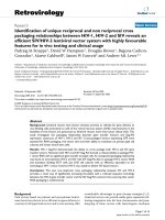
583 clinical samples (226 pulmonary and 357
extra-pulmonary) were analyzed during the
period of our study (Figure 1).
Number of Positive and Negative samples,
screened through Ziehl-Neelsen AFB Staining
procedure and culture by liquid media MGIT
320 are given in Table1. Out of these 583
samples, 257 were male patient and 326 were
females, in which 63 and 78 were positive
respectively, summarized in Table 2. There
was no much difference in gender distribution
among positive pulmonary samples whereas
females were predominant in case of Extra
pulmonary positive samples (Figure 2 and 3).
The results of age wise distribution among
positive cases in both pulmonary and Extra
pulmonary samples shows majority of case in
the age group of below 40 years, summarized
in Table 3. Distribution of various samples is
given in the Table 4.
The results show that, out of 226 pulmonary
samples, 81 were MGIT culture positive, of
which 47 were positive for AFB by ZN
staining and out of 357 Extra pulmonary
samples, 60 were MGIT culture positive, of
which 19 were positive for AFB by ZN
staining (Table 5 and 6). Out of these 81
culture positive isolates from pulmonary
samples 71 were MPT64Ag test positive and
9 were negative samples. This was considered
as Non-Tubercular Mycobacterium sp.
(Speciation not done).
Similarly, Out of these 60 culture positive
isolates from Extra pulmonary samples 44
were MPT64Ag test positive and 16 were
negative samples. This was considered as
Non-Tubercular
Mycobacterium
sp.
(Speciation not done) (Table 7).
The average TTD was 15 days for MGIT 320
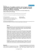
with the extremity from 6 to 38 days. Among
pulmonary positive cases, resistance to any
drug was found in16 cases (19.75%), to S in
5(6.17%), to I in 13(16.04%), to R in
7(8.64%) and to E in 2(2.46%). Multidrug
resistance rate was6 (7.40%) (Figure 4).
Similarly among Extra pulmonary positive
cases, resistance to any drug was found in 8
cases (13.3%), to I in 2(3.33%), to R in
3(5.00%) and to E in 1(1.66%) and no mono
resistance in S. Multidrug resistance rate
was1(1.66%) (Figure 5).
1175
Int.J.Curr.Microbiol.App.Sci (2019) 8(1): 1172-1182
Table.1 Distribution of culture positive cases
No. of cases studied
583
No. of positive
cases
141(24.18%)
No. of negative
cases
442(75.18%)
Table.2 Gender distribution of patients and percentage of positive samples
Gender
Male
Female
Total
No. Of collected
samples (%)
257 (44.08)
326 (55.92)
583
Positive isolates
(%)
63 (10.80)
78 (13.38)
141
Table.3 Age and Sex distribution of positive cases
Age
Distribution
20 and below
21 – 40
41 – 60
61 and above
Pulmonary
Male(n=41)
Female(n=40)
9(22%)
13(32%)
11(27%)
16(40%)
15(36%)
8(20%)
6(15%)
3(8%)
Extra Pulmonary
Male(n=22)
Female(n=38)
6(27%)
8(21%)
9(41%)
13(34%)
4(18%)
17(45%)
3(14%)
0
Table.4 Sample distribution in patients
Type of sample
Pulmonary
n=226 (38.77%)
Extra pulmonary
n= 357(61.23%)
Sputum
BAL
Pleural fluid
Pus
Ascitic fluid
ET Secretion
CSF
Urine
Pericardial fluid
Lymph Node Aspirates
Peritoneal fluid
Semen
Synovial fluid
Total
1176
No of cases
195
31
199
41
29
11
19
8
7
31
3
1
8
583
Positive
76
5
27
11
3
4
2
1
0
12
0
0
0
141
Int.J.Curr.Microbiol.App.Sci (2019) 8(1): 1172-1182
Table.5 Distribution of positive cases sample wise
Sample Type
Pulmonary
Extra
pulmonary
Total Samples
226
357
AFB Culture Positive
81
60
AFB Culture Negative
145
297
Table.6 correlation between stain and culture
Culture Positive
Culture Negative
Pulmonary
Stain +ve
47
0
Stain -ve
34
145
Total
81
145
Extra Pulmonary
Stain +ve
Stain -ve
19
41
0
297
Table.7 MTBC and NTM positive samples
Sample Type
Pulmonary
Extra Pulmonary
Total
MTBC
72
44
116
NTM
9
16
25
Fig 1 Distribution of sample type
Type of Samples(n=583)
Pulmonary
226
357,
61%
226,
39%
Extra
Pulmonary
357
1177
Total
60
297
Int.J.Curr.Microbiol.App.Sci (2019) 8(1): 1172-1182
Fig.2 Sex wise distribution of MTB positive pulmonary samples
Fig.3 Sex wise distribution of MTB positive extra pulmonary samples
Male 22
22,
37%
38,
63%
Fig.3 Results of Drug susceptibility testing in pulmonary samples
90
79
76
80
70
75
74
68
65
60
50
Resistant
40
Sensitive
30
20
16
10
13
7
5
6
2
0
Any drug
S
I
R
1178
E
MDR
Int.J.Curr.Microbiol.App.Sci (2019) 8(1): 1172-1182
Fig.4 Results of Drug susceptibility testing in extrapulmonary samples
70
60
60
58
59
57
59
52
50
40
Resistant
30
Sensitive
20
10
8
3
2
0
1
1
0
Any drug
S
I
R
Early
diagnosis
of
Mycobacterium
tuberculosis infection is pre-requisite to
achieve WHO’s target to end Global TB
epidemic. A definitive diagnosis of TB can
only be made by culturing Mycobacterium
tuberculosis organisms from a specimen
obtained from the patient. Therefore,
techniques which shorten the time for
detection of Mycobacterium deserve attention.
In our study, out of the 583 clinical samples
(both pulmonary and extra pulmonary),
141(24.18%) were culture positive. The
importance of early diagnosis and correct
etiological identification of Tuberculosis need
not be over-emphasised, since treatment is
different for Mycobacterium tuberculosis and
atypical
Mycobacteria
(non-tuberculous
Mycobacteria, NTM). In our study, out of 141
positive
isolates
116(82.2%)
were
Mycobacterium tuberculosis (MTBc) and
25(17.7%) isolates were NonTuberculous
Mycobacterium (NTM) using MPT64Ag test.
Similar results were given in various studies
like Kannade et al., (13) from Bombay
(Mumbai) who examined 165 isolates (125
MTB; 30 NTM; 10 Non-Mycobacterial
species) and observed sensitivity of 99.19%
and 100% values for specificity, positive
predictive value (PPV) and negative
E
MDR
predictive value (NPV) for the rapid MPT64
antigen detection kits in comparison to
conventional methods. Vadwai et al., (14)
from Bombay analysed 394 strains from 280
pulmonary and 114EPTB samples (388 MTB;
6 NTM) with similar result, i.e. 99.4%
sensitivity and 100% specificity. Kumar et al.,
(15) from Mysore, Karnataka, analysed 77
isolates (55 MTB; 10 NTM; 12 NonMycobacterial species) recorded 100% results
for all four parameters.
In our study majority of the pulmonary MTB
infected male patients were within the age
group of 20–40 years and female patients,
within the age group of 10-40 years. In the
case of extra pulmonary samples too both the
males and females were from the age group of
20 - 40 years. This is in correlation with the
study done by Kandhakumari et al., (16).
The prevalence of drug-resistant TB was
found variable in different studies from
around the world and in our country. In our
study, out of the 583 clinical samples, among
pulmonary samples the prevalence of
resistance to any drug was found in 16 cases
(19.75%), to S in 5(6.17%), to I in
13(16.04%), to R in 7(8.64%) and to E in
2(2.46%). Multidrug resistance rate was6
1179
Int.J.Curr.Microbiol.App.Sci (2019) 8(1): 1172-1182
(7.40%). Similarly among Extra pulmonary
positive cases, resistance to any drug was
found in 8 cases (13.3%), to I in 2(3.33%), to
R in 3(5.00%) and to E in 1(1.66%) and no
mono resistance in S. Multidrug resistance
rate was 1 (1.66%). Multidrug-resistance is
the independent factor for morbidity and
mortality due to tuberculosis (17) (18).
Treatment of MDR-TB is difficult and drugs
used for treatment are less potent, more toxic
and more expensive than firstline drugs (18)
(20). Many studies published from different
parts of India have reported high MDR-TB
prevalence, but mostly among first-time retreatment patients with relapse, treatment
after default, and treatment after failure (21)
(22). The possible reasons of a higher
prevalence of drug resistance in our study can
be, mixing of new as well as retreatment cases
and smaller sample size. Although many
Indian studies have reported lower prevalence
of Rifampicin mono-resistance from various
parts of the country, in our study the higher
rate can be due to a possible co-existence of
INH resistance and the rate may be acting as a
proxy to the local MDR-TB prevalence.
Various Indian studies have reported MDR
rates to be varying from 17.4% to 53% among
re-treatment
cases.(23,24)
World-wide
surveillance of MDR in re-treatment cases
ranged from 9.4% to 36.5%, from 1994-2000
across the world.(25) Previous exposure to
anti-tuberculosis agents is the most common
cause of developing MDR. In 2008, the WHO
reported a worldwide resistance rate to INH
of 5.9%. INH resistance rates higher than
10% can predict the development of MDR TB
according to the WHO (26). The higher
resistance rate of INH according to other first
line drugs may be resulted by both its wide
use in the chemoprophylaxis and latent TB
(27).
According to WHO in 2014, 220,000 people
died from TB in India, which is the highest in
the world. The same report says that 2.1%
cases in this emerging percentage are due to
MDR-TB.
Thus early detection of MDR-TB cases and
initiation of appropriate treatment based on
drug resistance testing can lower the burden
of this deadly disease.
In conclusion to conclude, globally the
prevalence of Tuberculosis is on the increase.
Due to prolonged time taken for positive
culture and drug susceptibility report by
conventional methods in suspected cases, the
clinicians in developing countries empirically
initiate anti-tuberculosis treatment (ATT)
with first-line drugs. However, if the etiology
happens to be NTM, this would be a burden
to the patients and can promote emergence of
drug resistance in Mycobacteria. The isolates
must be checked for drug sensitivity in this
era of increasing drug resistance. Thus rapid
isolation of Mycobacterium species using
automated MGIT320 system is more
beneficial when combined with rapid ICT kit
which detects MPT64 Ag in 15 minutes and
also differentiates MTBC from NTM isolates.
Notification of the DST results with clinical
data is a key element to get valid and
representative information on drug resistance.
As a study of prevalence of drug resistance in
TB from Hyderabad, we believe that this
study can help in the control of TB at the
national level and probably can help us in the
mapping drug resistant TB cases in this part
of the country.
References
1. Central Tuberculosis Division. Revised
National TB Control Programme: Annual
Status Report 2011. Available at: Http://
tbcindia.nic.in/pdfs/RNTCP%20TB%20In
dia%202011.pdf
2. Central Tuberculosis Division. Revised
National TB Control Programme: Annual
Status
Report
2013.
Availableat:
1180
Int.J.Curr.Microbiol.App.Sci (2019) 8(1): 1172-1182
3.
4.
5.
6.
7.
8.
Http://www.tbcindia.nic.in/pdfs/tb%20indi
a%202013.pdf
World Health Organization. Use of liquid
TB culture and drug susceptibility testing
(DST) in low and medium income
countries: Summary report of the expert
group meeting on the use of liquid culture
media. Geneva, Switzerland: WHO2007.
Available
from:
/>iquid_tb_culture_summary_report.pdf
World
Health
Organization.
The
WHO/IUATLD Global Project on
Anti‑ tuberculosis
Drug
Resistance
Surveillance. Anti‑ tuberculosis Drug
Resistance in the World. Fourth Global
Report.
Geneva,
Switzerland.
Availableat:Http://www.who.int/tb/publica
tions/2008/drs_report4_26feb08.pdf
Hasegawa, N., Miura, T., Ishii, K.,
Yamaguchi, K., Lindner, T.H., Merritt, S.,
et al., (2002) New Simple and Rapid Test
for
Culture
Confirmation
of
Mycobacterium tuberculosis Complex: A
Multicenter Study. Journal of Clinical
Microbiology,
40,
908-912.
/>Park, M.Y., Kim, Y.J., Hwang, S.H., Kim,
H.H., Lee, E.Y., Jeong, S.H., et al., (2009)
Evaluation of an Immunochromatographic
Assay Kit for Rapid Identification of
Mycobacterium tuberculosis Complex in
Clinical Isolates. Journal of Clinical
Microbiology, 47, 481-484.
Tortoli E, Cichero P, Piersimoni C,
Simonetti MT, Gesu G, et al., (1999) Use
of BACTEC MGIT 960 for recovery of
mycobacteria from clinical specimens:
multicenter study. J Clin Microbiol 37:
3578-3582.
Abe C, Hirano K, Tomiyama T. Simple
and
rapid
identification
of
the
Mycobacterium tuberculosis complex by
immunochromatographic assay using antiMPB64
monoclonal
antibodies.
J
ClinMicrobiol 1999; 37: 3693-7.
9. Maurya AK, Nag VL, Kant S, Kushwaha
RA, Kumar M,Mishra V, et al., Evaluation
of an immunochromatographic test for
discrimination between Mycobacterium
tuberculosis complex and non tuberculous
mycobacteria in clinical isolates from
extra-pulmonary tuberculosis. Indian J
Med Res., 2012; 135: 901-6.
10. Vadwai V, Sadani M, Sable R, Chavan A,
Balan
K,
Naik
A,
et
al.,
Immunochromatographic
assays
for
detection of Mycobacterium tuberculosis:
What is the perfect time to test? Diagn
Microbiol Infect Dis 2012; 74: 282-7.
11. Fraser Wares, Balasubramanian R, Mohan
A Sharma SK. Extra pulmonary
tuberculosis, management and control. In:
Agarwal SP, Chauhan LS, editors.
Tuberculosis Control in India. New Delhi:
Elsevier; 2005:95-114.
12. Correlation of Sputum Gram’s Stain and
Culture in Lower Respiratory Tract
Infections Anuradha Mokkapati, Madhavi
Yalamanchili. IOSR Journal of Dental and
Medical Sciences (IOSR-JDMS), 8, 6-9.
13. Kanade S, Nataraj G, Suryawanshi R,
Mehta P. Utility of MPT 64 antigen
detection assay for rapid characterization
of mycobacteriain a resource constrained
setting. Indian J Tuberc 2012;59:92-6
14. Vadwai V, Sadani M, Sable R, Chavan A,
Balan
K,
Naik
A,
et
al.,
Immunochromatographic
assays
for
detection of Mycobacterium tuberculosis:
What is the perfect time to test? Diagn
Microbiol Infect Dis 2012; 74: 282-7.
15. Kumar VG, Urs TA, Ranganath RR. MPT
64 Antigen detection for Rapid
confirmation of M. tuberculosis isolates.
BMC Res Notes 2011; 24: 79.
16. G Kandhakumari, S Stephen et al., Extra
pulmonary
tuberculosis:
Rapid
identification
of
Mycobacterium
tuberculosis grown in Mycobacterium
growth
indicator
tube
960
and
1181
Int.J.Curr.Microbiol.App.Sci (2019) 8(1): 1172-1182
Lowenstein-Jensen media, employing
Standard
diagnostics
Bioline
Mycobacterium
tuberculosis
protein
64antigen detection kit. Indian Journal of
Medical
Microbiology,
(2015)
33(Supplement 1): S122-25.
17. Petrini, B. and Hoffner, S. (1999) DrugResistant
and
Multidrug-Resistant
Tubercle Bacilli. International Journal of
Antimicrobial Agents, 13, 93-97
18. Pillay, M. and Sturm, A.W. (2007)
Evolution of the Extensively DrugResistant F15/LAM4/KZN Strain of
Mycobacterium tuberculosis in KwaZuluNatal, South Africa. Clinical Infectious
Diseases, 45, 1409-1414.
19. Tahaoğlu, K., Törün, T., Sevim, T., et al.,
(2001) The Treatment of MultidrugResistant Tuberculosis in Turkey. The
New England Journal of Medicine, 19,
170-174
20. Sevim, T., Aksoy, E., Ataç, G., et al.,
(2002) Treatment Adherence of 717
Patients with Tuberculosis in a Social
Security System Hospital in Istanbul,
Turkey. The International Journal of
Tuberculosis and Lung Disease, 6, 25-31.
21. Gupta, S., Bandyopadhyay, D., Gupta, S.,
Sadhukhan, S. and Banerjee, S. (2012) A
Sociodemographic Study of Multidrug
Resistant Tuberculosis Cases from DOTS
Clinics of Kolkata. Journal of the Indian
Medical Association, 110, 723-725.
22. Almeida, D., Rodigues, C., Udwadia, Z.F.,
Lalvani, A., Gothi, G.D., Mehta, P., et al.,
(2003) Incidence of Multidrug-Resistant
Tuberculosis in Urban and Rural India and
Implications for Prevention. Clinical
Infectious Diseases, 36, 152-154.
23. Ramachandran
R,
Nalini
S,
Chandershekhar V, Dave PV, Sanghvi AS,
Wares F, et al., Surveillance of drug
resistant tuberculosis in the state of Gujrat,
India. Int J Tuberc Lung Dis 2009; 134:
1154-60.
24. Parmasivan CN, Rehman F, Wares F,
Sundar Mohan N, Sundar S, Devi S, et al.,
First and second line drug resistance
patterns among previously treated
tuberculosis patients in India. Int J Tuberc
Lung Dis 2010; 14: 243-6.
25. Zignol M, van Gemert W, Falzon D,
Sismanidis C, Glaziou P, Floyda K, et al.,
Surveillance of anti-tuberculosis drug
resistance in the world: An updated
analysis 2007–2010. BullWorld Health
Organ 2012; 90: 111-9D.
26. WHO (2008) Anti-Tuberculosis Drug
Resistance in the World. Guidelines for
the Programmatic Management of Drug
Resistant Tuberculosis. World Health
Organization, Geneva.
27. Ndung’u, P.W., Kariuki, S., Ng’ang’a, Z.
and Revathi, G. (2012) Resistance Patterns
of Mycobacterium tuberculosis Isolates
from Pulmonary Tuberculosis Patients in
Nairobi. The Journal of Infection in
Developing Countries, 6, 33-39.
How to cite this article:
Qursheed Sultana, Ajaz Hussain, Mohammed Abdur Rab Ansari, Mohd Khaleel and
Maimoona Mustafa. 2019. Rapid Identification of Mycobacterium tuberculosis and Non
Tuberculous Mycobacterium Isolates from Pulmonary and Extra Pulmonary Samples using
MGIT320 Liquid Culture System and MPT64 Antigen Test. Int.J.Curr.Microbiol.App.Sci.
8(01): 1172-1182. doi: />
1182