In vitro efficacy of native trichoderma isolates against Pythium spp. and Rhizoctonia Solani (Kuhn.) causing damping-off disease in tomato (Solanum lycopersicum Miller)
Bạn đang xem bản rút gọn của tài liệu. Xem và tải ngay bản đầy đủ của tài liệu tại đây (503.82 KB, 14 trang )
Int.J.Curr.Microbiol.App.Sci (2019) 8(2): 566-579
International Journal of Current Microbiology and Applied Sciences
ISSN: 2319-7706 Volume 8 Number 02 (2019)
Journal homepage:
Original Research Article
/>
In vitro Efficacy of Native Trichoderma Isolates against Pythium spp. and
Rhizoctonia solani (Kuhn.) causing Damping-off Disease in Tomato
(Solanum lycopersicum Miller)
Markidahun Biam*, Dipali Majumder and Heipormi Papang
Department of Plant Pathology, College of Post Graduate Studies, Central Agricultural
University, Umiam-793103, Meghalaya, India
*Corresponding author
ABSTRACT
Keywords
Meghalaya,
Damping off,
Trichoderma spp.,
Pythium spp.,
Rhizoctonia solani,
Tomato, per cent
inhibition, PGPR
and biocontrol
potential
Article Info
Accepted:
04 January 2019
Available Online:
10 February 2019
The present study was conducted to evaluate the in vitro efficacy of native Trichoderma
spp. against Pythium spp. and Rhizoctonia solani (Kuhn.) causing damping-off disease in
Tomato. One hundred eighty soil samples were collected from 11 districts of Meghalaya
and ninety seven Trichoderma isolates were obtained. Rapid screening against damping
off pathogens (Pythium spp. and R. solani) of tomato revealed that 20 isolates showed
maximum antagonism and subsequently 20 best isolates were selected for further analysis.
Isolate TR 55 isolated from tomato rhizosphere was found to be the most effective isolate
against both Pythium spp. and R. solani Kuhn, showing an inhibition percentage of 89.26%
and 87.41% respectively, followed by other isolates like TR 66, TR 122 and TR 136.
Screening for the PGPR and biocontrol potential of the twenty potential isolates revealed
that all isolates were found positive for IAA production, ACC deaminase production and
phosphorous solubilization, whereas 17 positive for chitinase production, 16 isolates were
found positive for siderophore and ammonia production and 13 isolates were positive for
HCN production.
field or under greenhouse conditions.
Tomatoes are a good source of potassium,
vitamin C, vitamin A and excellent
antioxidant lycopene. They help in fighting
cancer, reducing heart disease and are also
good for eye health and digestion (Nahar and
Ullah, 2012).
Introduction
Tomato (Solanum lycopersicum Miller) is one
of the most important temperate vegetable
crops throughout the world and is also widely
cultivated in Meghalaya, India. Tomato
production has a major role in global
horticulture, ranking second in importance
next to potato in many countries (Sharma et
al., 2014). The main advantage of tomato
farming is that it can be grown either in the
China is leading producer among Asian
countries, followed by India. In India, tomato
is cultivated as one of the leading vegetable
566
Int.J.Curr.Microbiol.App.Sci (2019) 8(2): 566-579
crops, covering an area of 8.65 lakh hectares
with a total production of 168.26 lakh tonnes
having productivity of 19.60 tonnes ha-1
(Anon, 2014). Bihar, Karnataka, Odisha,
Maharashtra, Himachal Pradesh, West
Bengal, Tamil Nadu, Uttar Pradesh and
Gujarat are major tomato growing states
(Ghinaiya and Pandya, 2017) and among
them Karnataka is the largest producer state in
India. These States account for 91% of the
total production of the country. Meghalaya
accounts for 36.60 million tonnes of tomato
production, which is 0.16 % of country’s total
tomato production (Anon, 2018).
2018) and Rhizoctonia solani (Asad et al.,
2014; Abbas et al., 2017; Mangariello et al.,
2018). Trichoderma spp. are widely
distributed all over the world and found in all
natural habitats especially in those containing
forest humus layer (Wardle et al., 1993) as
well as in agricultural orchard soils (Roiger et
al., 1991) and soil consisting of organic
matter (Papavizas, 1985). It is known to be
one of the best candidates of BCA against
wide range of plant pathogens.
Therefore, in the present investigation major
emphasis was to study the bio-efficacy of
native Trichoderma isolates against damping
off caused by Pythium spp. and Rhizoctonia
solani Kuhn.
Although tomato is commercially grown
across the globe, there is no place where the
plant is free from diseases. One of the major
causes of seedling loss is damping-off, a
disease that is mainly caused by Pythium spp.
and Rhizoctonia solani Kuhn, which are
responsible for seed decay as well as preemergence and post-emergence damping-off
of tomato seedlings. Most of these fungi can
also cause cuttings to rot (Thakur and
Tripathi, 2015).
Materials and Methods
Isolation, identification and maintenance of
pathogen
Pythium spp. and R. solani Kuhn. were
isolated from naturally infected tomato,
showing damping off symptoms (soft rot and
wire stem symptom for Pythium spp. and R.
solani respectively) and pathogenicity test
was carried out in SCP, CPGS, CAU, Umiam,
Meghalaya. Diseased samples collected from
farmers’ fields were brought to laboratory and
isolations were done. With repeated
isolations, Pythium spp. and R. solani Kuhn
were consistently found with the infected
seedlings of tomato. Pythium spp. cultures
isolated from the infected tomato seedlings
were identified based on the types of fungal
mycelium and filamentous sporangia with
terminal, smooth and spherical oospores as
compared with the old cultures available in
the Laboratory. Also R. solani Kuhn was
identified based on the hyphae that tend to
branch at right angles and a septum near each
and a slight constriction at the branch are
diagnostic. The fungus was purified by hyphal
tip cut method. The purified culture was
Management of damping off by fungicides is
not the most desirable means of disease
management, for several important reasons.
Fungicides are heavily regulated and
additionally, they are expensive, cause
environmental pollution, and may induce
pathogen resistance (Lamichhane et al.,
2017). Since cultural practices alone are not
always sufficient to effectively manage the
disease, alternative strategies are needed.
Therefore, management of plant pathogens
using microbial bio inoculants has been
considered as a potential management
strategy for integrated disease management.
Many researchers have demonstrated the
potential of Trichoderma spp. in management
of damping-off diseases of crop plants caused
by Pythium spp. (Lamichanne et al., 2017;
Majeed et al., 2018; Al-shemamary et al.,
567
Int.J.Curr.Microbiol.App.Sci (2019) 8(2): 566-579
maintained on PDA slants at 4˚C in
refrigerator.
(90mm in dia) at equal distance from the
centre and incubated for 5-6 days at 28±1°C.
The relative growth rate of test antagonist and
the pathogen were observed and recorded.
The most efficient Trichoderma isolate was
sorted out as potent isolate against the
respective pathogen. These isolates were
multiplied and maintained as mentioned
earlier for long term preservation and
preserved at 4°C in PDA slants for subsequent
use.
Collection and isolation of Trichoderma
from different locations of Meghalaya
Soil samples from root rhizosphere, coal
mines, jhum fallows, manure compost and
submerged areas were collected from 11
districts of Meghalaya. Isolation of
Trichoderma was done by dilution plate
method using PDA (Dhingra and Sinclair,
1995). One gram of soil was suspended in 250
ml Erlenmeyer flasks with 100 ml sterilized
distilled water. Samples were shaked for 2030 minutes on a rotary shaker at 250 rpm and
dilutions of 10-1, 10-2, 10- 3, 10-4 and 10-5 were
made for each soil samples. An aliquot of 0.1
ml of substrate suspension was dispensed on
PDA. The Petriplates (90mm in dia) were
incubated at 28 ± 1ºC for 24 hours.
Morphologically distinct colony was isolated,
purified and grown in pure culture on PDA.
The obtained fungal isolates were grown on
PDA slants and kept at 4°C until being used.
Isolated Trichoderma was grown on Malt
Extract Agar (MEA) medium and identified
based on characters viz. conidiophores,
phialides and conidia (Rifai, 1969; Bisset,
1992). Microscopic examination was carried
out by mounting the culture in lactophenol
cotton blue.
The Trichoderma isolates were rated on the
basis of their ability to suppress the mycelia
growth of pathogen following the methods of
modified Bell’s scale (Bell et al., 1982).
S1: The antagonist completely overgrew the
pathogen (100% over growth)
S2: The antagonist overgrew at least 2/3rd
growth of the pathogen (75% overgrowth)
S3: The antagonized colonized half of the
growth of pathogen (50% overgrowth)
S4: The pathogen and antagonist (locked at
the point of contact)
S5: The pathogen overgrew the antagonist
The experiment was conducted with three
replicates per treatments.
Efficacy of Trichoderma isolates against
Pythium spp. and R. solani Kuhn
The potential Trichoderma isolates were
further evaluated for their antagonistic
potential in vitro against Pythium spp. and R.
solani Kuhn. through dual culture technique
(Ramanathan et al., 2013).
Rapid screening of Trichoderma isolates
Isolates tentatively identified as Trichoderma
were exposed to rapid screening of
Trichoderma isolates against Pythium spp.
and R. solani Kuhn. by dual culture technique
on PDA medium on the basis of their relative
growth rate measured as a function of
incubation period. Mycelial discs of 5mm
diameter was picked up from the margin of
young 3-4 days old culture of Trichoderma
and the respective pathogens were inoculated
at the peripheral region of the Petriplates
Dual culture technique
For mycelial growth inhibition of test plant
pathogens by the Trichoderma spp., both
pathogens (Pythium spp, R. solani Kuhn.) and
antagonists were inoculated at peripheral
region opposite to each other in sterilized
Petriplates (90 mm dia) containing 20 ml
568
Int.J.Curr.Microbiol.App.Sci (2019) 8(2): 566-579
sterilized PDA medium and incubated at
28±1°C. Plates inoculated with the pathogens
only served as the control. Observation for the
dual inoculation of the Trichoderma spp. and
the pathogen was taken till the growth of the
pathogen fully covered the plate. The
experiment was replicated three times. The
suppression effect of all Trichoderma spp.
isolates were evaluated in terms of Percentage
Inhibition in Radial Growth (PIRG) of
Pythium spp. and R. solani based on the
following formula (Gaigole et al., 2011).
°C until the smell of alcohol vanished. The
final colloidal chitin was stored at 4 °C until
further use.
Chitinase detection medium
The final chitinase detection medium per litre
comprised of 4.5 g colloidal chitin, 0.3 g
magnesium sulphate, 3.0 g ammonium
sulphate, 2.0 g potassium dihydrogen
phosphate, 1.0 g citric acid monohydrate, 15 g
agar, 0.15 g bromocresol purple and 200ul of
tween-80. The pH of the media was
maintained at 4.7 and autoclaved at 121 °C
for 15 min. The fresh culture plugs of
Trichoderma isolates to be tested for chitinase
activity were inoculated into the sterile plates
containing chitinase detection medium and
incubated at 28 ± 2 °C for 2–3 days and
observed for the coloured zone formation.
Formation of purple coloured zone was
observed and recorded.
PIRG = R1- R2 x 100%
R1
R1 = Radial growth of Pythium spp. and R.
solani in the absence of the antagonist in the
respective plate (control)
R2 = Radial growth of Pythium spp. and R.
solani in the presence of the Trichoderma
isolates (treatment)
Siderophores production test
Screening of isolated Trichoderma for In
vitro plant growth promoting and
biocontrol potential
The ability of Trichoderma spp. to produce
iron-binding compounds of siderophore-type
was detected in solid medium by universal
Chrome Azurol S (C.A.S) assay (Srivastava et
al., 2013)
Chitinolytic enzyme assay
The strains of Trichoderma isolates were
determined for chitinolytic activity on chitin
detection medium (Thakar and Saraf, 2015).
Preparation of the C.A.S. (Chrome Azurol
S) Blue Agar
One litre of C.A.S blue agar was prepared
using 60.5 mg C.A.S dissolved in 50 ml
distilled deionized water and mixed with 10
ml iron (III) solution (1 mM FeCl3.6H2O, 10
mM HCI). Under stirring, this solution was
slowly added to 72.9 mg Hexadecyl tri
methyl-ammonium
bromide
(HDTMA)
dissolved in 40 ml water. The resultant dark
blue liquid was autoclaved for 20 min. Also
autoclaved a mixture of 750 ml water, 15 g
agar, 30.24 g Pipes and 12 g of a solution of
50% (w/w) NaOH to raise the pH to the pKa
Preparation of colloidal chitin
5.0 g of chitin was added to 60 ml of conc.
HCl (acid hydrolysis) by constant stirring
using a magnetic stirrer at 4oC and kept in
refrigerator overnight. The resulting slurry
was then added to 200 ml of ice-cold 95%
ethanol and kept at 26 °C overnight (ethanol
neutralization). Then it was centrifuged at
3,000 rpm for 20 min at 4°C. The pellet was
repeatedly washed with sterile distilled water
by centrifugation at 3,000 rpm for 5 min at 4
569
Int.J.Curr.Microbiol.App.Sci (2019) 8(2): 566-579
of Pipes (6.8). The dye solution was finally
poured along the glass wall and agitated with
enough care to avoid foaming. Petri dishes
(9cm in diameter) were prepared with 30 ml
of PDA medium for culturing Trichoderma
spp. After solidification, the medium was cut
into halves, one of which was replaced by
C.A.S. blue agar (15 ml). The halves
containing culture medium were inoculated
with 5 mm discs of seven days old culture of
Trichoderma strains. The inoculum was
placed as far as possible, from the borderline
between the two media. The plates were
incubated at 28 ± 2°C for 7 days in the dark.
Colour-changed from blue to purple or dark
purplish- red (magenta) in the C.A.S.-blue
agar, starting from the borderline between the
two media was considered positive for
siderophore production. The experiment was
carried out in triplicates. The control plates of
C.A.S.-agar uninoculated were incubated
under the same conditions as described above.
The experiment was conducted with three
replicates per treatments.
and incubated at 25±1°C for seven days and
filtered with Whatman No. 2 filter paper, then
1 ml filtrate was mixed with 2 ml Salkowski
reagent (2% 0.5M FeCl3 in 35% perchloric
acid) in a test tube (Gravel et al., 2007). The
mixture was incubated at room temperature
for 20 minutes. Pink colour producing
samples was considered as positive reaction
and absorbance was measured at 540 nm by
spectrophotometer. A standard curve was
prepared using IAA and the presence of IAA
in the culture filtrate was quantified. The IAA
produced was compared to the standard graph
and expressed as μg/ml (Dixit et al., 2015).
Phosphate (P) solubilization
Production of HCN was detected by
inoculated different isolates of Trichoderma
spp. separately onto the PDA medium
amended with 4.4 g/ml glycine and lid of
plate was covered with the soaked Whatsman
no.1 filter paper in 0.5% picric acid and in 2%
sodium carbonate, then incubated for 5-7 days
at 28±1°C. Change in colour of filter paper
from deep yellow to orange and finally to
orange brown to dark brown indicated the
positive reaction. The experiment was
conducted with three replicates per treatments
(Dixit et al., 2015)
Solubilization of P was tested quantitatively
using 20 ml Pikovskaya’s broth medium
(PKV) amended with 5 g/l tricalcium
phosphate (17% P) then inoculated with a
mycelial disc of seven days old culture of
Trichoderma spp. and incubated at 28±1°C on
a shaker for 3-4 days. Uninoculaed PKV
broth served as control in each case. Each
experiment was done in triplicate set.
Mycelial growth was filtered through
Whatman No. 42 filter paper and 50 μl of
resultrant filtrate was added with 500 μl of
ammonium molybdate solution and shaked
well. An addition of 2ml distilled water, 13μl
chlorostannous acid and 2.5 ml distilled water
was made. Blue colour intensity was recorded
by spectrophotometer at 600nm. The
available phosphorus in the culture filtrate
was calculated from a standard curve
prepared using various concentration of
standard K2HPO4 solution and the results
were expressed in μg/ml (Rudresh et al.,
2005).
Indole-3-acetic acid (IAA) estimation test
ACC deaminase production test
Quantitative estimation of IAA was done
through addition of tryptophan in the potato
dextrose broth (PDB) for Trichoderma spp.
The ACC deaminase production of the
Trichoderma isolates was screened using the
methods described by Jasim et al., (2014). For
HCN production
570
Int.J.Curr.Microbiol.App.Sci (2019) 8(2): 566-579
this, the isolates were inoculated on to Difco
(DF) salts minimal medium (potassium
dihydrogen phosphate 4 g/L, disodium
hydrogen phosphate 6 g/L, magnesium sulfate
heptahydrate 0.2 g/L, ferrous sulfate
heptahydrate 0.1 g/L, boric acid 10 µg/L,
manganese(II) sulfate 10 µg/L, zinc sulphate
70 µg/L, copper (II) sulfate 50 µg/L,
molybdenum (VI) oxide 10 µg/L, glucose 2
g/L, gluconic acid 2 g/L, citric acid 2 g/L,
agar 12 g/L) amended with 0.2 % ammonium
sulphate (w/v). The fungal growth in this
media after 4-7 days of incubation was
considered as positive result. The experiment
was conducted with three replicates per
treatments.
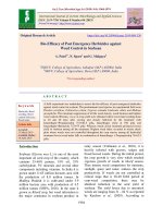
Rapid screening of Trichoderma isolates was
done against Pythium spp. and R. solani
Kuhn. by dual culture in potato dextrose agar
and the results showed that 77 isolates
attained S4 stage (the pathogen and antagonist
locked at the point of contact), 8 isolates
showed S3 stage (the antagonized colonized
half of the growth of pathogen i.e. 50%
overgrowth) and remaining 12 isolates
showed S2 stage (the antagonist overgrew at
least 2/3rd growth of the pathogen i.e. 75%
growth) from 4-7 days after inoculation on the
basis of modified Bell’s scale (Plate 1).
The study of antagonistic potential of 20
screened isolates of Trichoderma spp. against
damping-off pathogens which showed S2 and
S3 stage on the basis of modified Bell’s scale
revealed that all 20 isolates showed an
inhibition percentage of more than 65%.
Among which 4 Trichoderma isolates viz. TR
55, TR 66, TR 122 and TR 136 were most
effective in inhibiting Pythium spp. with
percent inhibition of 89.26%, 88.15%,
88.89% and 87.78% respectively, whereas
only 2 isolates viz. TR 55 and TR 122 were
found effective against R. solani with percent
inhibition of 87.41% and 86.48% respectively
(Table 1).
Ammonia production
Trichoderma isolates was tested for the
production of ammonia in peptone water.
Freshly grown cultures were inoculated in 10
ml peptone water separately and incubated for
48-72 h at 36 ± 2°C. Nessler’s reagent (0.5
ml) was added in each tube. Development of
yellow to brown colour indicated for positive
test (Thakkar and Saraf, 2015). The
experiment was conducted with three
replicates per treatments.
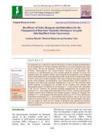
The result (Table 2, Plate 2) showed that out
of 20 screened isolates, only 16 isolates were
positive for siderophore production as
indicated by colour-changed from blue to
purple or dark purplish- red (magenta) in the
C.A.S.-blue agar. Also 16 isolates were
positive for ammonia production indicated by
development of yellow to brown colour in
Nessler’s reagent whereas 17 isolates were
positive for chitinase and 13 isolates were
positive for HCN production as evidenced by
the change in the colour of filter paper.
However all 20 screened isolates grew on
Difco (DF) salts minimal medium showing
their ability to produce ACC deaminase.
Results and Discussion
Soil samples collected from different
locations of Meghalaya were tested. The
isolates showing the lime green to greenish
colour sporulation with highly fluffy growth
and sparse to compact colony after 7-10 days
of incubation were selected. The selected
isolates were grown on Malt Extract Agar
(MEA) to observe conidiophores, phialides
and conidia. Based on taxonomic keys
provided by Rifai (1969) Bisset (1992), it is
evident that altogether ninety seven (97)
Trichoderma isolates were isolated from 180
soil samples collected.
571
Int.J.Curr.Microbiol.App.Sci (2019) 8(2): 566-579
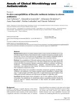
Table.1 In vitro efficacy of Trichoderma isolates against Pythium spp and R. solani
Sl.
No.
1.
Trichoderma
isolates
TR 12
2.
TR 24
3.
TR 36
4.
TR 40
5.
TR 55
6.
TR 64
7.
TR 66
8.
TR 74
9.
TR 78
10.
TR 82
11.
TR 87
12.
TR 88
13.
TR 106
14.
TR 109
15.
TR 112
16.
TR 116
17.
TR 122
18.
TR 136
19.
TR 143
20.
TR 171
21.
Control
SE(m)
CD (p=0.05)
Growth (cm)
Pythium spp
2.00±0.06d
(1.41)
2.43±0.07c
(1.56)
2.80±0.06b
(1.67)
2.23±0.07c
(1.49)
0.97±0.03h
(0.98)
1.97±0.07d
(1.40)
1.07±0.03h
(1.03)
2.00±0.00d
(1.41)
1.83±0.09de
(1.35)
1.93±0.09d
(1.39)
2.37±0.03c
(1.54)
1.57±0.03fg
(1.25)
1.70±0.12ef
(1.30)
2.33±0.03c
1.53)
1.63±0.09f
(1.28)
1.83±0.07de
(1.35)
1.00±0.00h
(1.00)
1.10±0.06h
(1.05)
1.47±0.03g
(1.21)
1.63±0.09f
(1.28)
9.00±0.00a
(3.00)
0.002
0.06
R. solani
1.90±0.06ij
(1.37)
2.07±0.09gh
(1.43)
2.33±0.03cde
(1.52)
2.07±0.03gh
(1.43)
1.13±0.03o
(1.06)
1.97±0.07hi
(1.40)
1.43±0.03l
(1.19)
1.73±0.03k
(1.31)
2.40±0.06c
(1.54)
2.23±0.07def
(1.49)
2.18±0.04efg
(1.47)
1.53±0.03l
(1.23)
2.30±0.00m
(1.51)
2.12±0.10fg
(1.45)
2.63±0.03b
(1.62)
2.35±0.05cd
(1.53)
1.22±0.02no
(1.10)
1.82±0.06jk
(1.34)
2.37±0.03cd
(1.53)
2.70±0.06b
(1.64)
9.00±0.00a
(3.00)
0.001
0.051
Per cent inhibition over control
Pythium spp
R. solani
e
77.78±0.64
78.89±0.64de
(61.88)
(62.65)
72.96±0.74i
77.04±0.98fg
(58.67)
(61.37)
68.89±0.64j
74.07±0.37jk
(56.10)
(59.39)
f
75.19±0.74
77.04±0.37fg
(60.12)
(61.37)
89.26±0.37a
87.41±0.37a
(70.87)
(69.22)
78.15±0.74e
78.15±0.74ef
(62.13)
(62.13)
88.15±0.37a
84.07±0.37b
(69.86)
(66.47)
e
77.78±0.00
80.74±0.37c
(61.87)
(63.97)
79.63±0.98de
73.33±0.64l
(63.18)
(58.91)
78.52±0.98e
75.19±0.74hij
(62.40)
(60.12)
h
73.70±0.37
75.74±0.49ghi
(59.14)
(60.49)
82.59±0.37bc
82.96±0.37b
(65.34)
(65.62)
81.11±1.28cd
74.44±0.00ijk
(64.26)
(59.63)
74.07±0.37fg
76.48±1.13gh
(59.39)
(61.00)
bc
81.85±0.98
70.74±0.37m
(64.79)
(57.25)
79.63±0.74de
73.89±0.56jk
(63.17)
(59.27)
88.89±0.00a
86.48±0.19a
(70.53)
(68.43)
a
87.78±0.64
79.81±0.67cd
(69.55)
(63.30)
83.70±0.37b
73.70±0.37jk
(66.19)
(59.14)
81.85±0.98bc
70.00±0.64mn
(64.79)
(56.79)
0.00±0.00k
0.00±0.00o
(0.36)
(0.36)
0.72
0.44
1.40
1.10
*Mean of three replications
Note: Figures in parentheses are square root transformed values for growth and arc sine transformed values for per
cent inhibition over control.
572
Int.J.Curr.Microbiol.App.Sci (2019) 8(2): 566-579
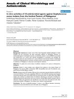
Table.2 PGPR and biocontrol efficacy of Trichoderma spp.
Sl.
No.
Trichoder
ma spp
Chitinase
production
Siderophore
Production
HCN
Production
Ammonia
production
ACC
deaminase
activity
IAA produce
(ug/ml)
Phosphorous
production
(ug/ml)
1.
2.
TR 12
TR 24
+
+++
++
++
-
+
+
+
1.47
0.84
0.68
0.21
3.
4.
5.
6.
7.
TR 36
TR 40
TR 55
TR 64
TR 66
+
++
++
+++
++
+
+++
+++
++
+
+
+
++
++
+++
+++
++
+
+
+
+
+
2.97
3.22
4.96
1.81
2.14
0.53
0.22
0.72
0.41
0.75
8.
TR 74
+
-
+
+
+
2.45
0.09
9.
TR 78
+
++
-
-
+
2.51
0.23
10.
TR 82
-
++
-
+
+
1.53
0.03
11.
TR 87
++
++
+
+
+
1.44
0.32
12.
TR 88
+
+
++
+
+
4.70
0.58
13.
TR 106
+++
+
+
+
+
0.48
0.98
14.
TR 109
+
-
+
+
+
0.48
0.74
15.
TR 112
+
+++
++
+
+
1.94
0.39
16.
TR 116
-
-
-
++
+
0.33
0.32
17.
TR 122
+++
++
++
+++
+
0.72
0.25
18.
TR 136
+
++
++
+++
+
0.99
0.79
19.
TR 143
++
-
+
-
+
0.58
0.59
20.
TR 171
_
++
+
+
+
0.87
0.29
*Mean of three replications
(+) indicates light color, (++) indicates dark color and (+++) indicates very dark color
(-) indicates absence
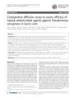
Plate.1 Rapid Screening of Trichoderma spp. following modified Bell’s scale
S
S3
2
Pathogen
573
S4
Int.J.Curr.Microbiol.App.Sci (2019) 8(2): 566-579
Plate.2 PGPR and biocontrol efficacy test of Trichoderma isolates
Chitinase production test
HCN production test
Ammonia production test
Siderophore production test
ACC deaminase production test
IAA production test
Phosphorous solubilization
test
574
Int.J.Curr.Microbiol.App.Sci (2019) 8(2): 566-579
Screening for plant growth promoting traits of
20 isolates revealed that all produced IAA
with TR 55 showing highest production (4.96
ug/ml) followed by others like TR 88 (4.70
ug/ml), TR 40 (3.22 ug/ml) and least in TR
116 (0.33 ug/ml). Also all produced
phosphorous with highest production in TR
106 (0.98 μg/ml) followed by TR 66 (0.75
ug/ml), TR 55 (0.72 ug/ml) and the least was
observed in TR 82 (0.03 ug/ml).
production and phosphate solubilisation
showed that all 20 screened isolates were
found positive for ACC deaminase production
and 17 positive for chitinase production. Out
of 20 screened isolates tested for other
functional attributes (determining antagonistic
potentials), 16 isolates were found positive for
siderophore and ammonia production,
whereas 13 isolates were positive for HCN
production. Screening for plant growth
promoting traits of 20 isolates revealed that
all produced IAA and Phosphorous with
values ranging from 0.33 to 4.96 μg/ml and
0.03 to 0.98 μg/ml respectively. The
production of lytic enzymes by Trichoderma
species is known as one of the major
mechanisms for biocontrol activity against
phytopathogenic fungi, involvement of
chitinase in control of phytopathogens was
reported (Harman et al., 2004a; Harighi et al.,
2007, Azad et al., 2015, Thakar and Saraf
2015). HCN, siderophores and ammonia are
produced by many Trichoderma spp. and are
believed to play a role in biological control of
pathogens (Rawat and Tiwari, 2011; Qi and
Zhao, 2013, Zhang et al., 2016). Phosphate
solubilizing efficiency of different isolates of
Trichoderma was observed by many workers
like Tallapragada and Gudimi (2011);
Sarawanakumar et al., (2013); Promwee et
al., (2014): Borges Chagas et al., (2015),
Franca et al., (2017). ACC deaminase
production of Trichoderma spp was reported
by several workers (Viterbo et al., 2010;
Hermosa et al., 2012; Aban et al., 2017).
Aban and his co-workers also reported IAA
production and phosphate solubilisation by
Trichoderma yunnanense and Trichoderma
simmonsil which is similar to the present
findings.
From 180 soil samples collected from all 11
districts of Meghalaya, 97 Trichoderma
isolates were obtained which showed that
Trichoderma isolates are predominant in
different habitat i.e., crop rhizosphere,
compost manure, sacred forest, coal mine and
lime
stone.
The
predomination
of
Trichoderma in natural soils, decaying wood,
plant materials, crop rhizosphere were
reported by several workers (Kredics et al.,
2012; Kumar et al., 2012; Rai et al., 2016;
Jaisani and Pandey, 2017).
Out of 97 isolates screened by dual culture
against Pythium spp. and R. solani Kuhn.,
only 20 isolates colonized more than half of
the growth of pathogen i.e. 50% overgrowth.
TR 55 isolated from tomato rhizosphere was
found to be the most effective isolate against
both Pythium spp. and R. solani Kuhn,
showing an inhibition percentage of 89.26%
and 87.41% respectively, followed by other
isolates like TR 66, TR 122 and TR 136. The
antagonism of Trichoderma spp. against
Pythium spp. and R. solani were widely
reported (Goud et al., 2015; Kotasthane 2015;
Waghunde et al., 2016; Kumari et al., 2016;
Naik et al., 2017; Rajendraprasad et al., 2017)
which supported the antagonism of
Trichoderma spp. against Pythium spp. and R.
solani Kuhn. during the present investigation.
In conclusion, many Trichoderma isolates
were obtained from the crop rhizospheric
soils in the 11 districts of Meghalaya, India.
Via screening test, 20 isolates showed the best
antagonism against damping off pathogens
All 20 isolates screened for their PGPR and
biocontrol potential such as HCN, ammonia,
siderophore, IAA, chitinase, ACC deaminase
575
Int.J.Curr.Microbiol.App.Sci (2019) 8(2): 566-579
(Pythium spp. and R. solani) of tomato
seedlings. Screening of 20 isolates for their
PGPR and biocontrol potential found that
almost all revealed their ability of HCN,
ammonia, siderophore, IAA, chitinase, ACC
deaminase
production
and
phosphate
solubilisation. However, out of the 20
isolates, 4 Trichoderma isolates viz. TR 55,
TR 66, TR 122 and TR 136 were found to be
the most effective, so these 4 potent isolates
need further evaluation in field condition to
develop effective bio-formulation against
damping off of tomato.
Asad, S.A., N. Ali, A. Hameed, S.A. Khan, R.
Ahmad, M. Bilal, M. Shahzad, and
Tabassum, A. 2014. Biocontrol
efficacy of different isolates of
Trichoderma against soil borne
pathogen Rhizoctonia solani. Pol J
Microbiol. 63(1): 95-103.
Bell, D.K., H.D. Wells, and Markham, C.R.
1982.
In-vitro
antagonism
of
Trichoderma spp. against six fungal
plant pathogens. Phytopathol. 72:379382.
Bissett, J. 1992. Trichoderma atroviride.
Canadian J. Botany. 70 (3): 639-641,
10.1139/b92-082.
Borges, C. L. F., J. A. F. Chagas, C. M.
Rodrigues, L. D. Miller, and Orozco,
C. B. S. 2015. Evaluation of the
phosphate solubilization potential of
Trichoderma strains (Trichoplus JCO)
and effects on rice biomass. Journal of
Soil Science and Plant Nutrition. 15
(3): 794-804.
Dhingra, O.P., and Sinclair, J.B. (1995). Basic
plant pathology methods, 2nd edn.
CRC press, Bocca Raton, America.
Dixit, R., R. B. Singh, and Singh, H.B. 2015.
Screening of antagonistic potential
and plant growth promotion activities
of Trichoderma spp. and fluorescent
Pseudomonas spp. isolates against
Sclerotinia sclerotiorum causing stem
rot of French bean. Legume Research.
38 (3): 375-381.
Franca, V. C. D., C.K. Kupper, M. RosaMagri, T. Gomes, and Rossi, F. 2017.
Trichoderma spp. Isolates with
potential of phosphate solubilization
and growth promotion in cherry
tomato.
Pesquisa
Agropecuaria
Tropical. 47: 360-368. 10.1590/198340632017v4746447.
Gaigole, A.H., 2011. Trichoderma viride are
having ability to inhibit soil borne
pathogen of different crops like
Rhizoctonia
solani
and
most
References
Aban, J. L., Barcelo, R. C., Oda, E. E., Reyes,
G. A., Balangcod, T. D., Gutierrez, R.
M, and Hipol, R. M. 2017. Auxin
Production, Phosphate Solubilisation
and ACC Deaminase Activity of Root
Symbiotic Fungi (RSF) from Drynaria
quercifolia L. Bull. Env. Pharmacol.
Life Sci. 6(5): 18-23.
Abbas, A., D. Jiang, and Fu, Y. 2017.
Trichoderma spp. as Antagonist of
Rhizoctonia solani. J. Plant Pathol.
Microbiol. 8:402. doi:10.4172/21577471.1000402.
Anonymous, 2014. http:// www. indiastat.
com/ agriculture/ 2/ vegetable/ 17427/
stat. Aspx.
Anonymous, 2018. Monthly Report Tomato.
Horticulture
Statistics
Division
Department
of
Agriculture,
Cooperation and Farmers Welfare,
Ministry of agriculture and Farmers
Welfare, New Delhi.
Asad, S. A., A. Tabassum, A, Hameed, F.
Hassan, A. Afzal, S.A. Khan, R.
Ahmed and Shahzad, M. 2015.
Determination of lytic enzyme
activities of indigenous Trichoderma
isolates from Pakistan. Brazilian
Journal of Microbiology. 46(4): 10531064.
576
Int.J.Curr.Microbiol.App.Sci (2019) 8(2): 566-579
commonly used fungal bio control
agent and have long been known as
effective antagonist against plant
pathogen. Asiatic J. Biotechnol.
Resources. 2: 461-465.
Ghinaiya, H.K., and Pandya, J.R. 2017.
Association and occurrence of seedborne fungal pathogens (SFP) in
tomato (Solanum lycopersicum L.)
Asian J. of Appl. Sci and Technol.
5(1): 06-07.
Goud, T. S., R. A. Raju, S. Karri, and Kumar,
Y.S.
2015.
Production
and
antagonistic effect of Trichoderma
spp. on pathogenic microorganisms
(Botrytis
cinerea,
Fusarium
oxysporium,
Macrophomina
phasealina and Rhizoctonia solani).
African J. Biotechnol. 14: 668-675.
10.5897/AJB2014.13904.
Gravel, V., H. Antoun, and Tweddell, R.J.
2007. Growth stimulation and fruit
yield improvement of greenhouse
tomato plants by inoculation with
Pseudomonas putida or Trichoderma
atroviride: possible role of indole
acetic acid (IAA). Soil. Biol. and
Biochem. 39: 1968-1977.
Harighi M.J., M.R. Zamani., Motallebi, M.
2007. Evaluation of antifungal activity
of purified chitinase 42 from
Trichoderma atroviride PTCC5220.
Biotechnol 6:28-33.
Harman, G.E., C.R. Howell, A. Viterbo, I.
Chet and M. Lorito. 2004a.
Trichoderma
species-opportunistic,
avirulent plant symbionts. Nature Rev.
Microbiol., 2: 43-56.
Hermosa, R., A. Viterbo, I. Chet, and Monte,
E. 2012. Plant-beneficial effects of
Trichoderma and of its genes.
Microbiol.
158:17–25.
DOI
10.1099/mic.0.052274-0
Jaisani, P., and Pandey, R.N. 2017.
Morphological
and
molecular
characterization for identification of
isolates of Trichoderma spp. from
rhizospheric soils of crops in middle
Gujarat. Indian Phytopath. 70(2): 238245.
doi:10.24838/ip.2017.v70.i2.71652
Jasim, B., A.A. Joseph, C.J. John, J. Mathew,
and Radhakrishnan, E.K. 2014.
Isolation and characterization of plant
growth promoting endophytic bacteria
from the rhizome of Zingiber
officinale. 3 Biotech. 4(2): 197–204.
doi: 10.1007/s13205-013-0143-3.
Kotasthane, A., T. Agrawal, R. Kushwah, and
Rahatkar,
V.
2015.
In-vitro
antagonism of Trichoderma spp.
against
Sclerotium
rolfsii
and
Rhizoctonia solani and their response
towards growth of cucumber, bottle
gourd and bitter gourd. European J. of
Plant Pathol. 141(3): 523-543.
Kredics, L., M. Laday, P. Kormoczi1, L.
Manczinger,
G.
Rakhely,
C.
Vagvolgyi, and Szekeres, A. 2012.
Genetic and biochemical diversity
among Trichoderma isolates in soil
samples from winter wheat fields of
the Great Hungarian Plain. Acta
Biologica Szegediensis. 56(2):141149.
Kumar, K., N. Amaresan, S. Bhagat, K.
Madhuri, and Srivastava, R.C. 2012.
Isolation and Characterization of
Trichoderma spp. for Antagonistic
Activity Against Root Rot and Foliar
Pathogens. Indian J Microbiol. 52(2):
137–144. doi: 10.1007/s12088-0110205-3.
Kumari, A., R. Kumar, S. Maurya, and
Pandey, P.K. 2016. Antagonistic
Potential of Different Isolates of
Trichoderma against Rhizoctonia
solani.
Lamichhane, J. R., C. Durr, A.A. Schwanck,
M. H. Robin, J. P. Sarthou, V. Cellier,
A. Messean, and Aubertot, J.N. 2017.
Integrated management of damping577
Int.J.Curr.Microbiol.App.Sci (2019) 8(2): 566-579
off diseases. A review. Agronomy for
Sustainable Development, Springer
Verlag/EDP Sciences/INRA, pp. 37
(2): 25.
Majeed, M., M.G. Hassan, M. Hassan, F.A.
Mohuiddin, S. Paswal, and Farooq, S.
2017. Damping Off in Chilli and Its
Biological Management-A Review. J.
Curr. Microbiol. App. Sci. 7(4): 21752185.
Manganiello, G., A. Sacco, M.R. Ercolano, F.
Vinale, S. Lanzuise, A. Pascale, M.
Napolitano, N. Lombardi, M. Lorito,
and Woo, S.L. 2018. Modulation of
Tomato Response to Rhizoctonia
solani by Trichoderma harzianum and
Its Secondary Metabolite Harzianic
Acid. Front Microbiol. 9: 1966.
doi: 10.3389/fmicb.2018.01966.
Nahar, K., and Ullah, S.M. 2012.
Morphological and physiological
characters of tomato (Lycopersicon
esculentum Mill) cultivars under water
stress. Bangladesh J. Agril. Res. 37(2):
355-360.
Naik, M.K., Y.S. Amaresh, Ravikiran, D.S.
Ashwathanarayan,
M.G.
Patil,
Mahadevaswami, D. Nair, and
Shruthi, P. 2017. Morphological,
Molecular
Characterization
of
Trichoderma species Isolated from
Different Rhizosphere Soils and Its
Anti-Pathogenic Properties. Imperial
J. of Interdisciplinary Research. Vol-3,
Issue-4.
Papavizas, G.C., 1985. Trichoderma and
Gliocladium: biology, ecology, and
potential for biocontrol. Annual Rev.
of Phytopathol. 23: 23-54.
Promwee, A., M. Issarakraisila, W. Intana, C.
Chamswarng, and Yenjit, P. 2014.
Phosphate Solubilization and Growth
Promotion of Rubber Tree (Hevea
brasiliensis
Muell.
Arg.)
by
Trichoderma Strains. J. Agricul.
Science. 6. 10.5539/jas.v6n9p8.
Qi, W., and Zhao, L. 2013. Study of the
siderophore-producing Trichoderma
asperellum Q1 on cucumber growth
promotion under salt stress. J Basic
Microbiol. 53(4): 355-64. doi:
10.1002/jobm.201200031.
Rai, S., P.L. Kashyap, S. Kumar, A.K.
Srivastava, and Ramteke, P.W. 2016.
Identification, characterization and
phylogenetic analysis of antifungal
Trichoderma
from
tomato
rhizosphere. SpringerPlus, 5(1): 1939.
doi:10.1186/s40064-016-3657-4.
Rajendraprasad, M., B. Vidyasagar, G.U.
Devi, and Rao, S.R.K. 2017.
Biological control of tomato damping
off caused by Pythium debaryanum.
International J. of Chemical Studies.
5(5): 447-452.
Ramanathan, G., M. Saran Sundar, and
Vinodhkumar T. 2013. Evaluation of
antifungal activity of metabolites from
Trichoderma species against fungal
phytopathogens. International J. of
Science Innovations and Discoveries,
3(5): 528-538.
Rawat, R., and Tewari, L. 2011. Effect of
abiotic
stress
on
phosphate
solubilization by biocontrol fungus
Trichoderma
sp.
Current
Microbiology, 62(5), 1521-1526.
/>Rifai, M.A., 1969. A revision of the genus
Trichoderma. Commonow. Mycol.
Inst. Mycol. pp. 116-156.
Roiger, D.J., S.N. Jeffer, and Caldwell, R. W.
1991. Occurrence of Trichoderma
species in apple orchard and woodland
soil. Soil Biology and Biochemistry
23, 353-359.
Rudresh, D.L., M.K. Shivaprakash, and
Prasad, R.D. 2005. Tricalcium
phosphate solubilizing abilities of
Trichoderma spp. in relation to P
uptake and growth and yield
578
Int.J.Curr.Microbiol.App.Sci (2019) 8(2): 566-579
parameters of chickpea (Cicer
arietinum L.). Can. J. Microbiol. 51:
217–222.
Saravanakumar, K., A.V. Shanmuga, and
Kandasamy, K. 2012. Effect of
Trichoderma on soil phosphate
solubilization
and
growth
improvement of Avicennia marina.
Aquatic
Botany.
104.
10.1016/j.aquabot.2012.09.001.
Sharma, P., M. Sharma, M. Raja, and
Shanmugam V. 2014. Status of
Trichoderma research in India: A
review. Indian Phytopath. 67 (1): 119.
Srivastava, M.P., R. Tiwari, and Sharma N.
2013. Effect of different cultural
variables on siderophores produced by
Trichoderma
spp.
International
Journal of Advanced Research. 1(7):
1-6.
Tallapragada, P., and Gudimi, M. 2011.
Phosphate solubility and biocontrol
activity of Trichoderma harzianum.
Turkish Journal of Biology. 35.
10.3906/biy-0911-4.
Thakkar, A., and Saraf, M. 2015. Role of
Volatile
Metabolites
from
T.
citrinoviride
in
Biocontrol
of
Phytopathogens. Int. J. Res. Chem.
Environ. 5(1): 86-95.
Thakur, N., and Tripathi, A. 2015. Biological
Management
of
Damping-Off,
Buckeye Rot and Fusarial Wilt of
Tomato (cv. Solan Lalima) under
Mid-Hill Conditions of Himachal
Pradesh. Agricultural Sciences. 6:
535-544.
Viterbo, A., U. Landau, S. Kim, L. Chernin,
and Chet, I. 2010. Characterization of
ACC deaminase from the biocontrol
and plant growth-promoting agent
Trichoderma
asperellum
T203.
FEMS-Microbiol. Lett. 305: 42–48
Waghunde, Rajesh, Shelake, Rahul, N.
Sabalpara, and Ambalal. 2016.
Trichoderma: A significant fungus for
agriculture and environment. African
J. of Agricultural Research. 11: 192196. 10.5897/AJAR2015.10584.
Wardle, D.A., D. Parkinson, and Waller, J.E.
1993.
Interspecific
competitive
interaction between pairs of fungal
species in natural substrates. Ecologia
94, 165-172.
Zhang, S., Y. Gan, and Xu, B. 2016.
Application
of
Plant-GrowthPromoting
Fungi
Trichoderma
longibrachiatum
T6
Enhances
Tolerance of Wheat to Salt Stress
through Improvement of Antioxidative
Defense System and Gene Expression.
Frontiers in plant science, 7, 1405.
doi:10.3389/fpls.2016.01405.
How to cite this article:
Markidahun Biam, Dipali Majumder and Heipormi Papang. 2019. In vitro Efficacy of Native
Trichoderma Isolates against Pythium spp. and Rhizoctonia solani (Kuhn.) causing Dampingoff Disease in Tomato (Solanum lycopersicum Miller). Int.J.Curr.Microbiol.App.Sci. 8(02):
566-579. doi: />
579