Molecular cloning and characterization of coat protein gene of banana bract mosaic virus affecting banana cv. Mysore poovan (Aab)
Bạn đang xem bản rút gọn của tài liệu. Xem và tải ngay bản đầy đủ của tài liệu tại đây (537.97 KB, 12 trang )
Int.J.Curr.Microbiol.App.Sci (2019) 8(2): 2539-2550
International Journal of Current Microbiology and Applied Sciences
ISSN: 2319-7706 Volume 8 Number 02 (2019)
Journal homepage:
Original Research Article
/>
Molecular Cloning and Characterization of Coat Protein Gene of
Banana bract mosaic virus Affecting Banana cv. Mysore Poovan (Aab)
G. Darshan1, C.K. Anitha2*, S. Manjesh1 and P.S. Abida3
1
Centre for Plant Biotechnology and Molecular Biology, College of Horticulture,
Kerala Agricultural University, Thrissur, India
2
Department of Plant Pathology, College of Horticulture, Kerala Agricultural University,
Thrissur, India
3
Department of Plant Breeding and Genetics, Regional Agricultural Research Station,
Pattambi, Kerala, India
*Corresponding author
ABSTRACT
Keywords
Banana bract
mosaic virus, Coat
Protein gene,
Mysore Poovan,
Cloning, ELI
Article Info
Accepted:
15 January 2019
Available Online:
10 February 2019
Banana bract mosaic disease (BBrMD) is one of the most important viral diseases of
banana which leads to a yield reduction. We have identified banana bract mosaic virus
(BBrMV) in banana plants growing in the regions of southern India based on
symptomatology, sequence homology, and Serodiagnostic assays. The viral sequence
encoding the coat protein was specifically amplified by RT-PCR (Reverse Transcriptase –
Polymerase chain reaction) using specific primers bordering the Coat Protein (CP) gene.
The unique amplified product thus obtained was cloned into the pGEM-T vector and the
authenticity of the cloned gene verified by colony PCR. The nucleotide sequences and the
deduced amino acid sequences were compared with the other BBrMV isolates and found
to be identical at both the nucleotide and amino acid sequence level of other isolates with
99 to 96 per cent and 95 to 83 per cent respectively. The phylogenetic analysis by the
alignment of CP gene sequences of selected 22 isolates also revealed that the present
isolate was more similar to KER2 (Kasaragod) isolate. The recombinant clones developed
in the present study could be applied in serodiagnosis and genetic engineering. This could
be also used as disease diagnostic probes for more sensitive molecular diagnostic
techniques like Nucleic acid spot hybridization.
Introduction
Banana (Musa spp.), identified as a ‘tropical
treasure’ is the most remunerative fruit crop
which plays a pivotal role in the income
security of farmers. The crop is adaptable to
diverse environmental condition, could be
cultivated throughout the year and suited for
homesteads as well as an inter-crop. Banana
is vulnerable to a number of pests and
diseases which limit its production and
productivity (Singh et al., 2011). Among
various diseases, the viral diseases caused by
Banana bunchy top virus (BBTV), Cucumber
2539
Int.J.Curr.Microbiol.App.Sci (2019) 8(2): 2539-2550
mosaic virus (CMV) Banana streak virus
(BSV) and Banana bract mosaic virus
(BBrMV) causes major significant yield loss
in banana (Kumar et al., 2015). The disease
caused by Banana bract mosaic virus
(BBrMV) is important one leads to average
yield loss of 30 per cent (Selvarajan and
Jeyabaskaran, 2006). This disease was first
reported from Kerala as ‘Kokkan’ disease
with unknown aetiology (Samraj et al., 1996).
Later, it was authentically reported that the
kokkan disease in Nendran banana is caused
by BBrMV (Rodini et al., 1997). Now the
disease has been recorded from many
growing states viz., Karnataka, Tamil Nadu,
Andra Pradesh and Kerala (Balasubramanian
et al., 2012; Rodini et al., 1997) and
identified as one of the diseases of national
importance in India. Mysore Poovan is an
important commercial cultivar grown
throughout the country with location specific
ecotypes like palayankodan in Kerala, Poovan
in Tamil Nadu, Karpura Chakkarakeli in
Andhra Pradesh and Alpan in North Eastern
Region. It is commonly cultivated as a
perennial crop. Tamil Nadu is the leading
producer of Poovan cultivar owing to its
climatic and marginal soil condition. Poovan
is also commercially cultivated for leaf
industry throughout Tamil Nadu and in
certain parts of Kerala. Fruit is slightly acidic,
firm and has typical sour-sweet aroma. Fruits
turn to attractive golden yellow on ripening.
Medium sized bunch, closely packed fruits,
good keeping quality and resistant to fruit
cracking is its plus points. In addition, the
recent studies revealed that presence of
phenols, flavonoids in major amounts in the
peals of Mysore Poovan and also shown AntiPsoriatic activity (Durga and Kumar, 2015).
But the variety is highly susceptible to
Banana Bract Mosaic Viral (BBrMV) disease
and Banana Streak Virus, (BSV), which cause
considerable reduction in yield. BBrMV is a
distinct member of the genus Potyvirus and
family Potyviridae, has flexuous filamentous
particles (660-760 x 12 nm) with single
stranded positive sense RNA genome
(Thomas et al., 1997) of length 9711 bp long
(Ha et al., 2008) coding for 3,125 amino acids
with yielding of ten functional protein.
BBrMV is transmitted in a non-persistent
manner by several aphid including
Rhopalosiphum maidis, Aphis gossypii
(Magnaye and Espino, 1990) and Pentalonia
nigronervosa Cocq (Bateson and Dale, 1995).
This virus spreads through vegetative planting
materials such as suckers and tissue cultured
plantlets but not soil-borne (Thomas and
Magnaye, 1996). In case of any viral disease,
early diagnosis is very important since
symptomless hosts carry the viral inoculum.
Development of molecular clones of viral
genome has immense application in the field
of disease diagnostics and management.
Hence, we developed the molecular clones of
coat protein (CP) gene of BBrMV and
characterized in Mysore Poovan. These
clones could use as diagnostic probes in
Nucleic acid spot hybridization (NASH) and
knowledge by characterisation would help in
development of disease resistant banana lines
through coat protein mediated resistant using
transgenic technology.
Materials and Methods
Naturally infected suckers of variety Mysore
poovan (Grown in Kerala and Karnataka)
showing the symptoms were collected from
the fields of Banana Research Station,
Kannara (Kerala). The collected suckers were
maintained under insect proof net house in the
Department of Plant Pathology, College of
Horticulture, Thrissur, Kerala (India). Healthy
tissue cultured plants were also maintained
separately. These were used for further study.
Symptomatology
The types of symptoms expressed on different
parts of the plant viz., leaves, pseudostem,
2540
Int.J.Curr.Microbiol.App.Sci (2019) 8(2): 2539-2550
male-bud (bract) and bunches associated with
Banana bract mosaic virus (BBrMV) infection
were monitored and documented under
natural field conditions.
Virus isolate
The infected suckers showing typical
symptoms of BBrMD were collected from
Banana Research Station (BRS), Kannara and
maintained in the insect proof net house.
Leave sample were collected and stored at 800 C.
Virus detection by serodiagnosis
Direct Antigen Coating-Enzyme linked
immunosorbent assay (DAC-ELISA)
Titre for monoclonal antibody (Agdia Inc.)
was determined using dilutions of 1:100,
1:200, 1:300 and 1:500 using procedure
described by the Clark and Adams (1997).
Determined the best one among four different
dilutions based on the highest absorbance
value. The result of the absorbance measured
at 405 nm VERSAMAX ELISA reader. Using
this titre, DAC- ELISA was performed and
absorbance values of test sample and healthy
sample were compared, if the absorbance
value of test sample is more than twice that of
healthy sample then the sample were
considered as positive for virus infection.
DIBA (Direct antigen binding assay)
DIBA was done using procedure described by
Banttari and Goodwin (1985) with slight
modification. A desired size of Nitrocellulose
membrane was cut and one cm2 drawn on it.
The membrane was washed with distilled
water and air dried. 2 µl of sample containing
crude antigen was spotted on appropriate
square and air dried it for 15 min. After
drying, membrane was immersed in blocking
solution with gently shaking for one hour.
Then it was washed three times with PBS-T
for 3 min interval each. Primary antibody
solution (Monoclonal antibody from Agdia
Inc.; with 1: 200 dilution) was added on blot,
incubated for 2 h at room temperature and
followed by washing with PBS- T buffer
thrice at 3 min interval each. Secondary
antibody (Agdia Inc.) conjugated with
alkaline phosphatase was added on the blot,
incubated for 1 h followed by three times (5
min each) wash with PBS-T buffer. Finally,
the membrane was rinsed in substrate solution
and incubated under dark condition for 15-20
min. Then the membrane was washed with
distilled water, air dried and observed for
color development.
Primer designing
Virus specific primers were designed using
coat protein sequences (Table 1) obtained
from NCBI genbank were aligned by the
program clustal omega ( />Tools/msa/clustalo/) and based on the
homology, conserved boxes of 18 to 24 bases
were selected throughout the sequence and
the forward and reverse primers were selected
from those conserved boxes based on ideal
primer parameters (Faruk, 2013). Selected
primer set was validated using in-silico tool
OligoAnalyzer
3.1
(Integrated
DNA
technologies; http://eu. idtdna.com/site),
named
as
BCF1
(Forward:
5'
GATGATGACCCAAGCCGC 3') and BCR1
(Reverse 5' GCAGAGAG GCATATCAC 3')
Preparation of total RNA and cDNA
synthesis
100 mg of leaf sample of infected plants were
frozen in liquid N2 and ground to a fine
finder. Total RNA was isolated using the
AmbionPureLink® Plant RNA reagent as per
manufacturer’s protocol (Thermo Scientific)
and the complementary DNA (cDNA) was
synthesized using RevertAid H Minus First
2541
Int.J.Curr.Microbiol.App.Sci (2019) 8(2): 2539-2550
Strand cDNA synthesis kit as described by
manufacturer (Thermo Scientific).First strand
cDNA was confirmed by amplification with
actin gene specific primers.
Reverse transcription PCR
The reverse transcription (RT)-PCR was
carried out in a reaction mixture containing
dNTP mix, BCPF1/R1 primer and 10× PCR
buffer with MgCl2 and TaqDNA Polymerase
(Thermo Fisher Scientific, USA) to obtain
amplified product of CP gene of BBrMV. The
PCR was carried out in Agilent Technologies
(USA) with PCR programme set with an
initial denaturation at 94°C for 3 min,
followed by 30 cycles of denaturation at 94°C
for 45 s, annealing at 58.2°C (Optimized by
gradient PCR) for one min. and extension at
72°C for one min. The final extension was
carried out at 72°C for 10 min.
Cloning
The synthesized RT-PCR products were
resolved in 1.5 % agarose gel electrophoresis,
and the fragments were eluted using GenElute
Gel Extraction Kit (Sigma, USA), ligated into
pGEM-T Vector (Promega, USA), and
transformed into competent E. coli DH5α
cells as per manufacturer’s instructions.
Transformed colonies (white colonies) were
selected based on blue / white selection were
resuspended in 20 l of distilled water, heated
at 98 C for 3 min. followed by centrifugation
at 12000 rpm for 2 min. The supernatant was
taken in fresh PCR tube and was used as
template DNA for colony PCR reaction to
confirm presence of insert in recombinant
plasmid. The colony PCR was carried out in a
reaction mixture containing dNTP mix,
plasmid primers (T7 and SP6), 10× PCR
buffer with MgCl2 and Taq DNA Polymerase
(Thermo Fisher Scientific, USA) to obtain
amplified product of cloned CP gene. The
thermo-cycling conditions were as follows: 2
min at 94 °C (1 cycle), 94 °C for 45 s, 55 °C
for 1 min. and 72 °C for 1 min (30 cycles),
and a final extension at 72 °C for 10 min.
Amplified PCR product from colony PCR
were Sequenced at the SciGenome Pvt. Ltd,
Kochi.
Sequencing analysis
The trimmed sequence was compared with
sequence available in the National Centre for
Biological Information (NCBI) database
using BLASTn tool (.
nih.gov/Blast.cgi; Altshul et al., 1997) to find
best aligned sequence. The CP gene
sequences of BBrMV isolates generated in
this study was aligned with 22 CP gene
sequences of BBrMV isolates of banana and
cardamom from India and Southeast Asia
were retrieved from NCBI for analysis (Table
2). Alignments of total 23 nucleotide (nt)
sequences were done using Clustal Omega
( />msa/clustalo)
and the phylogenetic relationship was inferred
using the Neighbor-Joining method (Saitou
and Nei, 1987) among BBrMV isolates from
different geographical region and conducted
using MEGA 7.0 software by constructing
phylogenetic tree.
Results and Discussion
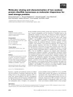
Symptomatology is very essential for early
detection of disease. The banana plants
infected with BBrMV showed different kinds
of symptoms on different parts of the plant
viz., leaves, pseudostem, male-bud, and
bunches. The primary symptoms of BBrMV
infected plants exhibiting longitudinal
irregular reddish streaks of varying sizes on
the psuedostem of Mysore Poovan (Fig. 1).
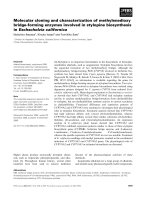
The orientation of infected leaves became fan
shaped which resembled travelers palm and
such symptoms were noticed (Fig. 2A).
Infected leaves showed spindle shaped lesions
running parallel to the veins (Fig. 2B) and
2542
Int.J.Curr.Microbiol.App.Sci (2019) 8(2): 2539-2550
mosaic pattern were visible on the lower side
of petiole which extended throughout the
midrib (Fig. 2C).
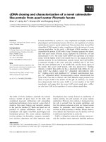
The symptoms seen on the bract were the
main indication of disease in almost all the
cultivars. The disease infected bracts showed
a distinct reddish streaks and mosaic pattern
(Fig. 3A). The infected bunches were under
sized with malformed fingers (Plate 3C) and
mosaic pattern observed on peduncle (Fig.
3B).
BBrMV best detected in 1:200 primary
antibody dilution along with 1:500 secondary
antibody
(alkaly-phosphate
conjugated)
dilution and can gave clear difference
between healthy and infected sample by
DAC-ELISA. DIBA also gave positive
reaction for infected leaf sample and this
could be detected by the purple coloured spots
on nitrocellulose membrane (Fig. 4). The
species specific primer pair was designed to
amplify of the coat protein gene of virus
based on the most favorable combination of
conserved regions in the multiple aligned
nucleotide sequences and named as BCPFl
and BCPRl respectively. RT-PCR analysis
with this primer yielded an amplicon of ~850
bp in infected samples (Fig. 5). The positive
samples were gel eluted and cloned into Ecoli DH 5 cells. Colony PCR using plasmid
specific primer (T7 and SP6) which yielded
amplicons of expected band size of 1150 bp
(Fig. 6). BLAST analysis of CP coding region
of the virus has maximum homology of 99
percent to KER2 isolate (Kasargod, Kerala;
accession
KF385491).
The
sequence
exhibited significant nucleotide identity (99 to
96 percent) and amino acid identity (95 to 83
percent) with other BBrMV nucleotide and
protein sequence of BBrMV in the database.
The Phylogenetic analysis (Fig. 7) by the
alignment of CP gene sequences of 23
isolates also revealed that the present isolate
was more similar to KER2 isolate which is
clustering with TN13 (KF385477; Tanjore,
Tamil Nadu) and the Indian isolates did not
show any relationship based on geographical
origin.
Table.1 Details of selected sequences from NCBI which were for primer designing
Sl.No.
1
Accession
Number
EU009210
Isolate
name
Trichy
Source
Size (bp)
Reference
Tamil Nadu
914
Selvarajan and
Balasubramanian, 2007
2
KF385480
AP7
Andra
Pradesh
900
3
KF385483
AS2
Assam
900
4
5
6
KF385491
KF385490
KF385473
KER 2
KAR 3
TN9
Kerala
Karnataka
Tamil Nadu
900
900
900
2543
Balasubramanian and
Selvarajan, 2014
Int.J.Curr.Microbiol.App.Sci (2019) 8(2): 2539-2550
Table.2 Details CP sequence used for phylogenetic analysis
SI.NO
Accession
Isolate
No
name
Source
Reference
1
HQ709165
Card1
Madikere, Karnataka
Siljo et al., 2012
2
HQ709166
Card2
Mudigere,
Karnataka
Siljo et al., 2012
3
HQ709164
Card3
Sirsi, Karnataka
Siljo et al., 2012
4
HQ709163
Card5
Idukki, Kerala
Siljo et al., 2012
5
HQ709162
Card6
Wynadu, Kerala
Siljo et al., 2012
6
EU009210
TN4
Trichy, Tamilnadu
Selvarajan, R and Balasubramanian, V., unpublished
data, 2014; unreferenced
7
KF385470
TN6
Pudukottai,
Tamilnadu
Balasubramanian and Selvarajan, 2014
8
KF385472
TN8
Theni, Tamilnadu
Balasubramanian and Selvarajan, 2014
9
KF385474
TN10
Karur, Tamilnadu
Balasubramanian and Selvarajan, 2014
10
KF385476
TN12
Cuddalore,
Tamilnadu
Balasubramanian and Selvarajan, 2014
11
KF385477
TN13
Tanjore, Tamilnadu
Balasubramanian and Selvarajan, 2014
12
KF385480
AP7
West godhavari, AP
Balasubramanian and Selvarajan, 2014
KAR2
Bangolore, KA
Balasubramanian and Selvarajan, 2014
13
KF385481
14
KF385488
KER3
Kayankulam, KER
Balasubramanian and Selvarajan, 2014
15
KF385490
KAR3
Arabhavi,KAR
Balasubramanian and Selvarajan, 2014
16
KF385491
KER2
Kasargod, KER
Balasubramanian and Selvarajan, 2014
17
AY953427
AP1
Kovur, AP
Ramesh, B., Sreenivasulu, P. and Krishna prasadji, J.
unpuplished data, 2005; unreferenced
18
EU414267
P5
Philippines
Iskra Caruana, M.L., Bringaud, C. and Bousalem, M.
unpuplished data, 2008; unreferenced
19
AF071585
P2
Philippines
Rodoni et al.,1999
20
AF071587
WS1
Western Samoa
Rodoni et al.,1999
21
AF071588
VT1
Vietnam
Rodoni et al.,1999
22
AF071589
TH1
Thialand
Rodoni et al.,1999
2544
Int.J.Curr.Microbiol.App.Sci (2019) 8(2): 2539-2550
Fig.1 Symptoms on pseudostem showing linear red lesions on Mysore Poovan
Fig.2 Symptoms on leaf (A). Fan like arrangement of leaves; (B). Spindle shaped lesions running
parallel to the veins on the leaf lamina; (C). Mosaic on leaf petiole
2545
Int.J.Curr.Microbiol.App.Sci (2019) 8(2): 2539-2550
Fig.3 Symptoms on Bract and fruits: (A). Reddish streaks and mosaic pattern on bract; (B).
Mosaic on peduncle
Fig.4 Detection of virus by dot immuno binding assay (DIBA). H- Healthy sample; B- Buffer
control; D- Infected sample
Fig.5 Amplification of CP region by BC F1/R1: M- Marker (1kb, Genei); Lane 1 - Healthy
control; Lane 2 and 3- Infected samples
2546
Int.J.Curr.Microbiol.App.Sci (2019) 8(2): 2539-2550
Fig.6 Analysis of recombinant clones using colony PCR: M- Marker (1kb, Genei); Lane 1 to 6Recombinant plasmid; Lane7-Positive control (DNA insert); Lane 8-Negative control
Fig.7 Phylogenetic analysis of coat protein gene of banana bract mosaic virus Isolates. Tree was
constructed by Neighbor-joining method using mega 7
2547
Int.J.Curr.Microbiol.App.Sci (2019) 8(2): 2539-2550
Among the solid phase serological detection
methods such as DIBA and ELISA, DIBA
was simple and convenient for field level
application since it does not require
sophisticated instruments such as ELlSAreader, PCR and could detect virus within 4
hrs. A field level diagnostic kit based on
DIBA was developed for detection of
Cassava mosaic geminivirus (CMG) and it
was able to obtain the result within 4 h (Nair,
2012). Both ELISA and DIBA could be
considered as efficient method for detection
of BBrMV, although probability of nonspecific reaction of antibody and components
of plant sap is higher in DIBA than in multi
well ELISA (Dhanya et al., 2007).
Molecular cloning of
CP
gene
has
been carried out and the main objective
of cloning plant
viruses
has
been the
improvement of virus detection and diagnosis
(Jelkmann et al., 1989).
Many potyvirus RNAs had been partially or
completely cloned (Nagel and Hiebert, 1985;
Rosner et al., 1986). The complete genome
sequences of isolates BBrMV- PHI from
Philippines (Ha et al., 2008) and BBrMVTRY from India (Balasubramanian and
Selvarajan, 2012) were determined using
cloning. In the family Potyviridae, species
demarcation criteria are based on genetic
information mainly based on CP gene (Berger
et al., 2005). Characterization of CP gene will
be used to establish evolutionary relationships
at both species and strain levels and used to
develop pathogen derived resistance against
potyvirus through coat protein mediated
resistance by means of genetic engineering.
Therefore, it could be a target of selection in
the present study.
In the present study, CP gene specific primer
was designed to amplify the product size of
850 bp and this could be utilized for detection
of BBrMV in the samples for routine
molecular detection purpose. Phylogenetic
analysis was used to study genetic diversity
BBrMV based on CP gene sequence
information. As CP gene sequences are
frequently used to develop pathogen derived
resistance against potyvirus by means of
genetic engineering, BBrMV diversity could
help in predicting the risk of breakdown of
resistance in the developed resistant
transgenic banana lines. Hence the efficient
long term management strategies could be
achieved by preventing the loss-of resistance
of CP mediated virus resistant due to the
evolution of new variants. In this study, we
compared CP gene sequence of BBrMV
isolates with 22 previously reported isolates
originating from different geographical
regions. Phylogenetic tree was constructed
from CP gene sequences showed two
monophyletic clusters in the world population
of BBrMV. However, the Indian isolates did
not show any relationship according to
geographical origins and the hosts from which
they were isolated. This finding is consistence
with result of Balasubramanian and
Selvarajan (2014) who reported, using
phylogenetic analysis based on CP coding
region of 49 BBrMV isolate. A probable
reason for the geographical distribution of
BBrMV is that the virus has moved as a
separate event. Perhaps through different
infected cultivars of banana and BBrMV exist
in India for a longer period of time (Rodoni et
al., 1999). BBrMV was first noticed in 1966
(Samraj et al., 1996); because of prolonged
presence, high divergence of BBrMV
populations might have occurred. However,
BBrMV was noticed first in southern parts of
Kerala, has moved to three neighboring states
viz., Andra Pradesh, Tamil Nadu and
Karnataka during the past five decades either
through infected planting material or through
aphid vector. There is no domestic quarantine
enforced to restrict the movement of banana
suckers between the states. This virus has
recently been reported to infect small
cardamom which is grown along with banana
2548
Int.J.Curr.Microbiol.App.Sci (2019) 8(2): 2539-2550
in Western Ghats region of Kerala and
Karnataka (Siljo et al., 2012).
Statement of author contributions
The project was initiated by Dr. Anitha
Cherian (Author 2) who is well known plant
pathologist and the project was funded by
Kerala Agricultural University, Thrissur.
Author 1 (Darshangowda M.R) did the main
work as a part of Master’s research for two
years. Author 3 (SaakreManjesh) did the gene
sequence studies of coat protein and primer
designing. Author 4 (Dr. Abida P. S) was the
member of this committee helped in various
ways such as cloning, standardization of PCR
profile for cDNA amplification. Author 5
(Mr. Ashwathappa Reddy) is from is a
background of both Agriculture and Plant
pathology was contributed in this project in
various ways.
Acknowledgement
We thank the Centre for Plant Biotechnology
and Molecular Biology, College of
Horticulture, (Thrissur) for lab facilities and
Department of Plant Pathology insect proof
net house. We also thank Banana Research
Centre, Kannara for planting materials and
Kerala Agricultural University for funding the
research.
References
Altschul, S. F, Madden, T. L., Schaffer, A.
A., Zhang, J., Zhang, Z., Miller, W., and
Lipman, D. J. 1997. Gapped BLAST
and PSI-BLAST: a new generation of
protein database search programs.
Nucleic Acids Res. 25(17): 3389-3402.
Balasubramanian, V. and Selvarajan, R. 2012.
Complete genome sequence of a banana
bract mosaic virus isolate infecting the
French plantain cv. Nendran in India.
Arch. Virol. 157: 397-400.
Balasubramanian, V., Sukanya, R. S.,
Anuradha, C., and Selvarajan, R. 2014.
Population structure of Banana bract
mosaic virus reveals recombination and
negative selection in the helper
component protease (HC-Pro) gene.
Virus Dis. 25(4): 460-466.
Banttari, C. E. and Goodwin, P. H. Detection
of potato viruses, S, X and Y by ELISA
on nitrocellulose membranes (DotELISA). 1985. Plant Dis. 69: 202-208.
Bateson, M. F. and Dale, J. L. 1995. Banana
bract mosaic virus: Characterization
using potyvirus specific degenerate
PCR primers. Arch. Virol. 140: 515527.
Berger, P. H., Adams, M. J., Barnett, O.W.,
Brunt, A. A., Hammond, J., Hill, J. H.,
Jordan, R. L, Kashiwazaki, S., Rybicki,
E., Spence, N., Stenger, D. C., Ohki, S.
T., Uyeda, I., van Jaayen, A., Valkonen,
J., and Vetten, H. J. 2005. Potyviridae.
In: Fauquet, C. M., Mayo, M. A.,
Maniloff, J., Desselberger, U., and Ball,
L. A. (eds), Virus taxonomy. VIIIth
Report of the International Committee
on
Taxonomy
of
Viruses.
Elsevier/Academic Press, London, pp.
819-841.
Clark, M.F. and Adams, A.N. 1977.
Characteristics of the Microplate
method of Enzyme-Linked Immuno
Sorbent Assay for the detection of plant
viruses. J. Gen. Virol. 3: 475-483.
Dhanya,
M.
K.,
Rajagopalan,
B.
Umamaheshwaran, K. and Ayisha, R.
2007. Comparison of detection methods
for Banana bract mosaic virus in
banana (Musa spp.). World J. Agric.
Sci. 3(5): 659-662.
Durga, N. and Kumar, M. 2015. AntiPsoriatic Activity of Musa Mysore Aab
(Poovan Banana) Peel Extract Using
Human Keratinocyte Cell Line. Int. J.
Sci. Res. 4 (3): 614-626.
2549
Int.J.Curr.Microbiol.App.Sci (2019) 8(2): 2539-2550
Ha, C., Coombs, S., Revil, P. A., Harding, R.
M., Vu, M., and Dale, J. L. 2007.
Design and application of two novel
degenerate primer pairs for the
detection and complete genomic
characterization of potyviruses. Arch.
Virol., 153: 25-36.
Jelkmann, W., Martin, R. R., and Maiss, E.
1989. Cloning of four plant viruses
from small quantities of doublestranded RNA. Phytopatholology, 79:
1250-1253.
Kumar, P. L., Selvarajan, R., Caruana, M. L. I
and Hanna M. C. R. 2015. Biology,
Etiology, and Control of Virus Diseases
of Banana and Plantain. Adv. Virus Res.
91(1): 229-269.
Magnaye, L. V. and Espino, R. R. C. 1990.
Note: banana bract mosaic: a new
disease of banana I. symptomatology.
Philipp. Agric., 73: 55-59.
Rodini, B. C., Ahlawat, Y. S., Varma, A.,
Dale, J. L., and Harding, R. M. 1997.
Identification and characterization of
Banana bract mosaic virus in India.
Plant Dis., 81: 667-672.
Saitou, N. and Nei, M. 1987. The neighborjoining method: A new method for
reconstructing phylogenetic trees. Mol.
Biol. Evol., 4: 406-425.
Samraj, K. S., Menon, M. R., and Christudas,
S. P. 1996. Kokkan a new disease of
banana. Agric. Res. J. Kerala. 4: 106113.
Selvarajan, R and Jeyabaskaran, K. J. 2006.
Effect of Banana bract mosaic virus
(BBrMV) on growth and yield of
cultivar Nendran (Plantain, AAB).
Indian Phytopath. 59(4): 496-500.
Siljo, A., Bhat, A. I., Biju, C. N., and
Venugopal, M. N. 2012. Occurrence of
Banana bract mosaic virus on
cardamom.Phytoparasitica40: 77-85.
Singh, H. P., Uma, S., Selvarajan, R and
Karihaloo, J. L. 2011. Micropropagation
for production of quality banana
planting material in Asia-Pacific. Asia
Pacific Consort. Agri. Biotech. 95 pp.
Thomas, J. E. and Magnaye, L.V. 1996.
Banana bract mosaic disease-Musa
Disease Fact Sheet No.7. INIBAP.
Montpellier. France.
Thomas, J. E., Geering, A. D. W., Gambley,
C. F., Kessling, A. F., and White, M.
1997. Purification, properties and
diagnosis of banana bract mosaic
potyvirus and its distinction from abaca
mosaic potyvirus. Phytopathology 87:
698-705.
How to cite this article:
Darshan, G., C.K. Anitha, S. Manjesh and Abida, P.S. 2019. Molecular Cloning and
Characterization of Coat Protein Gene of Banana bract mosaic virus Affecting Banana cv.
Mysore Poovan (Aab). Int.J.Curr.Microbiol.App.Sci. 8(02): 2539-2550.
doi: />
2550