Microbially induced sedimentary structures in evaporite–siliciclastic sediments of Ras Gemsa sabkha, Red Sea Coast, Egypt
Bạn đang xem bản rút gọn của tài liệu. Xem và tải ngay bản đầy đủ của tài liệu tại đây (3.3 MB, 10 trang )
Journal of Advanced Research (2014) 5, 577–586
Cairo University
Journal of Advanced Research
ORIGINAL ARTICLE
Microbially induced sedimentary structures in
evaporite–siliciclastic sediments of Ras Gemsa
sabkha, Red Sea Coast, Egypt
Amany G. Taher
*
Department of Geology, Faculty of Sciences, Cairo University, Giza, Egypt
A R T I C L E
I N F O
Article history:
Received 26 March 2013
Received in revised form 27 July 2013
Accepted 28 July 2013
Available online 2 August 2013
Keywords:
Biofilms
Coastal sabkha
Evaporites
Microbial mats
Siliciclastics
A B S T R A C T
The coastal sabkha in Ras Gemsa, Red Sea coast with its colonizing microbial mats and biofilms was investigated. The sabkha sediments consist mainly of terrigenous siliciclastic material
accompanied by the development of evaporites. Halite serves as a good conduit for light and
reduces the effect of intensive harmful solar radiation, which allows microbial mats to survive
and flourish. The microbial mats in the evaporite–siliciclastic environments of such sabkha display distinctive sedimentary structures (microbially induced sedimentary structures), including
frozen multidirected ripple marks, salt-encrusted crinkle mats, jelly roll structure, and petee
structures. Scanning electron microscopy of the sediment surface colonized by cyanobacteria
revealed that sand grains of the studied samples are incorporated into the biofilm by trapping
and binding processes. Filamentous cyanobacteria and their EPS found in the voids in and
between the particles construct a network that effectively interweaves and stabilizes the surface
sediments. In advanced stages, the whole surface is covered by a spider web-like structure of biofilm, leading to a planar surface morphology. Sabkha with its chemical precipitates is a good
model for potential preservation of life signatures. It is worthy to note that the available, published works on the subject of the present work are not numerous.
ª 2013 Production and hosting by Elsevier B.V. on behalf of Cairo University.
Introduction
Many studies on microbial mats, the oldest and most successful microorganisms, showed that metabolic activity of cyanobacteria and heterotrophic bacteria in carbonate marine
environments induces the precipitation of carbonates, which
* Tel.: +20 2 38379581; fax: +20 2 35727556.
E-mail address:
Peer review under responsibility of Cairo University.
Production and hosting by Elsevier
in turn form a microbial buildup named stromatolites [1–3].
Recent studies have shown that microbial mats are also of
paleoenvironmental significance in shallow siliciclastic shelf
settings through much of Earth history. Increasingly, microbial communities are recognized for playing a potentially
important role in defining and modifying surface sediment
characteristics in various settings, ranging from terrestrial,
through marginal marine to continental margins [4]. Siliciclastic microbially induced sedimentary structures MISS [5–10] is
adding to our knowledge about both present and past life. Systematic studies, leading from modern to increasingly older
deposits, have revealed that fossil MISS occur in tidal flat
and shelf sandstones of Phanerozoic, Proterozoic, and Archean ages and appear to have shown very little changes since at
2090-1232 ª 2013 Production and hosting by Elsevier B.V. on behalf of Cairo University.
/>
578
A.G. Taher
least 3.2 Ga [9,11–13]. The morphologies and paleoenvironmental distribution of such structures record the former presence of photoautotrophic microbial mats.
Sabkhas or ‘‘salt flats’’ are among the most saline natural
environments that form under arid or semiarid climate. Their
level is dictated by the local level of the water table and forms
an equilibrium geomorphological surface, which may be periodically inundated by water [14,15]. Capillary evaporation
leads to an increase in salinity of the interstitial waters and
thus favors the formation of evaporites. The study of sabkha
is important for several fields. Biologists were increasingly
interested in the study of hypersaline ecosystems as amazingly
high primary productivities are supported by such systems.
Geologists became aware of the fact that many metal–sulfide
deposits are associated with paleosabkha conditions [16,17].
Moreover, sabkhas have received many recent studies as they
form important permeability barriers in both aquifers and
hydrocarbon reservoirs [18].
In modern tidal flat environments, e.g. sabkhas, where high
salinity restricts metazoans grazing, microbial mats tend to
flourish. Siliciclastic sediments are widely overgrown by a great
variety of benthic microorganisms, especially cyanobacteria
which are most abundant in the upper intertidal and lower
supratidal zones [19]. Cyanobacteria are blossoming in wet
sandy environments and secrete sufficient extracellular polysaccharides (EPS). The EPS are adhesive mucilage that enables
the benthic microorganisms to attach themselves to solid substrates such as the surface of a quartz grain, to transport nutrients toward the cell, and to buffer the microbes against the
changing salinities in their microhabitat [20].
A biofilm is a collection of microorganisms, and their extracellular products bound to a solid surface termed as substra-
Fig. 1
tum [21]. It can be also regarded as microbially stabilized
water [22] that glue and fix the surface sediments in a process
known as biostabilization which increases stability against erosion [23–26].
The aim of the present work is to characterize different sedimentary structures induced by the microbial activity in evaporites and siliciclastic sediments in the coastal sabkha
hypersaline environment, also to discuss the fossilization potential of microbes in evaporites. It is worthy to note that these
structures are documented for the first time in this particular
area of Egypt. In this respect, the detailed characteristics of
their formation are given, especially in the absence of many
published works on the subject.
Study area
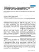
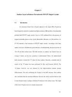
The study area is the coastal sabkha of little Gemsa on the
western side of the Gulf of Suez along the Red Sea coast
Fig. 1. The Gulf of Suez is a large elongated embayment which
is part of the rift system dividing the African and the European
Asian plates. The modern Gulf of Suez occupies the central
trough of the Suez rift which is only 50–70 m in depth [27].
A relatively wide gently sloping, coastal plain exists along this
Gulf. The shoreline comprises a low-angle siliciclastic carbonate ramp depositional system that passes onshore into an
extensive coastal sabkha environment. The sabkha extends
over a large area and displays a low slope with isolated patches
of high salt tolerant vegetation. It has no surface connection
with the sea but a subterranean one, fluctuating with the tide
in the open sea.
Ras Gemsa is located between 27°390 000 N and 33°340 6000 E
and covers an area of about 10 km2. Gemsa forms two tongues
Location map of the study area.
Microbially induced sedimentary structures
579
which are extending out in the sea. The eastern one is named
Great Gemsa and is 6.5 km long, while the western one is
named Little Gemsa and is 3.5 km long Fig. 1 [28]. They form
interbeds of evaporitic sulfates and marls of Miocene age.
Sand and gravel intercalations are found and marked by the
occurrence of an alluvial fan. The two tongues are separated
by a lagoon of about 4 km2. Numerous small wadis drain
the mountains of the area and dissect adjacent plains. These
are lined with scattered Acacia trees. In little Gemsa, wide flat
area of extensive intertidal mud and sand flats develops. The
intertidal sediment deposits are subjected to regular emergence
which leaves the surficial sediment layers exposed to extreme
temperatures, rain and wind erosion, subsequent drying and
compaction. Perennial and shallow ephemeral water bodies exist, and the formation of salt crusts depends on the annual
alternation of dry and rainy seasons. These are depositional
areas of no water current velocities. The sediments often experience large fluctuations in water content, salinity, and temperature resulting in extreme conditions that limit the range of
organisms able to inhabit such an environment. Lower areas
are submerged for longer periods of time. The sabkha deposits
consist mainly of brown sand and silt with evaporites including
gypsum, and halite. Non-evaporite components, mostly of
detrital origin, include quartz, feldspars, and clay minerals.
The sabkha region is dominated by deposition of clastics
accompanied by the development of evaporites.
Material and methods
The material used in the present study was collected during
June and December 2011 and October 2012. Thirty six surface
sediment samples were obtained from different zones of the
sabkha using a standard Ekman grap with a sampling dimension of 231 cm2. Sediment samples obtained were subjected to
granulometric and microscopic studies. The grain size analysis
of the sand (0.063–2 mm) and gravel (>2 mm) fractions was
carried out using dry–wet sieving techniques; silt and clay fractions (finer than 0.063 mm) were analyzed using the pipette
method [29].
Areas around the saline pools with luxuriant microbial
mats and biofilm-forming assemblages were selected for sampling. Selection was based on mat development and accessibility. Samples of the microbial surface were collected by
Table 1
inverting a Petri dish and pressing it into the mat. The dish
was then removed, and the shallow mat core carefully lifted
and placed right-side-up in the Petri dish. The samples were
studied and investigated under the binocular microscope.
For scanning electron microscopy (SEM), 10 samples were
placed in small glass tubes (diameter 0.5 cm) fixed immediately
in 4% glutaraldehyde solution diluted with water from the
sampling site. This treatment prevents osmotic shock and artifacts. The water was then removed in an ethanol series from
10% to 95% followed by two passages through absolute ethanol. Samples were then critical-point dried, gold-sputtered,
and studied under the SEM Jeol JSM 35 CF, Tokyo.
Climatic setting
The study area could be regarded as semiarid. The climate is
hot and dry, and rainfall is scarce. Data from Egyptian Meteorological authority, 2012 showed that the maximum temperature recorded in the summer season (June–September) is
27.5 °C, while the minimum, recorded in the winter season
(December–March), is 17.8 °C. Relative humidity varies between 43% and 55%. The annual mean rainfall is 3 cm, concentrated in a few showers in the winter season, whereas
there is almost no rainfall for the rest of the year. The area
is one of the intense evaporations, where annual mean evaporation rates along the coast are estimated to be 13.9 mm/
month. Northerly and northwesterly winds dominate the Gulf
of Suez. Stormy southern winds are much less frequent and occur mainly in February. These arid conditions keep the surface
water salinity above 220 g/L and allow the formation of the
sabkha along the coast. It also affects the formation of evaporites. The fresh water supply in the area is limited to the amount
which could account for all the detrital material in the sabkha
area. Strong onshore winds prevail in the area and could contribute some material from the Miocene outcrops and the basement rocks.
Results and discussion
Grain size distribution
The grain size distribution and related textural classification of
the surface sediments in the sabkha are summarized in Table 1.
Grain size distribution of the studied sediment samples (values in %).
Site
No. of samples
Average for each 3 samples
Textural classification
Gravel
Sand
Silt
Clay
Perennial saline lake
3
3
3
5.3
10.87
21.2
51.1
89
77.42
19.6
0.13
1.36
24
0.01
0.01
Slightly gravelly muddy sand
Gravelly sand
Gravelly sand
Ephemeral saline pools
3
3
3
6.75
19.49
10
82.71
80.43
34.85
2.00
0.06
33.4
8.54
0.01
21.75
Gravelly muddy sand
Gravelly sand
Gravelly sandy mud
Dry and capillary mud flat
3
3
3
0.86
5.95
4.90
89.00
37.5
36.5
2.13
33.55
30.22
8.01
23.00
28.38
Muddy sand
Gravelly sandy mud
Sandy mud
Salt crust
3
3
3
21.49
–
–
78.43
99.9
95.9
0.06
0.09
2.3
0.02
0.01
1.8
Gravelly sand
Sand
Sand
580
The surface sediments of the investigated area consist of a wide
variety of textural classes. In the majority of sites, sand and
gravel fractions constitute the bulk of the sediment fractions
Table 1. The increase in gravel content in some samples reflects
the abundance of transported terrigenous sediments and biogenic materials. The high mud content is most probably due
to the terrigenous flux of wadies.
Depositional subenvironments in Ras Gemsa sabkha
Geomorphological and Sedimentological features enable identification of four well-defined zones located at different topographical levels (1) perennial saline pools; (2) ephemeral
saline pools; (3) dry and capillary mud flat; and (4) supratidal
flat of efflorescent halite crusts.
Perennial saline pools
Perennial saline pools extend along the deepest part of the sabkha area Fig. 2a, with a depth that varies between 20 and
50 cm. The maximum surface area of the pools occurs during
winter season when several water bodies form one single pool.
Ground and surface waters flow into the pools during flooding. Since the pools are closed and evaporation is high, water
levels and salinity fluctuate seasonally, and the pools are much
A.G. Taher
diminished in summer, to be subdivided into a group of completely smaller remnants. Windblown sand barriers, are common in the area, and influence to a considerable extent the
subdivision of the pools. Such barriers are colonized by vegetation, which is in distinct association with a tendency for salt
tolerance, along the periphery of the pools. The peripheral
parts of the pools are gently elevated and are surrounded by
concentric channels which are partially water-filled. The minerals in the pools are dominated by evaporites. Halite is the
essential mineral with a subordinate amount of gypsum. A
very thin film of halite precipitates on the brine surface, which
tightly connected, mostly flattened euhedral crystals, of translucent halite develop later at the air–water interface as floating
rafts Fig. 2b. They are held by surface tension until they are
large enough to sink to the bottom of the brine forming a
crust. Many crusts form as overgrowths on cumulated layers.
Small biscuits may result from crystal growth diagenesis at local nucleation centers.
Cohesive microbial mats of gelatinous appearance grow to
a notable thickness at the margin of the perennial saline pools.
Samples taken from the upper surface of the sediments show
that the growth of microbial mats reaches up to 1.5 cm in overall thickness. This photosynthetic layer consists of a green zone
that is dominated by filamentous cyanobacteria and unidentified algae with their EPS Fig. 2c. At depths greater than 2 cm,
the microbial mat becomes increasingly black in color.
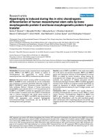
Fig. 2 Depositional subenvironments in Ras Gemsa sabkha: (a) Perennial saline pools with halite floating crusts that grow at the
periphery of the pools, (b) floating crusts of euhedral halite crystals. Scale is 15 cm, (c) SEM photomicrograph of the upper layer of
microbial mat formed of filamentous cyanobacteria and algae with their EPS. Scale is 5 lm, (d) patches of oxidized reddish brown organic
material at the surface followed in depth by black color of the anoxic part of the mat. Length of the match box is 5 cm, (e) ephemeral saline
pools fringing the perennial ones and formed of halite raft texture, (f) dry sand flat with thin efflorescent salt crust, a few cm in thickness
and the capillary mud flat is wet with the lack of interstitial saline minerals (marked as d and c).
Microbially induced sedimentary structures
In some parts around the saline pools, irregular patches of
oxidized organic material of reddish brown color are observed
Fig. 2d. Sediments with black iron sulfide are a distinctive feature in the coastal microbial mats, where sulfate reducing bacteria are active. The sediments contain high amounts of iron as
evidenced by the black color of the anoxic part of the mature
mats and the brown oxidized layer of the oxic part of the
freshly colonized sediment. The black color of iron reduction
is recorded for a few mm below the surface and could reach
the surface. This is probably due to the disturbance of the surface cyanobacteria layer by current action which removes the
surface layer, leaving the iron-rich layer. According to Stal
[30], some species of filamentous cyanobacteria are capable
of binding iron to the polysaccharide sheaths where the iron
is reduced concomitant with its binding. Krumbein et al. [31]
explained that iron oxides may function as a redox buffer between anaerobic and aerobic organisms in the sediment, which
helps in maintaining low sulfide concentrations, and may contribute to sediment stabilization.
Ephemeral saline pools
Ephemeral saline pools are mainly restricted to the southwestern
part of the area. They fringe also the perennial saline pools
Fig. 2e. Field observations during the years 2011 and 2012 indicate that most of the pools in the western area are of the ephemeral type, since they dry up once a year during the hot summer.
However, water bodies may persist without drying up for a few
months, as was observed in the late 2011. They have relatively
flat, vegetation-free surfaces, and lack surface outflows. The
pools area, range from 2 to 4 km2 during winter, being reduced
to 1–3 km2 during summer. The pools normally reach a maximum water depth of 10 cm. During winter months, the sediments are submersed due to the slightly higher water table of
the pools. In summer, the water level decreases and leaves white
halite crusts. The pools have a characteristic asymmetrical morphology most probably due to their migration following the
dominant wind direction (NW). When the brine reaches the halite saturation point, crystallization starts at the brine-air interphase as rafted textures. With time, crystals sink to the floor
where syntaxial overgrowth may take place resulting in chevrons
and cornet halite. Whenever the pools have completely dried up,
a salt layer of variable thickness forms and cover the whole area
of the pools. The salt layer typically reaches a maximum thickness of 5 cm; it is composed almost exclusively of cubic halite
with minor quantities of gypsum. The duration of the evaporative concentration period is mostly governed by the hydrological
balance, where crystallization is very high in summer and where
dilution occurs in winter.
Dry sand and capillary mud flats
The shallow hypersaline pool surface water is widely surrounded by air-exposed saline flats with a general scarcity of
water bodies Fig. 2f. The groundwater table stands closer to
the surface (approximately 0.8 m), allowing the development
of thinner efflorescent salt crusts (up to 1 cm thick). The efflorescence is composed almost exclusively of halite (over 90%)
but is limited, almost entirely, to the surface. This high abundance of halite indicates the total evaporation of upward moving marine water at the sediment–air interface. The sediments
581
are mainly terrigenous which consist of poorly sorted medium
gravelly-sized siliciclastics sand. Halite crusts rarely exceed a
few cm in thickness, which generally dissolve during winter.
In the capillary mudflat, sediment surfaces are permanently
wetted by capillary movement of the groundwater and lack
any interstitial saline minerals Fig. 2f. Both dry and capillary
mud flats are defined by the total absence of vegetation.
Supratidal flat of efflorescent halite crusts
The supratidal area is the marginal elevated zone that borders
the saline pools; it is located inland from the pools border to a
distance of about 200 m, so that it is not affected by tidal currents
or even strong storms. There is no significant presence of microbial material in this zone. The sediments that may reach 60 cm in
thickness are mainly clastics represented by sand, silt, and minor
clay with halite and disseminated gypsum. The clastics can be regarded as a matrix for the host evaporites. The supratidal area
acquires variable morphological shapes which pass abruptly
from one to another over distances of a few meters. They are
not exclusive to particular areas, but tend to have a patchy distribution around most of the pools. Polygonal crusts are thick,
with small surface relief, mostly <10 cm high and develop into
a distinctive pattern of ridges that are polygonal in plane form
Fig. 3a. Due to scarcity of rainfall, the crusts form with clearly
defined uplifted polygon margins. The diameter of the polygons
usually ranges from 15 to 20 cm and varies with the thickness of
the salt crust, which affects the mechanical strength of the crust
[32]. White, efflorescent halite commonly forms along the margins of some of the polygons, also inside them, caused by evaporation of the groundwater brine that is drawn up to the surface
by capillary pressure. The continuous growth and development
of polygonal crusts without interruption for a long time produces a high-relief blocky surface, mostly >50 cm high
Fig. 3b. Imitative salt crusts were also recognized and may be
completely filled with sandy muddy sediments of the same grain
size as the sediments underlying the crust Fig. 3c. They form
when the surface was previously affected by wind. Smoot and
Lowenstein [33] recorded similar crusts and mentioned that imitative efflorescent crusts rarely form on completely flat sediment
surface, and that the crystallization of efflorescent salt partially
preserves the preexisting surface morphology by leaving a cast of
its original form. Other well recognized crusts often exhibit an
irregular network of puffy or dome-like blisters Fig. 3d. They
are rounded to oval in shape; the diameter varies between 2
and 3 cm. They include a higher proportion of underlying sediment and adhering eolian dust. Goodall et al. [32] have documented a similar structure in modern salt flats in SE Arabia
and revealed that in blisters to form, the halite must be precipitated in an irregular manner and at different rates, which causes
the salt crust surface to be characterized by a random network of
rounded pressure ridges rather than the more organized polygonal pattern. They also added that the close proximity of the pressure ridges to each other interrupts growth and causes the
polygonal network to lose its integrity and become more
dome-like.
Microbially induced surface sedimentary structures
Because the studied site is relatively isolated anthropogenic
activity is low, and in the absence of metazoans grazing,
582
A.G. Taher
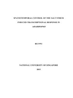
Fig. 3 Supratidal flat of efflorescent halite crusts: (a) thick polygonal crusts with small surface relief. Halite is commonly growing along
the margin and inside the polygons, (b) high-relief blocky crusts. N.B. Growth of halite along the fractures, (c) imitative salt crusts
completely filled with sandy muddy sediments, (d) an irregular network of dome-like blisters.
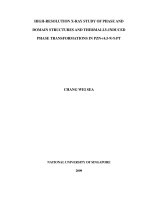
Fig. 4 Frozen multidirected ripple marks: (a) two different generations of ripple marks (I and II, scale is 1 m), (b) thick biofilm binding
and trapping sand grains leading to stabilization of the sediment surface, arrow points to M. chthonoplastes. Scale is 10 lm.
microbial mats can grow freely without being disturbed.
Microbial films and mats occur mainly along the margin of
the saline pools. Field observations in such areas revealed a
variety of structures induced by microbial mats and biofilm
including frozen multidirected ripple marks, crinkle structures,
jelly roll and petee structures.
Frozen multidirected ripple marks in the sense of Gerdes
et al. [34] are patterns of consolidated ripple marks of various
directions Fig. 4a. They are characteristic features of the upper
intertidal to lower supratidal zones [5]. This ripple pattern
arises from a set of subsequent storms each of which forms a
generation of ripple marks. During the periods in between
the storm events, the newly formed ripple marks are overgrown and biostabilized by microbial mats and thus cannot
be reworked by the later events [10]. Field observations revealed that frozen multidirected ripple marks form during
the spring growth season where a homogenous microbial
mat veneer was observed over the sediment surface. SEM
study of samples taken from these mats revealed that EPS of
the common Microcoleus chthonoplastes was seen to bind sediment grains in the samples collected from the upper 2 mm of
the surface layer. The processes of trapping, where particles are
glued to the mat surface and binding where the mat gradually
incorporates grains as it grows upwards are discussed by Gerdes et al. [34]. Sand grains of the studied samples that had been
transported by wind are seen to be incorporated into the biofilm by the trapping and binding process Fig. 4b. Filamentous
cyanobacteria, particularly M. chthonoplastes, have great effect
in modifying the sediment surface as they form thick sheaths
consisting of EPS which is very tough and resistant to degradation [21]. In addition, Gerdes and Krumbein [19] in their study
of the tidal flats of the North Sea have revealed that the
biomass production is increased in places characterized by
the predominance of M. chthonoplastes. Accumulation of
Microbially induced sedimentary structures
sedimentary particles by binding and trapping is a typical feature associated with this species [24]. Besides this, the binding
meshwork of thick EPS effectively interweaves and stabilizes
the surface sediments [35,26].
Salt-encrusted crinkle mats are confined to the periphery of
the perennial saline pools. They appear as gently meandering
uplifted microbial ridges with crests up to 2–5 cm. When
viewed from up, such crests show more or less planar surfaces
Fig. 5a. Crinkle structures result from the burial of microbial
mats by freshly deposited sand [9]. High evaporation rates
on the highest intertidal portion have resulted in the precipitation of gypsum and halite crystals within and beneath the
microbial mat. The crystallization of halite and gypsum results
in load pressure over the sediments which squeezes water out
of the organic layers and probably enhance lithification processes. SEM study of representative samples taken from such
structures showed that biofilms colonize first the deepest parts
of the sediment surfaces as they provide greater moisture and
protect the organisms against erosion [6]. They are found to fill
the voids in and between the particles Fig. 5b and c. In advanced stages, the whole surface is covered by spider web-like
structure of biofilm leading to a planar surface morphology
Fig. 5d. Noffke et al. [35] documented similar features during
their study in the tidal flats of the North Sea, where biomass
production is high and named such microbial activity leveling.
They revealed that filamentous mat-builders shelter their substrate against erosion by entangling sand and silt grains along
the sediment surface. The microbes also secrete extracellular
polymers that further increase the cohesion of surficial sediments [36]. In the fossil record, crinkle structures are documented in Archean sandstone [7], in Proterozoic [37], in the
lower and middle Cambrian as well as the Silurian of Sweden
[11].
583
Another feature of particular interest, which is confined to
the very shallow parts of the pools where the thickness of the
mat is about 0.5–0.7 cm, is the jelly roll structures Fig. 6a.
They indicate the high production of biofilm and reflect the
cohesive behavior of soft and jelly-like surfaces of microbial
origin [38]. They are formed of 0.5–1 cm diameter separated
and rounded burst open bubbles reflecting the flexible and
cohesive behavior of the soft mat. Cohesive behavior during
erosion, transport, and deposition is observed in both sandstones and mudstones and can be a very useful indicator of
microbial mat colonization [38]. Additionally, experimental
flume studies by Hagadorn and McDowell [25] revealed that
microbial communities characterized by a thicker surface film
provided greater erosional resistance and can inhibit the
growth of ripples entirely, so that the bed shear stresses result
in roll-up structures. They found that at flow velocities that
produce ripples, no grain movement occurred, but at higher
flow velocities (over ca 35 cm sÀ1), the entire surface and subsurface part of the mat was folded and then curled up on itself
to form a roll-up structure. Cyanobacteria, even when they are
not abundant, significantly affect the critical shear stress required for initiation of grain motion in medium sand [25].
During late summer, when the mat dries, it tends to break
apart into overthrust, saucer-like petee structures Fig. 6b. The
petees are thin crusts with small, mostly <10 cm high surface
relief and develop into a distinctive pattern of polygonal
rounded ridges in plane view. During wet seasons, microbial
mats were found to colonize the outer petee surfaces, while
during the dry season halite precipitates and leads to expansion and overthrust of the surface crust. The diameter of the
polygons usually ranges from 25 to 50 cm and the relief is directly related to the groundwater level; the deeper the water
table, the higher the relief of the petee structure is.
Fig. 5 Salt-encrusted crinkle mats: (a) meandering uplifted microbial ridges with salt-encrusted crinkles. Length of Marker pen is 15 cm,
(b) thin section of microbial mats of filamentous cyanobacteria coated the sand and gypsum crystals. Scale is 500 lm, (c) biofilm
colonizing the deepest parts of the sediment surfaces and fills the voids in and between the particles. Scale is 10 lm, (d) spider web
structure of biofilm coating the whole particles of sand and gypsum crystals and leads to planar surface morphology. Scale is 2 lm.
584
A.G. Taher
Fig. 6 (a) Jelly roll structure formed of burst open bubbles reflecting the flexible and cohesive behavior of the soft mat. Scale is 5 cm, (b)
polygonal overthrust petee structures in halite-encrusted siliciclastic sediments. Scale is 50 cm.
Preservation potential of microbes with evaporites
It is highly accepted that evaporites could be preserved over
geological times where surface hydrological cycles are absent.
They would be able of preserving traces of life independent
of their producers. Evaporites, particularly halite, mineral precipitation permits the passage of photosynthetically active
radiation and acts as UV-light scatterers so they can provide
protection from cosmic radiation and allow certain life forms
to survive in salt fluid inclusions for more than 100 million
years [39,40]. Moreover, the interior of halite crusts seems to
have unique microhabitats whose microenvironmental conditions cannot be found in soils or other lithic substrates [41].
This particular microhabitat is determined essentially by its
hygroscopic nature which enhances the moisture conditions.
A study by Wierzchos et al. [42] has shown that cyanobacteria
which, grow within the pore spaces of rocks or below translucent rocks can retain more moisture than ambient conditions.
In some cases however, they are endoevaporitic and found
only within the halite rocks other than colonizing the surface
of quartz grains. The fine halite and its intercrystalline spaces
are occupied by air and/or high salt solution, a habitat that will
not only aid in the retention of moisture because of capillary
effects [43], but also might play an important role in conditioning the distribution and survival of microbial colonies [44].
Also cyanobacteria can carry out their metabolic activities under the stressed conditions of high salt and even low water [45].
EPS could act as a shield, slowing down desiccation and ameliorating the extreme external conditions [46]. Rothschild et al.
[47] demonstrated that cyanobacteria inhabiting wet evaporite
crusts of halite and gypsum were metabolically active for a
long time after the dryness of the rock. The survival of archaea
halophiles in dry salt over geological time scales has been reported by McGenity et al. [48].
Modern coastal sabkhas are widely colonized by microbial
mats and biofilms. The abundance of a biofilm of EPS reflects
the behavior developed by microbial mats living in such hypersaline systems. EPS layers allow cyanobacteria to increase their
fossilization potential through early diagenesis of crystallization and cementation by gypsum and halite of these soft
shaped layers. Preservation of EPS and biofilms depends on
their rapid lithification before degradation [19]. Once lithified,
these gel layers retain their biologic related morphology, which
can be recognized in the fossil record. Furthermore, in carbonate environment the formation and persistence of ancient stro-
matolites depend on the binding and stabilizing of sediments
by microbial mats and biofilms [31]. The initial biogenic stabilization of depositional systems may be an essential requirement to allow or enhance future lithification of the sediments
[49].
The preservation potential of microbial mats with ancient
evaporites has been documented by many authors. Barbieri
et al. [50] studied the Upper Pleistocene evaporite deposits of
a wide continental sabkha in southern Tunisia and found that
biosignatures, with an intimate association with mucilaginous
slime, are mostly contained in gypsum lithofacies precipitated
from high salt-concentrated waters. These biosignatures include gypsified microfibers formed after the partial degradation of bacterial mucilaginous secretions. Recently, Noffke
et al. [51] studied the Archean stromatolites in the Pilbara area
of Western Australia which include a mixed carbonate–evaporite–siliciclastic coastal sabkha. The microbial mats generate a
plethora of well-preserved MISS arising from interaction with
sedimentary processes such as erosion or evaporite crystal
growth. Upon comparison of fossil and modern biogenic structures and their facies-related distribution in sabkha settings,
they strongly addressed the presence of microbial communities
in the Paleoarchean of such hypersaline and extreme
ecosystem.
Conclusions
Sabkhas, with its easier recognition and sampling is a good
model not only for evaporites deposition in shallow marine
environments, but also for preserving potential of traces of life
within chemical precipitates. The climate of Ras Gemsa is hot
and dry and solar radiation is intensive. These conditions favor
the deposition of halite, which serves as a good conduit for
light, reduces the effect of intensive harmful solar radiation,
and provides protection from high cosmic radiation, which allows microbial mats to survive and flourish. The area is characterized by a low sedimentation rate, little wave action, lack
of bioturbation, and is protected by vegetated patches of sea
grasses, which results in an optimal development of microbial
mats and biofilms. The microbial mats of Ras Gemsa sabkha
produce distinctive sedimentary structures such as frozen multidirected ripples, salt-encrusted crinkle mats, jelly roll structures and petee structures. Most of these structures were
found to be encrusted with halite. Thus within the halite rich
rocks there will continually exist conditions suitable for the
Microbially induced sedimentary structures
survival of microbes, particularly cyanobacteria. If the right
balance is met between absence of surface hydrological cycles
and rapid sealing provided by precipitating evaporite minerals,
an ultimate lithification process with good preservation of
microbially induced sedimentary structures can form. Intensive
researches on microbial processes occurring in modern coastal
sabkhas, which are extensively inhabited by microbial mats
and biofilms, are needed to open the path of fully and better
understanding of ancient microbial biota.
Conflict of interest
The author has declared no conflict of interest.
Compliance with Ethics Requirements
This article does not contain any studies with human or animal
subjects.
Acknowledgments
The author wishes to express her thanks to the anonymous referees for their constructive criticisms of an earlier version of
the manuscript. Dr. G. Phillip, Cairo University, is highly
acknowledged for his critical reading of the manuscript. I am
much indebted to General Petroleum Company (GPC) for
its virtuous hospitality during the trip. Dr. A. Abdel-Motelib
and Dr. A. Wagdi, Cairo University, are highly acknowledged
for their help in the field work.
References
[1] Gerdes G, Dunaftschik-Piewak K, Riege H, Taher AG,
Krumbein WE, Reineck HE. Structural diversity of biogenic
carbonate particles in microbial mats. Sedimentology
1994;41:1273–94.
[2] Olveri E, Neri R, Bellanca A, Riding R. Carbonate stromatolites
from a Messinian hypersaline setting in the Caltanissetta Basin,
Sicily: petrographic evidence of microbial activity and related
stable isotope and rare earth element signatures. Sedimentology
2010;57:142–61.
[3] Spadafora A, Perri E, Mckenzie JA, Vasconcelos CG. Microbial
biomineralization processes forming modern Ca:Mg carbonate
stromatolites. Sedimentology 2010;57:27–40.
[4] Schieber J. Microbial mats in terrigenous clastics: the challenge
of identification in the rock record. In: Hagadorn JW, Pflueger
F, Bottjer DJ, editors. Unexplored microbial worlds. Palaios
1999; 14: 3-13.
[5] Noffke N, Gerdes G, Klenke Th, Krumbein WE. Microbially
induced sedimentary structures––Examples from modern
sediments of siliciclastic tidal flats. ZBL fu¨r Geologie und
Pala¨ontologie 1996;1(2):307–16.
[6] Noffke N, Gerdes G, Klenke T, Krumbein W. Microbially
induced sedimentary structures––a new category within the
classification of primary sedimentary structures. J Sediment
Res 2001;71:649–56.
[7] Noffke N. Microbially induced sedimentary structures in
Archean sandstones: a new window into early life. Gondwana
Res 2007;11:336–42.
[8] Schieber J, Bose PK, Eriksson PG, Banerjee S, Sarkar S,
Altermann W, Catuneanu O. Atlas of microbial mat
features
preserved
within
the
siliciclastic
rock
record. Amsterdam: Elsevier; 2007.
585
[9] Noffke N. Geobiology – microbial mats in sandy deposits from
the Archean Era to today. Berlin: Springer; 2010.
[10] Cuadrado DG, Carmona NB, Bournod C. Biostabilization of
sediments by microbial mats in a temperate siliciclastic
tidal flat, Bahia Blanca estuary. Sediment Geol 2011;237:
95–101.
[11] Calner M, Eriksson ME. Microbial mats in siliciclastic
depositional systems through time. In: Noffke N, Chafetz H,
editors. The record of microbially induced sedimentary
structures (MISS) in the Swedish paleozoic. SEPM Spec,
Publication; 2012. p. 29–36.
[12] Gamper A, Heubeck C, Demskec D. Microbial mats in
siliciclastic depositional systems through time. In: Noffke N,
Chafetz H, editors. Composition and microfacies of Archean
microbial mats (Moodies Group, ca. 3.22 Ga, South
Africa). SEPM Spec, Publication; 2012. p. 65–74.
[13] Wehrmann A, Gerdes G, Ho¨fling R. Microbial mats in
siliciclastic depositional systems through time. In: Noffke N,
Chafetz H, editors. Microbial mats in a lower Triassic
siliciclastic playa environment (Middle Buntsandstein North
Sea). SEPM Spec, Publication; 2012.
[14] Khedr ES. Recent coastal sabkhas from the Red Sea: a model of
sabkhaization. Egypt J Geol 1991;33(2):87–120.
[15] Schreiber BC, Lugli S, Babel M. Evaporites through Space and
Time. Geol Soc London 2007;285:470, Special Publication.
[16] Renfro AR. Genesis of evaporite associated-stratiform
metaliferous deposits – a sabkha process. Econ Geol 1974;69:
33–45.
[17] Mossman DJ. Stratiform barite in sabkha sediments, WaltonCheverie, Nova Scotia. Econ Geol 1986;81(8):2016–21.
[18] Leeder MR. Sedimentology and sedimentary basins from
turbulence to tectonics.2nd ed. Wiley-Blackwell 2011.
[19] Gerdes G, Krumbein WE. Biolaminated deposits. Lect Notes
Earth Sci. Berlin: Springer; 1987, 9.
[20] Noffke N. Turbulent lifestyle: microbial mats on Earth’s sandy
beaches-Today and 3 billion years ago. GSA Today 2009;18:
1–4.
[21] Decho AW. Microbial exopolymer secretions in ocean
environments: their roles in food webs and marine processes.
Oceanogr Mar Biol Annual Rev 1990;28:73–154.
[22] Krumbein WE. Paracelsus und die mucilaginischen Substanzen500 Jahre EPS-Forschung. DGM-Mitt Jg 1993:8–14.
[23] Taher AG. Contribution of microbial mats and biofilms to
biogenic stabilization of sediments. In: 3rd Int. Conf. Geology of
the Arab World, Cairo University, Egypt; 1996: 235–54.
[24] Noffke N, Krumbein WE. A quantitative approach to
sedimentary surface structures controlled by the interplay of
microbial colonization and physical dynamics. Sedimentology
1999;46:417–26.
[25] Hagadorn JW, McDowell C. Microbial influence on erosion,
grain transport and bedform genesis in sandy substrates under
unidirectional flow. Sedimentology 2012;59:795–808.
[26] Taher AG. Abdel Motelib A. Microbial stabilization of
sediments in a recent Salina, Lake Aghormi, Siwa Oasis,
Egypt. Facies 2014;60(1):45–52.
[27] Sneh A, Friedman GM. Hypersaline Ecosystems. In: Friedman
GM, Krumbein WE, editors. Hypersaline Sea-marginal
flats of the Gulfs of Elat and Suez. Berlin: Springer; 1985. p.
104–24.
[28] Aref MA. Petrological and sedimentological studies on some
sulfur-bearing evaporite deposits on the western side of the Gulf
of Suez, Egyp. Ph.D Thesis, Fac. Sci. Cairo Univ. Egypt.
[29] Folk
L.
Petrology
of
sedimentary
rocks. Austin,
Texas: Hemplill; 1974, p. 182 [1992 P. 7].
[30] Stal LJ. Biostabilization of sediments. In: Krumbein WE, Stal L,
Paterson D, editors. Ecophysiological interactions related to
biogenic sediment stabilization. Oldenburg: Germany; 1994. p.
41–53.
586
[31] Krumbein WE, Paterson DM, Stal L. Biostabilization of
sediments. Germany: Oldenburg; 1994, p. 525.
[32] Goodall M, North CP, Glennie KW. Surface and subsurface
sedimentary structures produced by salt crusts. Sedimentology
2000;47:99–118.
[33] Smoot JP, Lowenstein TK. Evaporites, petroleum and mineral
resources. In: Melvin JL, editor. Depositional environments of
non-marine evaporates. Amsterdam: Elsevier; 1991.
[34] Gerdes G, Krumbein WE, Noffke N. Microbial sediments. In:
Riding RE, Awramik SM, editors. Evaporite microbial
sediments. Berlin: Springer; 2000.
[35] Noffke N, Gerdes G, Klenke T. Benthic cyanobacteria and their
influence on the sedimentary dynamics of peritidal depositional
systems (siliciclastic, evaporitic salty and evaporitic carbonatic).
Earth Sci Rev 2003;62:163–76.
[36] Decho AW. Microbial sediments. In: Riding R, Awramik S,
editors. Exopolymer microdomains as a structuring agent for
heterogeneity within microbial biofilms. Berlin: Springer; 2000.
p. 9–15.
[37] Hagadorn JW, Bottjer DJ. Restriction of a late Neoproterozoic
biotope; Suspect-microbial and trace fossils at the Vendian–
Cambrian transition. Palaios 1999;14:73–85.
[38] Gerdes G. Structures left by modern microbial mats in their host
sediment. In: Schieber J, Bose P, Eriksson PG, Banerjee S,
Altermann W, Catuneanu O, editors. Atlas of microbial mat
features preserved within the siliciclastic rock record. Elsevier;
2007. p. 5–38.
[39] Kminek G, Bada JL, Pogliano K, Ward J. Radiation dependent
limit for the viability of bacterial spores in halite fluid inclusions
and on Mars. Radiat Res 2003;159:722–9.
[40] Cockell SA, Raven JA. Zones of photosynthetic potential on
Mars and the early Earth. Icarus 2004;169:300–10.
[41] De Los Rios A, Wierzchos J, Sancho LG, Ascaso C.
Acid microenvironments in microbial biofilms of Antarctic
endolithic microecosystems. Environ Microbiol 2003;5:
231–7.
[42] Wierzchos J, Ascaso C, Mckay CP. Endolithic Cyanobacteria in
halite rocks from the hyperarid core of the Atacama Desert.
Astrobiology 2006;6:1–8.
A.G. Taher
[43] McKay CP, Friedmann EI, Go´mez-Silva B, Ca´ceres-Villanueva
L, Andersen DT, Landheim R. Temperature and moisture
conditions in the extreme arid regions of the Atacama Desert:
four years of observations including the El Nin˜o of 1997–1998.
Astrobiology 2003;3:393–406.
[44] De Los Rios A, Valea S, Ascaso C, Davila A, Kastovsky J,
McKay CP, Gomez-Silva B, Wierzchos J. Comparative analysis
of the microbial communities inhabiting halite evaporites of the
Atacama Desert. Int Microbiol 2010;13:79–89.
[45] Rothschild LJ. Earth analogs for Martian life: microbes in
evaporites, a new model system for life on Mars. Icarus
1990;88:246–60.
[46] Grilli CM, Ocampo-Friedmann R, Friedmann EI. Cytology of
long-term
desiccation
in
the
desert
cyanobacteria
Chroococcidiopsis (Chroococcales). Phycologia 1993;32:315–22.
[47] Rothschild LJ, Giver LJ, White MR, Mancinelli RL. Metabolic
activity of microorganisms in evaporites. J Phycol
1994;30:431–8.
[48] McGenity TJ, Gemmell RT, Grant WD, Stan-Lot-ter H.
Origins of halophilic microorganisms in ancient salt deposits:
mini review. Environ Microbiol 2000;2:243–50.
[49] Paterson DM, Aspden RJ, Visscher PT, Consalvey M, Andres
MS, Decho AW, Stolz J. Reid RP Light-dependent
biostabilization of sediments by stromatolite assemblages.
PLoS ONE 2008;3:1–10.
[50] Barbieri R, Stivalettaa N, Marinangelib L. Gabriele Orib G.
Microbial signatures in sabkha evaporite deposits of Chott el
Gharsa (Tunisia) and their astrobiological implications. Planet
Space Sci 2006;54:726–36.
[51] Noffke N, Christian D, Wacey D, Hazen RM. A microbial
ecosystem in an ancient sabkha of the 3.49 GA Pilbara, Western
Australia and comparison with Mesoarchean, Neoproterozoic
and Phanerozoic examples. GSA, annual meeting and
exposition; 2013. No. 190–8.