Toxicity of non-microcystin producing Microcystis wesenbergii isolated from the Tri An reservoir
Bạn đang xem bản rút gọn của tài liệu. Xem và tải ngay bản đầy đủ của tài liệu tại đây (1.34 MB, 6 trang )
Environmental Sciences | Ecology
Doi: 10.31276/VJSTE.61(4).70-75
Toxicity of non-microcystin producing
Microcystis wesenbergii isolated from the Tri An reservoir
Pham Thanh Luu1, 2*
Graduate University of Science and Technology, VAST, Vietnam
2
Institute of Tropical Biology, VAST, Vietnam
1
Received 5 August 2019; accepted 18 November 2019
Abstract:
Introduction
Harmful cyanobacterial blooms have become a global
threat to human health and aquatic biota around
the world. While the ecotoxicity of cyanobacterial
toxins such as microcystins (MCs) has been studied
extensively, the toxicity of non-toxin producing
cyanobacteria has not been evaluated to the same
extent. In this study, five strains of Microcystis
wesenbergii were isolated from the Tri An reservoir
and cultured under laboratory conditions. Microscopic
observation was used for morphological identification.
The MCs concentration was measured by highperformance liquid chromatography (HPLC). The
microcrustacean (Daphnia magna) was exposed to
different concentrations of crude extracts in a series
of acute (48 h) and sub-chronic (15 day) toxicity
experiments. The acute assay showed that crude
extract from all isolated strains of M. wesenbergii
generated toxic effects on D. magna, but no variant of
MCs was detected in the cultures of M. wesenbergii.
The 48 h EC50 values of crude extracts of M.
wesenbergii on D. magna ranged from 307.2-491.5 mg
dry weight (dw)/l. Sub-chronic exposure of D. magna to
the crude extract of M. wesenbergii at concentrations
of 1, 10, 40, and 120 mg dw/l resulted in a decline of
survival rates with dose dependence. Both maturation
and reproduction of parent D. magna were inhibited
with increasing concentrations of crude extract. This
finding indicated that crude extracts from non-MCproducing M. wesenbergii isolated from the Tri An
reservoir had significant acute and chronic toxic effects
on D. magna.
Cyanobacterial blooms in eutrophic freshwater
ecosystems have become an environmental concern
worldwide [1]. Microcystis is one of the most common
planktonic freshwater cyanobacterium, which frequently
causes bloom-forming and toxin-producing genus in
continental aquatic ecosystems. Microcystis is known to
produce various toxins such as microcystins (MC), other
bioactive peptides, alkaloid groups, and lipopolysaccharides
(LPS) [2], all of which may generate toxic effects on
aquatic organisms as well as human beings. In its natural
environment, Microcystis blooms may contain either MCproducing or non-MC-producing strains [3]. However,
previous studies have primarily focused on isolated MC or
cyanobacterial extracts containing MC, but the roles of other
toxic compounds present within a complex cyanobacterial
extract have not been studied to the same extent [4]. There
has been some evidence that various other cyanobacterial
metabolites, including LPS, alkaloid groups or unknown
secondary metabolites, and non-specific factors also
contribute significantly to the adverse effects associated
with blooms [4, 5].
Keywords: acute, cyanobacteria, Daphnia,
non-microcystins producing, Sub-chronic.
Classification number: 5.1
In their natural environment, aquatic animals may be
directly exposed to toxic cyanobacteria via feeding on toxic
cyanobacterial cells or be indirectly exposed via ingestion
of water contaminated with dissolved cyanotoxins [6].
Microcrustaceans play critical roles in aquatic ecosystems,
serving as both feeders and consumers. As a filter-feeder,
microcrustacean Daphnia spp. are potential consumers
of planktonic cyanobacteria. These filter feeders are
therefore seriously affected by the presence of toxic
second metabolites released into the water column during
cyanobacterial blooms or after the collapse of toxic
cells at the end of the blooms [7]. Because of relatively
*Email:
70
Vietnam Journal of Science,
Technology and Engineering
DECEMBER 2019 • Vol.61 Number 4
Environmental Sciences | Ecology
high sensitivity to toxicants, rapid reproduction, and
short lifetime, D. magna has been used extensively for
ecotoxiclogical studies. Several studies [4, 7, 8] have
examined the toxic effects of cyanobacterial bloom and
MCs on D. magna in laboratory conditions. Acute exposure
of Daphnia to cyanotoxins resulted in inhibition of filtration
rate, decrease in swimming movements, and even death
[4, 8]. Among chronic effects, literature reports decreased
fecundity and population growth rate [7, 9]. Despite not
producing MCs, some non-toxic cyanobacteria caused a
significant increase of biotransformation enzyme activities
from the exposed D. magna after a longer incubation [10].
However, the adverse effects of non-microcystin producing
on microcrustaceans species in natural environments, where
they may be exposed to a complex cyanobacterial biomass,
remain somewhat unclear.
The non-microcystin producing M. wesenbergii
are proliferate in many lakes, rivers and reservoirs in
Vietnam. Little is known about their toxicity in the aquatic
environment. In this study, we isolated five strains of the M.
wesenbergii from the Tri An reservoir and maintained them
in laboratory conditions. Microscopic observation was used
for morphological identification. The MCs concentration
was measured by HPLC. In addition, the toxic effects of a
crude extract of M. wesenbergii on the freshwater D. magna
were investigated.
Materials and methods
Blooms collection and isolation of cyanobacteria
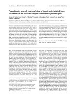
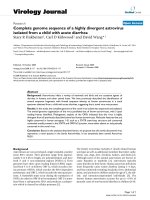
Bloom samples from the Tri An reservoir were collected
on surface water during July of 2017 (Fig. 1). Samples
were then brought to the laboratory. Observation under
microscope indicated several cyanobacteria dominant in the
samples including Microcystis, Oscillatoria, and Anabaena.
The colonies of the M. wesenbergii was identified under a
microscope (Olympus CK40-F200) equipped with a digital
camera (Olympus, Tokyo, Japan). Taxonomic classification
was based on the system of Komárek and Anagnostidis
(2005) [11]. For isolation, single colonies of M. wesenbergii
were picked out by micro-pipetting. After several times
washing in milli-Q water, the colonies were transferred into
tubes with Z8 medium and grew at a temperature of 280C
under a 12:12 h light:dark cycle at an intensity of 50 µmol
photons/m2/s. The biomass of M. wesenbergii was collected
onto GF/C fiberglass filters at stationary phase. After drying
completely at 450C, the samples were kept at -200C prior to
the experiment.
Fig. 1. Collection of water bloom samples in the Tri An reservoir.
Crude extract preparation and analysis
The crude extracts of M. wesenbergii were prepared
according to the method of Pietsch, et al. (2001) [12].
Briefly, a 1.0 g dry weight (dw) biomass of M. wesenbergii
was dissolved into 100 ml milli-Q water and frozen at
-700C, then thawed at room temperature. Then, the samples
were sonicated for 3 min. This freeze-thaw-sonicate cycle
was repeated five times. After centrifugation at 4000 rpm
for 10 min, the supernatant was collected and kept at
-200C. Subsamples of the crude extract were used for the
measurement of MC by HPLC according to the methods
reported previously by Pham, et al. (2015) [13]. Briefly, 100
μl of the supernatants was centrifuged at 4000 rpm for 15
min. The supernatant was collected into new glass tubes
and dried completely. The MC’s content in the samples
were collected by re-constitute in 500 μl of 100% MeOH.
MC concentrations were analyzed by an HPLC system with
UV-visible photodiode array (PDA) detector (Shimadzu
10A series, Kyoto, Japan). Three commercial MCs included
MC-RR, -LR, and -YR from Wako company (Osaka, Japan)
were used as internal standards.
Acute and sub-chronic bioassays
D. magna Straus purchased from the MicroBioTests Inc,
Belgium has been permanently maintained for more than 3
years under controlled conditions: temperature 25±10C and
14:10 h light:dark cycle in the ISO medium. The animals
were fed by a mixture of viable green algae Chlorella sp.
and Scenedesmus sp. Neonates less than 24 h were isolated
for toxicity experiments.
Acute toxicity bioassays were performed according to
the Protocol 202 of the Organization for the Economical
Cooperation and Development (OECD) [14]. Briefly, D.
magna neonates (<24 h old) were maintained in an ISO
DECEMBER 2019 • Vol.61 Number 4
Vietnam Journal of Science,
Technology and Engineering
71
Environmental Sciences | Ecology
medium with crude extracts of M. wesenbergii. At least
six different concentrations with a dilution factor of 0.5
were tested in triplicate with 10 neonates per replicate.
Test containers were placed at a controlled temperature of
25±10C and a 14:10 h photoperiod during 48 h. The 48 h
immobility of cladocerans was used to determine the half
maximal effective concentration (EC50) values with the 95%
confidence interval by using the SPSS software.
Sub-chronic tests were performed with the crude
extract of M. wesenbergii at four sublethal concentrations
of supernatant (equal to 1, 10, 40, and 120 mg dw/l) and a
control. Sub-chronic tests were done by using 50 ml beaker
cups with 20 ml of ISO medium or exposure solutions.
Neonates of D. magna less than 24 h in age were used. Each
treatment contained 15 replicates (n=15). Test solutions
were renewed every two days. A mixture of Scenedesmus sp.
and Chlorella (approximately 1X10⁶ cells/ml) was provided
as food for Daphnia in a two-day period. The mortality,
maturation and production of live offspring were observed.
The parent daphnid was checked daily for numbers of
neonates per clutch. Reproduction was calculated by using
the average of the number of neonate per female. The subchronic test was examined for 15 days. One-way ANOVA
and Kruskal-Wallis test (Sigma Plot, version 12) was
applied for calculations of the significant difference of the
maturation and reproduction of D. magna between control
and treatments.
Results and discussion
Isolation and morphological characteristics
Microscopic observation of the cyanobacterial bloom
samples revealed the dominance of Microcystis spp. (mainly
M. aeruginosa, M. wesenbergii, and M. botrys) and the less
frequent occurrence of other genera (Dolichospermum,
Arthrospira,
Planktothrix,
Pseudanabaena,
and
Cylindrospermopsis). Results of the present study are in
accordance with previous observations that Microcystis
was dominant group and the most common bloom-forming
species in Vietnamese waters [7, 13, 15].
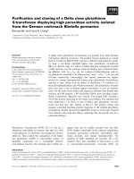
From bloom samples, five strains of M. wesenbergii
were isolated and cultured in Z8 medium. Morphology
examination from both field and isolated samples indicated
that M. wesenbergii species forms elongate, often lobate, and
sometimes spherical colonies that are commonly composed
of sub-colonies with a distinct refractive mucilage edge.
In nature, a sub-colony of this species is 15-100 µm in
diameter and contains from a few (2-3) to many (>150)
cells, depending on the age of the colony. Cells are more
or less evenly spread throughout the colony, are 5.5-8.5
72
Vietnam Journal of Science,
Technology and Engineering
µm in diameter, and have many spherical aerotopes. Cells
are slightly dark under a high magnification microscope.
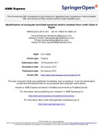
Colonies of this species forms an agar-like substrate that is
soft and often breaks up in culture (Fig. 2).
Fig. 2. Morphology of M. wesenbergii (scale bar: 20 µm).
M. wesenbergii is a common species of phytoplankton
and sometimes forms surface blooms in many lakes and
rivers worldwide. In Vietnam, the water bloom of M.
wesenbergii has been reported from the Dau Tieng and Tri
An reservoirs [7, 13], Hoan Kiem, and Nui Coc lake [15].
Measurement
cultures
microcystins
concentration
from
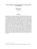
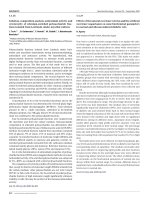
Results of HPLC analysis indicated that the water bloom
samples contained two variants of MCs including (MC-RR
and MC-LR) with the highest concentration ranged from
778.2±12.6 µg/g dw (Fig. 3B and Table 1). But none of the
isolated strains of M. wesenbergii produced microcystins
(Fig. 3A). Three variants of MC, including MC-RR,
MC-LR, and MC-RR from water blooms and isolated
Microcystis species with the maximum concentration
of 2130 µg/g dw have been reported from the Dau Tieng
reservoir. Many strains of M. aeruginosa isolated from
the Dau Tieng reservoir were reported to produce MC
[13] but none of the strains of M. wesenbergii were MCproducing. It is possible that M. aeruginosa was the main
toxin producer in the Dau Tieng reservoir. From the Tri An
reservoir, Dao, et al. (2010) [7] reported four variants of
MC, including MC-LR, MC-RR, MC-LA, MC-LY, and one
unknown variant in the scum samples but none were found
in the cultures. This may be a small number of strains of
Microcystis were included in the examination. In this study
several MCs variants were detected from the water bloom
which indicated that the cyanobacterial community from
the Tri An reservoir contained MC-producing and nonMC-producing cyanobacteria. Probably, the M. wesenbergii
species is a non-toxic species. Further study is needed to
determine the MC producers from the Tri An reservoir.
DECEMBER 2019 • Vol.61 Number 4
Environmental Sciences | Ecology
Fig. 3. HPLC-chromatograms of (A) M. wesenbergii, (B) water
bloom samples, and (C) microcystin standards.
Acute bioassays with D. magna
During the acute test, the survival of D. magna in the
control was higher than 90%, and the highest concentration
of crude extract (1.5 g/l) caused 100% mortality of Daphnia
daphnids after 48 h. Therefore, the test met the requirement
of the OECD (2004) [14] guideline for the acute test. The
calculated 48 h EC50 for the crude extracts of M. wesenbergii
and water bloom samples are shown in Table 1. Although
MCs were not detected in crude extracts of M. wesenbergii,
all samples caused acute toxicity on D. magna. The EC50
values of crude extracts of M. wesenbergii on D. magna
after 48 h ranged from 307.2-491.5 mg dw/l (Table 1).
The water bloom samples containing high concentrations
of MCs also caused the highest toxicities to cladocerans.
The calculated 48 h EC50 value is 279.4 mg dw/l at 95%
confidence interval (Table 1).
Previous studies [16-18] have demonstrated various
toxic effects on feeding behaviour and reproduction of
daphnids after exposure to cyanobacterial cells or their
purified toxins. However, the toxicity of the complex
extract from non-MC-producing cyanobacteria is not
examine in the same extent. Herrera, et al. (2014) [19]
reported that the EC50 values 48 h of a cyanobacteria
bloom contained MC-LR (538 µg/g dw) collected from a
reservoir in Colombia on Daphnia sp. were from 175-336
mg dw/l. The results of this study indicated that the toxic
effects of non-MC-producing M. wesenbergii are somewhat
lower than the water blooms samples. Probably, these
water blooms samples contained other toxic compounds
that contribute toxic effects other than MCs. The present
results confirmed the toxic effect of non-MC-producing
strains of M. wesenbergii on D. magna. In a recent study,
Pawlik-Skowrońska, et al. (2019) [20] compared the toxic
effects of purified MC and the extracts from Microcystis,
Planktothrix and Dolichospermum on Daphnia pulex, and
found that the toxicity of the crude extracts to D. pulex was
higher than that from pure cyanotoxins. Authors reported
that other toxic compounds present in the cyanobacterial
extracts such as non-ribosomal oligopeptides and LPS may
contribute to the toxic effects on cladocerans. The findings
of this research are consistent with previous studies that
natural extract from cyanobacteria contains various toxic
compounds that may even be more toxic than cyanotoxins
[4, 5, 9]. Further study is needed to understand the toxic
effects of these compounds in cyanobacteria from the Tri
An reservoir.
Sub-chronic toxicity and reproduction bioassay
Sub-chronic toxic effects of crude extracts of M.
wesenbergii (strain MW2) on D. magna over a period of 15
days revealed that the crude extracts of non-MC-producing
M. wesenbergii had dose-dependent toxic effects on the
survival of D. magna (Fig. 4).
Table 1. List of samples used for acute test with microcystin
concentration and 48 h EC50 values.
Strain name
Samples name
MW1
MW2
MW3
M. wesenbergii
MW4
MW5
BL-TA
Water bloom samples
MC (µg/g dw)
48 h EC50 (mg dw biomass/l)
ND
491.5
ND
307.2
ND
386.3
ND
383.1
ND
311.6
778.2±12.6
279.4
ND: no detectable microcystins.
Fig. 4. Effects of crude extracts of M. wesenbergii on survival
of D. magna.
DECEMBER 2019 • Vol.61 Number 4
Vietnam Journal of Science,
Technology and Engineering
73
Environmental Sciences | Ecology
No deaths of daphnids was recorded in the control
treatment. But a mortality rate of 13% of the exposed
daphnids was recorded in the treatment with 1 mg/l. The
survival decreased to 80% in the 10 mg/l treatment by the end
of the experiment. Only about 50% of daphnids survived in
the 40 mg/l treatment. At the highest crude extract treatment
(120 mg/l), mortality occurred quickly starting from day 2
and all the daphnids died after 13 days of exposure (Fig. 4).
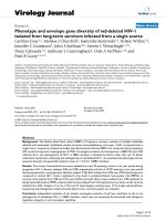
Results of the maturation age and average number
of offspring per female of D. magna exposed to different
concentration of crude extracts of M. wesenbergii are
shown in the results indicate that crude extracts of nonMC-producing M. wesenbergii at a concentration of 10
mg/l or higher inhibited the maturation and reproduction of
parent daphnids. In the control and 1 mg/l treatments, there
was no significant difference of maturation age between
the two groups, where the maturation age of the daphnid
was 5.4±0.3 days and 5.2±0.5 days, respectively. But the
maturation ages of the exposures with 10 mg/l, 40 mg/l and
120 mg/l are significantly longer than the CT (Fig. 5A).
wesenbergii significantly delayed maturity age and caused
a decline in the number of offspring of the parent daphnids.
Smutná, et al. (2014) [4] exposed D. magna to both MCcontaining and non-MC-containing cyanobacterial water
bloom samples in a series of acute (48 h) and chronic (21
day) toxicity experiments. Results showed that high acute
toxicity was observed for 6 of the 8 crude biomass samples.
The chronic exposure assays indicated the complex biomass,
the crude aqueous extract, and the microcystin-free extract
all elicited similar and significant lethal effects on D.
magna. The authors confirmed that cyanobacterial water
blooms are highly toxic to zooplankton (both acutely and
chronically) at environmentally relevant concentrations.
Dao, et al. (2010) [7] reported malformation of neonates
and cessation of the eggs/embryos of D. magna caused by
cyanobacterial toxins from crude extract. In addition, the
production of nonviable eggs and reduced fertility in D.
magna were observed after exposure to toxic cyanobacteria
[21]. The present study supports the previous findings that
both toxic and non-toxic cyanobacteria exert significantly
toxic effects on cladocerans.
Fig. 5. Maturation age (A) and number of offspring per female (B) of D. magna exposed to different concentration of crude extracts
of M. wesenbergii. Asterisks indicate significant difference between control (CT) and exposures.
*: p<0.05; **: p<0.01; ***: p<0.001.
During 15 days of the experiment, one parent D. magna
in the CT treatment produced about 33±2 offspring, which
was not significantly different than the 1 mg/l treatment.
However, the treatment with 10, 40, and 120 mg/l resulted in
significant decreases of the number of offspring per female
(Fig. 5B). The reproduction results indicated that there is
a concentration-response pattern in the parent daphnids
exposed to crude extracts of non-MC-producing M.
wesenbergii. Previous studies have confirmed the adverse
effects of toxic cyanobacteria on D. magna. The toxicity
includes inhibition of filtration rate, decrease in swimming
movements, and fecundity or reduction population growth
rate [7-9]. However, little is known about the chronic effects
of non-MC-producing on cladocerans. The findings of this
study revealed that crude extracts of non-MC-producing M.
74
Vietnam Journal of Science,
Technology and Engineering
Conclusions
This study demonstrated that the bloom samples from
the Tri An reservoir contained MCs, but no MC variant
was detected from the cultures of five isolated strains of M.
wesenbergii. The crude extracts from non-MC-producing
M. wesenbergii isolated from the Tri An reservoir had
significant acute and chronic toxic effects on D. magna. The
present findings indicate that metabolites other than MC are
likely to be responsible for the observed toxic effects, and
that toxins produced from cyanobacteria may play only
a minor role in the overall ecotoxicity of cyanobacterial
blooms. MC producers and the toxicity mechanism of
these unknown metabolites remain to be explored and need
further investigation.
DECEMBER 2019 • Vol.61 Number 4
Environmental Sciences | Ecology
ACKNOWLEDGeMENTS
This research was founded by Vietnam National
Foundation for Science and Technology Development
(NAFOSTED) under grant number 106.04-2018.314.
The author declares that there are no conflicts of interest
regarding the publication of this article.
REFERENCES
cyanobacteria affect the biochemical responses and behavior of D.
magna”, Int. Rev. Hydrobiol., 98(5), pp.235-244.
[11] J. Komárek, K. Anagnostidis (2005), “Cyanoprokaryota 1.
Teil: Oscillatoriales”, Phycologia, 38(6), p.544.
[12] C. Pietsch, et al. (2001), “The effects of a cyanobacterial crude
extract on different aquatic organisms: evidence for cyanobacterial
toxin modulating factors”, Environ. Toxicol., 16(6), pp.535-542.
[1] T.L. Pham, M. Utsumi (2018), “An overview of the
accumulation of microcystins in aquatic ecosystems”, J. of Environ.
Manage., 213, pp.520-529.
[13] T.L. Pham, T.S. Dao, K. Shimizu, D.H. Lan-Chi, M. Utsumi
(2015), “Isolation and characterization of microcystin-producing
cyanobacteria from Dau Tieng reservoir, Vietnam”, Nova Hedwigia,
101(1-2), pp.3-20.
[2] E. Dittmann, et al. (2015), “Natural product biosynthetic
diversity and comparative genomics of the cyanobacteria”, Trends
Microbiol., 23(10), pp.642-652.
[14] OECD (2004), Daphnia sp. Acute immobilization test,
OECD Guideline for Testing of Chemicals No.202.
[3] S. Le Manach, et al. (2018), “Physiological effects caused
by microcystin-producing and non-microcystin producing M.
aeruginosa on medaka fish: a proteomic and metabolomic study on
liver”, Environ. Pollut., 234, pp.523-537.
[4] M. Smutná, et al. (2014), “Acute, chronic and reproductive
toxicity of complex cyanobacterial blooms in D. magna and the role
of microcystins”, Toxicon, 79, pp.11-18.
[15] T. Duong, et al. (2014), “The occurrence of cyanobacteria
and microcystins in the Hoan Kiem lake and the Nui Coc reservoir
(North Vietnam)”, Environ. Earth Sci., 71(5), pp.2419-2427.
[16] A. Ghadouani, et al. (2004), “Effects of M. aeruginosa and
purified microcystin-LR on the feeding behavior of D. pulicaria”,
Limnol. Oceanogr., 49(3), pp.666-679.
[5] B. Burýšková, et al. (2006), “Toxicity of complex
cyanobacterial samples and their fractions in Xenopus laevis embryos
and the role of microcystins”, Aquat. Toxicol., 80(4), pp.346-354.
[17] W. Chen, L. Song, D. Ou, N. Gan (2005), “Chronic toxicity
and responses of several important enzymes in D. magna on exposure
to sublethal microcystin‐LR”, Environ. Toxicol., 20(3), pp.323-330.
[6] T.L. Pham, et al. (2016), “Microcystin accumulation and
biochemical responses in the edible clam Corbicula leana P. exposed
to cyanobacterial crude extract”, J. Environ. Sci., 44, pp.120-130.
[18] R. Ortiz-Rodríguez, C. Wiegand (2010), “Age related
acute effects of microcystin-LR on D. magna biotransformation and
oxidative stress”, Toxicon, 56(8), pp.1342-1349.
[7] T.S. Dao, L.C. Do-Hong, C. Wiegand (2010), “Chronic effects
of cyanobacterial toxins on D. magna and their offspring”, Toxicon,
55(7), pp.1244-1254.
[19] N. Herrera, et al. (2014), “Effects of a cyanobacterial bloom
sample containing microcystin-LR on the ecophysiology of D.
similis”, Toxicol. Rep., 1, pp.909-914.
[8] A.S. Ferrão-Filho, et al. (2009), “Biomonitoring of cyanotoxins
in two tropical reservoirs by cladoceran toxicity bioassays”,
Ecotoxicol. Environ. Saf., 72(2), pp.479-489.
[9] N.A. Herrera, L.F. Echeverri, A.S. Ferrão-Filho (2015),
“Effects of phytoplankton extracts containing the toxin microcystinLR on the survival and reproduction of cladocerans”, Toxicon, 95,
pp.38-45.
[10] T.S. Dao, R. Ortiz-Rodríguez, L.C. Do-Hong, C. Wiegand
(2013), “Non-microcystin and non-cylindrospermopsin producing
[20] B. Pawlik-Skowrońska, M. Toporowska, H. Mazur-Marzec
(2019), “Effects of secondary metabolites produced by different
cyanobacterial populations on the freshwater zooplankters Brachionus
calyciflorus and Daphnia pulex”, Environ. Sci. Pollut. Res., 26(12),
pp.11793-11804.
[21] S. Gustafsson, K. Rengefors, L.A. Hansson (2005),
“Increased consumer fitness following transfer of toxin tolerance to
offspring via maternal effects”, Ecology, 86(10), pp.2561-2567.
DECEMBER 2019 • Vol.61 Number 4
Vietnam Journal of Science,
Technology and Engineering
75