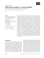
Báo cáo khoa học: Phaiodotoxin, a novel structural class of insect-toxin isolated from the venom of the Mexican scorpion Anuroctonus phaiodactylus pdf
Bạn đang xem bản rút gọn của tài liệu. Xem và tải ngay bản đầy đủ của tài liệu tại đây (287.2 KB, 9 trang )
Phaiodotoxin, a novel structural class of insect-toxin isolated from
the venom of the Mexican scorpion
Anuroctonus phaiodactylus
Norma A. Valdez-Cruz
1
, Cesar V. F. Batista
1
, Fernando Z. Zamudio
1
, Frank Bosmans
2
, Jan Tytgat
2
and
Lourival D. Possani
1
1
Department of Molecular Medicine and Bioprocesses, Institute of Biotechnology, National Autonomous University of Mexico,
Cuernavaca, Mexico;
2
Laboratory of Toxicology, University of Leuven, Leuven, Belgium
A peptide called phaiodotoxin was isolated f rom the venom
of the scorpion Anuroctonus phaiodactylus. It is lethal to
crickets, but non toxic to mice at the doses assayed. It has 7 2
amino acid residues, with a molecular mass of 7971 atomic
mass un its. Its covalent structure was determined by Edman
degradation and mass spectrometry; it contains four disul-
fide-bridges, of w hich one of the pairs is formed between
cysteine-7 and cysteine-8 (positions Cys63–Cys71). The
other three pairs a re formed between Cys13–Cys38, Cys23–
Cys50 and Cys27–Cys52. Comparative sequence analysis
shows that phaiodotoxin belongs to the long-chain sub-
family of scorpion peptides. S everal genes coding for t his
peptide and similar ones were cloned by PCR, using cDNA
prepared from the RNA of venomous glands of this scor-
pion. Electrophysiological assays conducted with this toxin
in several mammalian cell l ines (TE671, COS7, rat GH3 and
cerebellum g ranular cells), showed no effect on Na
+
cur-
rents. However, it shifts the voltage dependence of activation
and inactivation of insect Na
+
channels (para/tipE) to more
negative and positive potentials, respectively. Therefore, the
ÔwindowÕ current is increased by 225%, which is th ought to
be the cause of its t oxicity t oward insects. Phaiodotoxin is the
first toxic peptide ever purified from a scorpion of the family
Iuridae.
Keywords: Anuroctonus phaiodactylus; disulfide bridges;
insect toxin; Na
+
-channel; scorpion.
Most of the biochemical work performed with scorpion
venom has been reported using scorpions of the family
Buthidae, probably because they are dangerous to humans.
A large number of different protein and polypeptides have
been isolated and c haracterized from this family. Among
the most important findings are four different groups of
peptides, which specifically interact w ith ion channels: Na
+
channels [1], K
+
channels [2,3], Cl
–
channels [4] a nd Ca
2+
channels [5,6]. The scorpion Anuroctonus phaiodactylus
belongs to t he family Iuridae. Human accidents with these
scorpions have not been reported to cause symptoms of
intoxication. However, they are toxic to insects and other
arthropods from which they prey on. Scorpion toxins
affecting Na
+
channels are polypeptides with 61–76 amino
acid residues long, showing two basic different pharma-
cological a ctivities, either a or b according to their mode
of action and binding properties [7–9]. The a-scorpion
toxins (a-ScTxs) slow Na
+
current inactivation in v arious
excitable preparations, upon their binding to site 3, but
they show vast differences in p reference f or insect and
mammalian Na
+
channels. Accordingly, they are divided
into classical a-toxins that are highly active in mammalian
brain, a-toxins that are very active in insects and a-like
toxins that are active in both the mammalian and the insect
central nervous system [10]. b-Toxins shift the activation
voltage of sodium channels to more negative membrane
potentials upon binding to receptor site 4 [ 11]. This class
includes two types o f toxins, excitatory and depressant [7,8].
Na
+
channels specific ScTxs present a conserved core
formed by a-helix and three strands of b-sheet structural
motifs. The helix motif is linked to the b3strandbytwoof
the four d isulfide bonds. T he cysteine pair of the a-he lix
motif is spaced by a tripeptide CXXXC (where C stands for
cysteine and X for any amino a cid), whereas the pair of
cysteine residues of the b3 s trand is separated by only one
amino acid residue (CXC), usually linking the C3 (third
cysteine of the s equence) to C6 and C4 t o C7 [12]. A t hird
structurally conserved d isulfide bridge occur s between t he
C2 of the N-terminal segment with C5 of the b2 stran d [ 9].
The fourth disulfide bond is established between C1 and C8,
of the N- with t he C-terminal region. The excitatory insect
toxins lack the equivalent position of C1, present in most
scorpion toxins, a nd the fourth disulfide bridge is formed
between C5¢ (contiguous to C5) w ith C 8 [ reviewed in 9].
This last disulfide bridge is not present in birtoxin, which has
only three disulfide bridges, but functionally shows a b-like
activity and shares homology with the Centruroides’
b-toxins [13]. Recently, the functional surface of three
different toxins w as mapped. Analysis of the t hree-dimen-
sional models suggests that the functional differences reside
Correspondence to L. D. Possani, Instituto de Biotecnologı
´
aUNAM
Avenida Universidad, 2001 Apartado Postal 510–3 Cuernavaca 62210
Mexico. Fax: +52 777 3172388, Tel.: + 52 777 3171209,
E-mail:
Abbreviations: a.m.u., atomic mass unit; CD-immobilon, cationic,
hydrophilic, charged polyvinylidene fluoride membrane; COS7,
monkey kidney cell line 7; CNBr, cyanogen bromide; GH3, rat
pituitary cell line; ScTX, scorpion toxin; TE671, human cerebellar
medulloblastoma cell line 671.
(Received 13 August 2004, accepted 14 Octo ber 2004)
Eur. J. Biochem. 271, 4753–4761 (2004) Ó FEBS 2004 doi:10.1111/j.1432-1033.2004.04439.x
at the C -tail section of the toxins [14–16]. The authors
propose that evolutionary events occurred at the C-terminal
region, which plays an important role in determining
functional d iversification and constitute an important site
for Na
+
-channel recognition [16,17].
Here we describe the isolation and characterization of an
insect specific toxin from the scorpion Anuroctonus phaiod-
actylus, collected in Baja California, Mexico. We have
isolated and chemically and functionally characterized th is
peptide. The gene that codes for the toxin and several
isoforms were obtained. The three major characteristics of
phaiodotoxin are: its lethal effect on crickets, but non toxic
to mice; its different arrangement of the disulfide bridges,
and its pharmacological effect on para/tipE Na
+
channel
expressed on Xenopus laevis oocytes, where it causes an
important increment on the window of Na
+
currents. It is
worth mentioning that the unusual disulfide bridge is
situated at the C-terminal tail of the molecule.
Materials and methods
Venom collection and purification procedure
The sco rpions were collected in Maneadero Baja California,
Mexico. Their venom was obtained by electrical stimulation,
dissolved in double d istilled water, centrifuged at 15 000 g
for 15 min and the supernatant lyophilized and kept at
)20 °C. The s oluble venom was applied t o a Sephadex G-50
column (0.9 · 190 cm) in 20 m
M
ammonium acetate buffer
pH 4.7, resolving six fractions. The second fraction contains
the phaidotoxin which was obtained in a homogeneous
form after two independent steps of purification. Initially,
the separation was performed in a semipreparative C18
reverse phase column (Vydac, H isperia, CA, USA), using a
Waters 600E HPLC, equipped with a Photodiode Array
Detector 996 from Millipore (Milford, MA, USA). The
second HPLC was carried out in an analytical C18 reverse
column. In both c ases, a linear g radient was run f or 60 min,
from solution A (0.12% trifluoroacetic acid in water) to
60% solution B (0.10% TFA in acetonitrile).
Lethality tests
Lethality tests were carried out on female albino mice (CD1
strain) of approximately 20 g bodyweight. The various
samples dissolved in 100 lLNaCl/P
i
(phosphate buffered
saline; 0.15 m
M
NaCl in 0.1 m
M
sodium phosphate buffer,
pH 7.4) were injected intraperitoneally. These assays were
conducted using a minimum number of animals required to
validate t he experimental data, according to the guidelines
for animal usage of our Institute (the protocols were
approved by the Institutional Committee for Animal
Welfare). U sually, injection on two or three animals i s
considered enough to see if there is a visible effect on mice.
Lethality t ests on crickets weighing approximately 100 mg
were performed injecting 3 lL of variable amounts of
venom and/or fractions at the intersegments of the right leg.
Phaiodotoxin in amounts of 0.2, 0.5, 0.8 and 1.0 lgof
peptide per animal were injected, using two crickets at a time
and repeating the same procedure four times. The main
symptoms of intoxication were: flaccidity, impairment of
movements, paralysis and death.
Primary structure determination of phaiodotoxin
The amino acid sequence o f the N-terminal portion of
phaiodotoxin was obtained by Edman degradation carried
out with an automatic apparatus Beckman LF 3000 Pro-
tein Sequencer (Palo Alto, CA, USA), using the peptide
adsorbed on CD Inmmobilon m embranes (Beckman part
number 290110). A sample of the toxin was also sequenced
from its N-terminal region, after reduction and alkylation
in situ with acrylamide by the method described in [18]. In
order to c omplete the full sequence several fragments of the
peptide w ere obtained after cleavage of phaiodotoxin with
cyanogen bromide (CNBr), than ks to the presence o f two
methionine residues in the molecule. An eight-fold excess of
CNBr over toxin ( w/w) in 70% formic acid was used
according to t he technique described by B iedermann [19].
After o vernight reactio n, the pr oducts were reduced with
dithiothreitol for 30 min, at 56 °C and separated b y HPLC.
The sub peptides were used for Edman degrad ation a nalysis.
The molecular mass determination of pure phaiodotoxin
and the additional sequencing work w as performed by
mass spectrometry, using an LCQ
Duo
Finnigan mass
spectrometer, as described previously [20]. All spectra were
obtained in the positive-ion m ode. For sequence d etermin-
ation, MS/MS s pectra pr oduced were analyzed manually
and automatically by
SEQUEST
software. The acquisition
and deconvolution of data were performed with the
XCALI-
BUR
software on a Windows NT PC data system.
Determination of disulfide bridges
Native toxin was digested with several specific endo-
peptidases and their products were separated by HPLC
(same conditions as described above). The purified dimeric
peptides were directly used for Edman degradation and
mass spectrometry an alysis. It i s worth noting that for
these sequences no reduction of the peptides was per-
formed. Initially, 100 lg o f phaidoto xin was digested with
lysine-C endopeptidase (Lys-C). Subsequently, another
sample was treated with two enzymes chymotrypsin and
aspartic-N (Asp-N), all from Boehringer (Mannheim,
Germany), using the conditions described by the manu-
facturer. In order to confirm the disulfide pairs found, an
independent sample was processed using CNBr cleavage
[19]. T he products were s eparated by HPLC and directly
sequenced.
Sequence analysis
Nucleotide sequence similarities were searched with the
BLAST
program using the databases of GenBank (National
Center for Biotechnology Information). The sequences
obtained were edited and aligned using
CLUSTAL
-
X
[21].
Gene cloning of phaiodotoxin
Total R NA was isolated from venomous glands situated at
the last postabdominal segment (telson) of one Anuroctonus
phaiodatylus scorpion, by the method of Chirgwin et al.
[22]. Total RNA (500 n g) was u sed as template t o gener-
ate cDNA using the oligonucleotide poliT22NN
[23]. For gene amplification two primers were used:
4754 N. A. Valdez-Cruz et al.(Eur. J. Biochem. 271) Ó FEBS 2004
5¢-AARTTYATHCGRCAYAAG-3¢ and poliT22NN. We
cloned t he product o f the amplification in EcoRV site of
phagemid p KS(–) ( Stratagene, L a J olla, C A, USA). This
construct was used to transform Escherichia coli DH5-a
cells. Clone selection and DNA sequencing were p erformed
as described by C orona et al.[23].Inordertocompletethe
nucleotide sequence, the m ethod for rapid amplification of
the 5¢-region (RACE 5¢) was applied, using RLM-RACE
(RNA ligase mediated rapid amplification of cDNA ends)
protocol, according t o the instructions of the kit fr om
Ambion (Austin, TX, USA). The cDNA mix was synthes-
ized from poly(A)+ mRNA u sing M-MLV reverse tran-
scriptase. The cDNA was joined with the a daptor provided
by the kit (5 ¢-gcugauggcgaugaaugaacacugcguuugCUGG
CUUUGAUGAAA-3¢) using T4 DNA ligase. The modi-
fied cDNA was used as t emplate for PCR amplification.
Two rounds of amplification with the primers from the
Ambion kit were performed.
Expression in
Xenopus
oocytes
For t he expression in Xenopus oocytes, the para/pG H19-
13–5 vector [24] and tipE/pGH19 vector [25] were linearized
with Not I and transcribed with the T7 mMESSAGE-
mMACHINE kit ( Ambion). The harvesting of oocytes
from anaesthetized female Xenopus laevis frogs was as
described previously [26]. Oocytes were injected with 50 nL
of cRNA at a concentration of 1 ngÆnL
)1
using a Drum-
mond microinjector (Broomal, P A, USA). The solution
used for incubating the oocytes contained (in m
M
): NaCl,
96;KCl,2;CaCl
2
,1.8;MgCl
2
,2andHepes,5(pH7.4),
supplemented with 50 mgÆL
)1
gentamycin sulfate.
Electrophysiological recordings in
Xenopus
oocytes
Two-electrode vo ltage-clamp recordings were performed at
room temperature (18–22 °C) using a GeneClamp 500
amplifier (Axon Instruments, Union City, CA, USA)
controlled by a pClamp data acquisition s ystem (Axon
Instruments). Whole-cell currents from oocytes were recor-
ded 4 days after injection. Voltage and currents electrodes
were filled with 3
M
KCl. Resistances of both electrodes
were kept as low as possible ( < 0.5 MX). Bath solution
composition was (in m
M
): NaCl, 9 6; KCl, 2; CaCl
2
,1.8;
MgCl
2
, 2 and Hepes, 5 (pH 7.4). Using a four-pole low-pass
Bessel filter, currents were fi ltered at 2 kHz and sampled at
10 kHz. Leak and capacitance subtraction w ere performed
using a P/4 protocol. Current traces were evoked in an
oocyte expressing the cloned sodium channels by depolari-
zation between )70–40 mV, using 10 mV increments, from
a holding potential of )90 mV.
The window current was estimated following the des-
cription of Attwell et al. [27] using the weighing method.
Electrophysiological recordings with mammalian
cell lines
The e ffect of phaiodotoxin was also assayed i n several
mammalian cell lines: TE671 (from human cerebellar
medulloblastoma), COS7 (from monkey kidney fibro-
blasts), GH3 and cerebellum granular ce lls from r at, using
the technique described [28].
Results and Discussion
Purification, bioassays and chemical characterization
of phaiodotoxin
Figure 1 shows the results of the chromatographic steps
used for purification of phaiodotoxin. In short, a gel
filtration system with Sephadex G-50 column (Fig. 1A) and
two additional separations on HPLC (Fig. 1B) provided a
homogeneous peptide. Toxicity tests showed that it was non
toxictomiceusingadoseupto100lg per 20 g mouse
weight, but causing flaccidity and paralysis in crickets.
Crickets injected with little as 0.5 lg per animal showed
symptoms of intoxication such as: impairment of move-
ments and mild paralysis. A 0.8 lg per animal dose causes
a clear flaccid paralysis, but at 1.0 lg per animal all the
crickets die, within the first 2 h after injection. These
bioassays were repeated four times with phaiodotoxin, given
identical results. T his is s imilar to w hat was described by
Zlotkin et al. [29] for the insect toxin LqhIT2 of the
scorpion Leirus quinquestriatus hebraeus.
Despite the fact that phaiodotoxin was not toxic to mice,
using in vivo experiments at high doses (100 lg per mouse),
several cell l ines in culture (see Materials and methods) were
tested for possible electrophysiological effects on mamma-
lian Na
+
channels. It is w orth mentioning that scorpion
toxins such as Cn2 (toxin 2 from the scorpion Centruroides
noxius), specific for m ammals, have LD
50
values in the
range of 0.25 lg per 20 g m ouse bodyweight [30]. T hus,
mice injected with 400-fold more phaiodotoxin than that
required by other scorpion toxins, did not show any toxicity
symptoms, from which we assumed this peptide is not t oxic
to mice. E lectrophysiological tests conducted with micro-
molar concentrations of phaiodotoxin in the cell culture
systems mentioned (COS7, TE671, GH3 and cerebellum
granular cells) showed no effect (data not shown), from
which we surmised that this peptide was rather specific for
insects.
The primary structure of phaiodotoxin was obtained by a
combination of direct Edman degradation and mass
spectrometry analysis, as shown in Fig. 1C. Alkylated toxin
permitted to identify the first 39 residues (underlined with
the word ÔdirectÕ in the fi gure). Two subsequent p eptides
(corresponding to residues in positions M41 to R59 and
M62 to K70) were s equenced after cyanogen bromide
cleavage (underlined by CNBr). The C-terminal residues of
each peptide were identified by mass spectrometry frag-
mentation of the same purified subpeptides (underlined MS
in the Fig. 1 C). The full sequence w as also confirmed by
mass spectrometry. The molecular mass o f native phaiod-
otoxin was s hown to b e 7971.0 atomic mass units, whereas
the theoretical expected value based on the sequence
obtained was 7970.3 atomic m ass units (within the experi-
mental error). The correct overlapping segments were
further aligned, after c loning the g ene that codes f or the
toxin, as it will be discussed below.
cDNA clone of phaiodotoxin
Figure 2A shows t he nucleotide sequence obtained for the
cloned g ene o f phaiodotoxin. In total 372 nucleotide pairs
were identified. They code for the 72 amino acid residues of
Ó FEBS 2004 Phaiodotoxin, an insect-toxin (Eur. J. Biochem. 271) 4755
the mature toxin (capital letters below the nucleotide
codons), and for 18 amino acids of the corresponding signal
peptide (underlined sequence). At the most 5 ¢-untranslated
region, 71 nucleotide bases were identified, just before the
signal peptide; w hereas at the 3¢-end, after the stop codon,
28 nucleotide bases were determined. Figure 2 B shows the
Fig. 2. Nucleotide sequence of the g ene coding for phaiodotoxin. (A) The deduced amino acid sequence corres ponding to the gene of phaio dotoxin is
indicated below each codon, starting from th e signal peptide (u nderlined). T he seque nce co rresponding to the mature peptide is indicated i n bold. A
segment corresponding to the 5¢-untranslated region is s hown on the fi rst line (first 71 base pairs). The stop codon is indicated, followed by 28 base
pairs of untranslated s equence. Numbers on the right side indicate both the nucleotide se quenc e and the amino acid sequence. (B) Two additional
putative isoforms of phaiodotoxin were cloned and sequenced. The first line labelled PhTx contain s the amino a cid sequence of phaiodotoxin, the
second and third lines show two isoforms: PhTx2, and PhTx3, respectively. Residue in position 1 6 for PhTx2 is Ser instead o f Leu, and residue 25
for PhTx3 is Asn instead of Glu. The sequences are deposited into GenBank, accession numbers AY781122–AY781124.
Fig. 1. Phaiodotoxin purification. (A) Soluble venom (30 mg o f protein) was separated by Sephadex G-50 column. Frac tion s of 1.0 mL ea ch were
collected. Fraction II was toxic to insects and was further separated. (B) This fraction was applied to a semipreparative C18 reverse-phase column of
the H PLC system an d eluted with a linear gradient f rom solvent A (0.12% t rifluoroacetic acid in water) t o B (0.10% TFA in acetonitrile), run
during 60 min. The major component (asterisk) is the one with toxic activity. The inset shows the second HPLC separation of this component using
an analytical C18 column, eluted with similar gradient (pure to xin i ndicated b y aste risk). (C ) Full a mino acid sequence of phaiodotoxin a s describe d
in text. The numbers on top of th e s equenc e indic ate po sit ion o f the residu es. U nde rlined a mino acids with the word di rect me an s direct s equ ence b y
Edman degradation; those with CNBr were determined from peptides obtained by cyanogen bromide cleavage and those underlined by MS/MS
were determined by mass spectrometry fragmentation (some are overlapping sequence s). The pep tide G40–Y51 was obtained after chymotryptic
cleavage. This sequence is deposited into the SwissProt databank, accession number P84207.
4756 N. A. Valdez-Cruz et al.(Eur. J. Biochem. 271) Ó FEBS 2004
deduced amino acid sequences of two additional clones,
corresponding to putative isoforms of the toxin, labeled
PhTx2 and PhTx3. In these two peptides there is only one
amino acid change in each (L15S and D15N, respectively).
The s ignal p eptide is rich in hydrophobic residues, as
expected, and the amino acid length is similar to other
insect-toxin gene cloned [31–33].
Determination of the disulfide bridges
The digestion of native phaiodotoxin with endopeptid ase
Lys-C produced five peptides ( data not shown). The one
eluting at 27.05 min was sequenced and allowed the
identification of the heterodimeric peptide correspondent
to the C -terminal region o f the toxin (residues M62 to A72).
The automatic sequencer showed Met for amino acid o f
position 1; the Cys71 was not seeing, because it was bond to
Cys63. The amino acids in posit ion 2 were Ala72 and
cystine, confirming that the d isulfide bridge was b etween
Cys63-Cys71. The molecular mass found was 1175 atomic
mass units The expected theoretical value was 1159.39
(about 16 atomic mass units more than expected, due to the
oxidation of t he methionine, i n this p articular preparation).
These results showed that in phaiodotoxin, a new structural
arrangement of disulfide pairs occurs between non expected
cysteinyl residues. Because of t his fact, this experiment was
repeated with another a liquot of toxin, but the final results
were identical. Still another sample was analyzed (from the
cyanogen bromide cleavage) a lso c onfirming this unusual
disulfide pairing. From the other four peptides obtained
after endopeptidase Lys-C cleavage (mentioned before), the
one elutin g a t 3 3.08 min (data not shown) turned out t o
contain a mixture of the three remaining disulfide bridges
linked all together. This peptide was further digested with
chymotrypsin and Asp-N. The mixture was separated by
HPLC (data not shown), from which a peptide eluting at
25.20 m in was found to cor respond t o t he segments that
links the C ys13 with Cys38, i.e. disulfide pair: C 2–C5. The
peptide eluted at 26.15 min allowed the identification of
Cys23 with C ys50, corresponding to the pair: C3–C6. The
last disulfide pair was assumed to be between Cys28 and
Cys52, as the molecular mass of the native peptide was
consistent with the oxidation of the corresponding thiol
groups, in order t o form the last missing disulfi de bridge.
Furthermore, this is one of the c onstant disulfide pairs
found in all the scorpion toxins described to data.
In this way, as shown in F ig. 3, the structural arrange-
ment of the disulfide bridges of phaiodotoxin constitutes a
novel example of disulfide pairing for scorpion toxins.
Sequence comparison with other ScTXs
Figure 3 shows a comparative sequence analysis of phai-
odotoxin with representative examples of a-andb-ScTXs,
Fig. 3. Amino acid sequence comparison. T his figure shows the alignment of selected amino acid sequence of toxins and t heir disulfide bridge
arrangements. Phaiodotoxin is shown in the first line (PhTx) and t wo additional groups of sequences are shown thereafter. The first group
(11 sequences) is from the a-ScTXs, t he second is from the b-ScTXs. Birtoxin is the sho rtest. The de pressant and the long-chain ex citatory are in the
last two lines. T he right columns indicate percentage of similarities ( S) and identities (I). The brackets indicate h ow the disulfide patterns are
arranged. Solid lines indicate the disulfide bridges common to all of them, whereas broken lines are special disulfide pairin g. Dashes (–) were
introduced to increase similarities. Toxins sequences were obtained from data bank and the abbreviations stand for: AaH, Androctonus australis
Hector; Amm, Androctonus mauretanicus mauretanicus;Bj,Buthotus judaicus;Bot,Buthus occitanus tunetanus;Cn,Centruroides noxius;Lqh,
Leiurus quinq ues tria tus hebra eus ;Lqq,L. q. quinquestriatus;Me,Mesobuthus eupeus;Bo,Buthus occitanus;Bm,Buthus martensi Karsch; Ts, Tityus
serrulatus. The alignments were obtained with the program
CLUSTAL
-
X
, with best scores. Similarities and identities were calculated using the
pairwaise alignment algorithms by EMBOSS (www.ebi.a c.uk/emboss/align/).
Ó FEBS 2004 Phaiodotoxin, an insect-toxin (Eur. J. Biochem. 271) 4757
Fig. 4. Electrophysiological effects of phaiodotoxin on para/tipE expressed in Xenopus oocytes. In all panels, h represents control conditions a nd
n represents the effect of 2 l
M
phaiodotoxin after an application of 2 min . (A) Current traces were evoked from a n oocyte expressing para/tipE by
a 25 ms depolarization to )10 mV fro m a holdin g poten tial of )90 mV. On the left, an averaged trace (n ¼ 5) is shown before and afte r add ition of
2 l
M
phaiodotoxin (indicated). On th e right, a curren t–voltage relationship of p ara/tipE expressed in oocytes is shown before and after addition of
2 l
M
phaiodotoxin (n ¼ 5). A small increase i n current is noticed and changes in the activation process are presen t. Current traces w ere evoked by
10 mV depolarization steps from a holding poten tial of )90 mV. E ach point represents the mean ± SEM. (B) Phaiodotoxin shifts the voltage
dependence of activation of para/tipE. The left figure represents the normalized conducta nce/ voltag e relatio nship of para/tipE in th e absence
(h,V
1/2
¼ )20.5 ± 0.7 mV) and in the presence (n,V
1/2
¼ )23.1 ± 0.6 mV) of 2 l
M
phaiodotoxin. Data are presented as a Boltzmann
sigmoidal fit. The right figure shows the s teady-state inactivation of para/tipE channels in the absence (V
1/2
¼ )49.6 ± 0.4 mV) and presence
(V
1/2
¼ )43.8 ± 0.4 mV) of 2 l
M
phaiodotoxin. Data are prese nted as a Bo ltzm ann sigm oidal fit. Each p oint rep resents the mean ± SEM of data
from five e xperiments. (C) Superimposed graphics of th e activation and steady-state inactivation curves without toxin (left) and with phaiodotoxin
(right). The window current of para/tipE with p haiodoto xin is 225% larger than witho ut the toxin. Th e inset below the grap hs shows the
superimposed enlarged window currents without (black) and with phaiodotoxin (black + grey).
4758 N. A. Valdez-Cruz et al.(Eur. J. Biochem. 271) Ó FEBS 2004
chosen and modified from a n earlier publication by G ordon
and G urevitz [34]. The amino acid similarities of phaiod-
otoxin are closer to those of a-ScTXs, showing variable
scores of 30–49% similarity and only 22–3 2% of identity.
The similarities and identities are even lower when com-
paredtotheb-ScTXs (21–38% and 15–28%, respectively).
The cysteine residues are all aligned, although the length of
phaiodotoxin is longer (72 amino acid residues), only
surpassed by the insect-toxin Bj’xtrIT from Butothus
judaicus [35]. The insect toxin 1 from Androctonus australis
(AshIT1) has 71 amino acids [36]. These t wo last toxins were
described as insect-excitatory toxins [34–36]. Phaiodotoxin
as mentioned earlier is a toxin that causes flaccidity and/or
paralysis when injected into insects, rather than excitation.
All these toxins have a conserved core of three disulfide
bridges a s shown in F ig. 3 . However, t he fourth disulfide
pair of the excitatory toxins shown in this figure has a
distinct disulfide pattern. Thus, phaiodotoxin is a novel,
third different type of arrangement for the fourth disulfide
bridge. Exceptions to all of them are birtoxin and ikitoxin,
which have only three disulfide bridges [13,37], and are the
shortest ones.
The data reported here for phaiodotoxin supports the
proposition of Froy and Gurevitz [38], that the C-terminal
tail of the S cTXs are playing an important role in the
biological activity of these toxins, and should constitute an
important point of diversification of the interacting surfaces
with Na
+
channels [16,17].
Phaiodotoxin affects voltage-gated Na
+
channels
of insects
The activity o f the phaiodotoxin was electrophysiologically
tested on the cloned insect voltage-gated Na
+
channel,
para, coexpressed i n Xenopus l aevis oocytes with the in sect
Na
+
channel subunit, tipE. Current traces were evoked
using 25 m s step depolarizations of 5 or 10 m V to a voltage
range between )70 and 40 mV from a holding potential of
)90 mV. I n Fig. 4A, an averaged trace and I–V curve (n ¼
5) are shown before and after addition of 2 l
M
of
phaiodotoxin. An increase in current is noticed
(9 ± 0.3%) and the activation process is mildly shifted to
more negative potent ials (DV
1/2
¼ 2.6±0.9mV). In
Fig. 4B (left), this shift in activation is shown more clearly
(n ¼ 5). On the right, the steady-state inactivation of para/
tipE channels in the absence and presence of phaiodotoxin
is shown (n ¼ 5). Here, a s hift towards more positive
potentials w as seen. Current traces shown were evoked by
50 ms depolarizations of 5 mV from )120 mV t o )15 mV
followed by a 50 ms pulse to )10 mV, from a h olding
potential of )90 mV.
When the activation and inactivation curves o f control
conditions on the one hand and toxin conditions on the
other han d a re superimposed, we were able to determine the
window current for control conditions and toxin conditions
(Fig. 4 C) using the weighing method [27]. When this is
performed, it is noticeable that the window current in toxin
conditions (2 l
M
) is about 225% that of control conditions.
It is probable that this e vent causes toxicity in insects. For
comparison, in 2001, Cannon reported that voltage-
gated sodium channel mutations which resulted in a gain-
of-function defect lead to either enhanced excitability
(myotonia) or inexcitability (periodic paralysis) in heart,
skeletal muscle or brain [ 39]. Most often this phenomenon is
caused by a partial impairm ent of inactivation or shifted
voltage dependence. Moreover, Cannon [39] showed that
even a subtle disruptio n of inactivation (on average, about
2% of channels fail to inactivate) is sufficient to cause
myotonia. If an increase in the window current can result in
action potential prolongation, a reduced window current
will contribute to shortening of the action potential. A 60%
reduction in window current is reported to be responsible
for ventricular arrhythmias in Brugada syndrome [40].
These results highlight the importance of the window
current.
For the first time, we describe a toxin that causes an
alteration of window current in insects. As phaiodotoxin
causes an increase in window current of about 225% in
insect voltage-gated sodium chan nels, i t i s most probable
that this will have drastic effects on the insect itself (as
shown in the bioassays).
Phylogenetic considerations on phaiodotoxin
As phaiodotoxin is the first Na
+
channel-specific t oxic
peptide ever isolated from a scorpion of the family Iuridae,
it was tempting to analyze possible e volutionary aspects of
this peptide in the context of other known examples. The
great majority of known Na
+
channels specific scorpion
toxins were isolated from the Buthidae family [reviewed in
9,34,38]. As s hown i n Fig. 3, the amino acid sequence
similarities of phaiodotoxin are l ower than 49%, when
compared with the a-ScTx and less than 38% when
compared with the b-ScTx. We have enlarged this analysis
by generating a phylogenetic tree encompassing a ll known
scorpion toxins or genes coding for similar peptides
[9,34,38], but the final results clearly indicate that it is
phylogenetically closer to the a-ScTxs (data not shown).
However, due to the uniqueness of its sequence, it branches
independently of the other a-ScTxs. Figure 3 also shows
that the core o f t he three disulfide bridges of phaiodotoxin is
conserved similarly to the others, but as discussed in
[16,34,38], the fourth pair is differently positioned. Actually,
it is worth noticing that it is also different from the
b-e xcitatory toxins. Unfortunately thus far, the three
dimensional s tructure and the genomic sequence of phai-
odotoxin are not know, w hich could add some insight
concerning the evolutionary links with other peptides
isolated from the B uthidae scorpions. T he only p lausible
indication emerging from this analysis is that the C -terminal
arrangement o f this novel toxin might be responsible for its
specific novel pharmacological actions: toxic to insects,
where it e nlarges the ÔwindowÕ currents of N a
+
channels,
but non toxic to mammals.
Acknowledgements
Supported in part by grants 40251-Q from the National Council of
Science and Technology (CONACyT), Mexican Government, and
IN206003-3 from D ireccio
´
n G eneral de Asuntos del Personal Acad-
emico (DGAPA), UNAM to L.D.P. The authors are grateful to
Dr Martin S. Williamson, IACR- Rothamsted, UK, for sharing the
para and tipE clone; C. Maertens and R. Rodriguez de la Vega for the
discussions and Dr Alexei Licea for helping with the capture of
Ó FEBS 2004 Phaiodotoxin, an insect-toxin (Eur. J. Biochem. 271) 4759
scorpions. Experiments with COS7 and TE671 cells were kindly
performed by P rofessor Enzo Wanke and Rita R estano-Cassulini, from
the University of M ilano at Biccoc a, Italy, and those w ith GH3 and
cerebellum g ranular cells were performed by Dr Gianfranco Prestipino
from the I nstitute of Cybernetics and Biophysics, C.N.R. in Genova,
Italy. N.A.V C. was a recipient of a scholarship from CONACyT and
DGAPA-UNAM.
References
1. Catterall, W.A. (1980) Neurotoxins that act on voltage sensitive
sodium channels in excitable membranes. Annu.Rev.Pharmacol.
Toxicol. 20, 15–43.
2.Carbone,E.,Wanke,E.,Prestipino,G.,Possani,L.D.&
Maelicke, A. (1982) Se lective blockage of v oltag e-depen dent
K
+
channels by a novel scorpion toxin. Nature 296, 90–91.
3. Possani, L.D., Ma rtin, B . & Svendsen, I. ( 1982) T he pr imary
structure o f Noxiustoxin: a K
+
channel blocking peptide from the
venom o f the scorp ion Centruroides noxius Hoffmann. Carlsberg
Res. Comm. 47, 285–289.
4. Debin, J.A., Maggio, J.E. & Strichartz, G.R. (1993) Purification
and c haracterization of chlorotoxin, a chloride channel ligand
from the venom of the scorpion. Am.J.Physiol.264, C361–C369.
5. Chuang, R.S., Jaffe, H., Cribbs,L.,Perez-Reyes&Swartz,K.J.
(1998) Inhibition of T-type voltage-gated calcium channels by a
new scorpion toxin. Nat. Neurosci. 1, 668–674.
6. Olamendi-Portugal, T., Garcı
´
a, B.I., Lo
´
pez-Gonza
´
lez, I., Van Der
Walt, J., Dyason, K., Ulens, C., Tytgat, J., Felix, R., Darzon, A . &
Possani, L.D. (2002) Two new scorpion toxins that target voltage-
gated Ca
2+
and Na
+
channels. Biochem. Biophys. Res. Commun.
299, 562–568.
7. Martin-Eauclaire, M.F. & Couraud, F. (1995) Scorpion neuro-
toxins: effects and mechanisms. In Handbook on Neurotoxicology
(Chang, L.W. & Dyer, R.S., eds), pp. 683–716. Marcel Dekker,
New York.
8. Gordon, D., Savarin, P., Gurevitz, M. & Zinn-Justin, S. (1998)
Functional anatomy of scorpion toxins affecting sodium channels.
J. Toxicol. Toxin. Rev. 17 , 131–158.
9. Possani, L .D., Becerril, B., Delepierre, M. & Tytgat, J . (1999)
Scorpion toxins specific for Na
+
-channels. Eur. J . Biochem. 264,
287–300.
10. Gordon, D., Gilles, N., Bertrand, D., Molgo, J., Nicholson, G.M.,
Sauviat, M.P., Benoit, E., Shichor, I., Lotan, I., Gurevitz, M.,
Kallen, R.G. & He inemann, S.H. (2002) Scorpion toxins differ-
entiating among neuronal sodium channel subtypes: nature’s
guide for design of selective drugs. In Perspectives in Molecular
Toxinology (Menez, A., ed.) pp. 215–238. Wiley, Chichester, UK.
11. Catterall, W.A. (1992) Cellular and molecular biology of voltage-
gated sodium channels. Physiol. Rev. 72, S15–S48.
12. Kobayashi, Y., Takashima, H., Tam aoki, H., K iogoku, Y.,
Lambert, P., Kuroda, H., Chino, N., Watanabe, T.X., Kimura,
T., Sakakibara, S. & Moroder, L. (1991) The cystine-stabilized
alpha-helix: a common structural m otif of ion-channel blocking
neurotoxic peptides. Biopolymers 31, 1213–1220.
13. Inceoglu, B., Lango, J ., Wu, J., H awkins, P., So uthern, J. &
Hammock, B.D. (2001) Isolation and c haracterization of a novel
type of neurotoxic peptide from the venom of the South African
scorpion Par abuthus transvaalicus (But hidae). Eur. J. Biochem.
268, 5407–5413.
14. Zilberberg, N ., Froy, O ., Loret, E., Cestele, S., Arad, D., Gordon,
D. & Gurevitz, M. (1997) Identification of structural elements of a
scorpion alpha-neuro toxin importa nt for r ecepto r site r ecogn ition.
J. Biol. Chem. 272, 14810–14816.
15. Froy, O., Zi lberberg, N., Gordon, D., Turkov, M., Gilles, N.,
Stankiewicz, M., Pe lhate, M ., Loret, E., Oren, D.A., Shaanan, B.
& Gurevitz, M. (1999) The putative bioactive surface of
insect-selective scorpion e xc itatory ne urotoxin s. J. Biol. C hem.
274, 5769–5776.
16. Gurevitz, M., Gordon, D., Ben-Natan, S., Turko v, M. & Froy, O.
(2001) Diversification of neurotoxins by C-tail ÔwigglingÕ:ascor-
pion recipe for survival. FASEB J. 15, 1201–1205.
17. Cohen,L.,Karbat,I.,Gilles,N.,Froy,O.,Corzo,G.,Angelovici,
R., Gordon, D . & Gurevitz, M. (2004) Dissection o f t he fu nctional
surface of an a nti-insect excitatory toxin illuminates a putative hot
spot common to all scorpion beta-toxins affectin g Na
+
channels.
J. Biol. Chem. 279, 8206–8211.
18. Brune, D .C. (1992) Alkylation of c ysteine with a crylamide for
protein sequence analysis. Anal. Biochem. 207, 285–290.
19.Biedermann,K.,Montali,U.,Martin,B.,Sevendsen,I.&
Ottesen, M. (1980 ) T he amino acid s equence o f proteinase A
inhibitor 3 from bakers yeast. Carlsberg Res. Commun. 45, 225–
235.
20. Batista, C.V.F., del Pozo, L., Zamudio, F.Z., Contreras, S.,
Becerril,B.,Wanke,E.&Possani,L.D.(2003)Proteomicsofthe
venom from the Amazonian scorpion Tityus cambridgei and th e
role of prolines on mass spectrometry analysis of toxins.
J. Chromatogr. B 803, 55–66.
21. Thompson, J.D., G ibson , T.J., Plewniak, F ., Jeanmougin, F. &
Higgins, D.G. (1997) The
CLUSTAL
X windows interface: flexible
strategies fo r multiple s equence alignment aided by quality ana-
lysis tools. Nucleic Acids Res. 25, 4876–4882.
22. Chirgwin, J.M., Przybyla, A.E., McDonald, R.J. & Rutter, W.J.
(1979) Isolation of biologically active ribonucleic acid from source
enriched in ribonuclease. Biochemistry 18, 5294–5299.
23. Corona, M., Valdez-Cruz, N.A., Merino, E., Zurita, M. &
Possani, L.D. (2001) Genes and peptides from the scorpion
Centruroides sculpturatus Ewing, that re cognize N a
+
-channels.
Toxicon 39, 1893–1898.
24. Warmke, J.W., Reenan, R.A.G., Wang, P., Qian, S., Arena, J.P.,
Wang, J ., Wunderler, D., Liu, K., Kaczorowski, J.P., Van Der
Ploeg, L. H.T., Ganetzky, B. & C ohen, C.J. (1997) Functional
expression of Drosophila para sod ium channels. Modulation by
the membrane protein tipE and toxin pharmacology. J. Gen.
Physiol. 110, 119–133.
25. Feng, G., Dea
´
k, P., Chopra, M. & Hall, L. (1995) Cloning an d
functional analysis of tipE, a novel membrane protein that
enhances Drosophila para so dium channel f unction. Cell 82,
1001–1011.
26. Liman, E.R., Tytgat, J. & Hess, P. (1992) Subunit s toichiometry of
a mammalian K
+
channel d etermi ned b y construction of m ulti-
meric cDNAs. Neuron 9, 861–871.
27. Attwell,D.,Cohen,I.,Eisner,D.,Ohba,M.&Ojeda,C.(1979)
The steady state TTX-sensitive (ÔwindowÕ) s odium current in car-
diac Purkinje fibres. Pfl u
¨
gers Arch. 37 9 , 137–142.
28. Pisciotta, M., Coronas, F.I., Bloch, C., Prestipino, G. & Possani,
L.D. (2000) Fast K
+
currents from cerebellum granular cells are
completely blocked b y a peptide purified fro m Androctonus
australis Garzoni scorpion veno m. Biochim. Biophys. Acta 1468,
203–212.
29. Zlotkin, E., Gurevitz, M., Fowler, E. & Adams, M.E. ( 1993)
Depressant insect selective neurotoxins from scorpion venom:
chemistry, action, and gene cloning. Arch. Insect Biochem . Physiol.
22, 55–73.
30. Licea, A.F., Becerril, B. & Possani, L.D. (1996) FAB fragments of
the monoclonal antibody BCF2 are capable of neutralizing the
whole soluble venom from t he scorpion C entruroides noxius
Hoffmann. Toxicon 34, 843–847.
31. Froy, O., Zil berberg, N., Gordon, D., Turkov, M., Gilles, N.,
Stankiewicz, M., Pe lhate, M., L oret, E., Oren, D.A., Shaanan, B.
& G urevitz, M. (1999) The putative bioactive su rface of insect-
selective scorpion excitatory neurotoxins. J. Biol. Chem. 274,
5769–5776.
4760 N. A. Valdez-Cruz et al.(Eur. J. Biochem. 271) Ó FEBS 2004
32. Corona, M., Zurita, M., Possani, L.D. & Becerril, B. (1996)
Cloning and characterization of the genomic region encoding
toxin IV-5 from the scorpion Tityus serrulatus LutZ and Mello.
Toxicon 34, 251–256.
33. Ye, J G., Chen, J., Zuo, X P. & Ji, Y H. (2001) Cloning and
characterization of cDNA sequ ences en coding two novel a-like-
toxin precurso rs from the Chinese scorpion Buthus martensii
Karsch. Toxicon 39, 1191–1194.
34. Gordon, D . & Gurevitz, M. (2003) The selectivity of scorpion
alpha-toxins for sodium channel subtypes is determined by subtle
variations at the interacting surface. Toxicon 41, 125–128.
35. Froy, O., Amit, E., Kleinberger-Doron, N., Gurevitz, M. &
Shaanan, B. (1998) An excitatory scorpion toxin with a distinctive
feature: an additional alpha helix at the C terminus and its
implications for interaction with insect sodium channels. Structure
6, 1095–1103.
36. Loret, E.P., Mansuelle, P., Rochat, H. & Granier, C. (1990)
Neurotoxins active on i nsects: amino acid sequences, c he mical
modifications, and secondary structure estimation by circular
dichroism o f toxins from the scorpion Androctonus a ustralis
Hector. Biochemistry 29 , 1492–1501.
37. Inceoglu, A.B., Hayashida, Y., Lango, J., Ishida, A.T. &
Hammock, B.D. (2002) A single charged surface residue modifies
the activity of ikitoxin, a beta-type Na
+
channel toxin from
Parabuthus transvaalicus. Eur. J. Biochem. 269, 5 369–5376.
38. Froy, O. & Gurevitz, M. (2003) New insight on scorpion diver-
gence inferred from c omparative analysis of tox in structure,
pharmacology and distribution. Toxicon 42 , 549–555.
39. Cannon, S.C. (2001) Volta ge-gated ion channelopathies of t he
nervous system. Clin.Neurosc.Res.1, 104–117.
40. Mok, N.S., Priori, S.G., Napolitano, C., Chan, N.Y., Chahine, M.
& Baroudi, G. (2003) A newly characterized SCN5A mutation
underlying Brugada syndrome unmasked by hyperthermia.
J. Cardiovasc. Electrophysiol. 14, 4 07–411.
Ó FEBS 2004 Phaiodotoxin, an insect-toxin (Eur. J. Biochem. 271) 4761