Tissue specific structural variations of mitochondria of fish ectoparasite Argulus bengalensis Ramakrishna, 1951 (Crustacea: Branchiura): Functional implications
Bạn đang xem bản rút gọn của tài liệu. Xem và tải ngay bản đầy đủ của tài liệu tại đây (3.79 MB, 10 trang )
Journal of Advanced Research (2014) 5, 319–328
Cairo University
Journal of Advanced Research
ORIGINAL ARTICLE
Tissue specific structural variations of mitochondria
of fish ectoparasite Argulus bengalensis
Ramakrishna, 1951 (Crustacea: Branchiura):
Functional implications
Anirban Banerjee, Samar K. Saha
*
Fish Biology Research Unit, Department of Zoology, School of Life Sciences, Visva-Bharati, A Central University, Santiniketan 731
235, West Bengal, India
A R T I C L E
I N F O
Article history:
Received 6 January 2013
Received in revised form 13 April
2013
Accepted 14 April 2013
Available online 25 April 2013
Keywords:
Argulus bengalensis
Fish ectoparasite
Mitochondrial diversity
Functional correlation
A B S T R A C T
We studied the fine structure of some classical and six variant mitochondria from different tissues viz. proboscis gland, spinal gland, ovary, testis, and muscle of a fish ectoparasite, Argulus
bengalensis. In the proboscis gland and spinal gland, mitochondria are protected within vesicle
to preserve their structure and activity from exposure to glandular synthesis for its parasitic
mode of feeding. In the oocytes, mitochondria are larger and cylindrical in appearance. Oocyte
mitochondria are highly dynamic and exhibit frequent fission and fusion. Those are clustered in
the cytoplasm of previtellogenic oocytes which prepare for different synthetic activities for successful reproductive investment. In contrast, mitochondrial abundance is less in the male
gametic lineage. The spermatocytes and the nurse cells in the testis have an unusual type of
mitochondria, nebenkern which is formed by the fusions of number of mitochondria. A completely different type of mitochondrion is discovered in the flagellum of the spermatozoa. It
is provided with fifteen numbers of singlet microtubules at its outer periphery which is a salient
feature of the flagellum of this Branchiuran genus. This unique mitochondrion uses the microtubule tract for its movement to distribute energy efficiently along the axoneme. Such mitochondrion and microtubular association provide evidence in favor of phylogenetic relationship
between Argulus and pentastomid Raillietiella. In striated muscle of thoracic appendages, mitochondria maintain tight junctions with the endoplasmic reticulum and remain in close apposition of the myofibrils which helps in Ca2+ uptake for stimulating continuous muscular activity
required for ventilation of respiratory structures of the parasites.
ª 2013 Production and hosting by Elsevier B.V. on behalf of Cairo University.
* Corresponding author. Tel.: +91 9434 182141; fax: +91 3463
261268.
E-mail addresses: ,
(S.K. Saha).
Peer review under responsibility of Cairo University.
Production and hosting by Elsevier
Introduction
Recent studies have antiquated the classical structure of mitochondria as floating sausages of similar size with sheet-like baffles of cristae extending from the inner membrane as it was first
proposed by Palade [1]. Rather, mitochondria in most tissues
exist as a dynamic network, constantly undergoing fission
2090-1232 ª 2013 Production and hosting by Elsevier B.V. on behalf of Cairo University.
/>
320
A. Banerjee and S.K. Saha
and fusion [2,3]. Electron tomographical analyses of mitochondria show the cristae are originated from the inner membrane
as collections of folds ranging from tubes to lamellae [4]. Mitochondria perform a number of cellular functions in ATP synthesis, ion homeostasis, lipid metabolism, cell fate
determination, apoptosis, and aging [5–7]. Argulus bengalensis
is an obligatory parasite which has a specialized feeding apparatus and a curious type of respiratory structure. Its parasitic
fitness largely involves its efficient reproductive investment.
To encompass their diverse functions, the mitochondria often
establish specific numbers and locations, maintain specialized
shapes as well as make unique associations with other structures in different cell types [2,8]. In the course of a comparative
investigation of different cell types from those structures directly involved to its parasitic mode of life, some unusual mitochondrial forms along with the typical forms were observed.
Those are reported and described here to elucidate the underlying strategies of ultrastructural variations in mitochondrial
morphology which may focus our attention on some functional aspects of mitochondria not ordinarily considered.
with Adobe Photoshop CS4 software, and a grid was selected
from the menu bar and superimposed on it. The grid was used
as quadrate for sampling. Four chambers of the grid were selected randomly at each of five different sites, four at the corner and one at the center of the image. The number of
mitochondria from four chambers was counted by putting
individual marking to each with the eraser tool. Total number
of the mitochondria was computed considering total number
of chambers covering the entire area of the oocyte. An average
number of mitochondria of four oocytes are presented here.
Material and methods
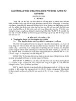
In the proboscis gland cell (Fig. 1a), the mitochondria are organized in two different forms (Fig. 1b and Table 1). Immediately
surrounding the nucleus, there is a cluster of small mitochondria. Each of those mitochondria appears oval in cross section
and provided by condensed cristae. Only very few mitochondria with orthodox cristae are distributed outside the cluster.
Material
A. bengalensis were collected from ‘‘Barasagar Dighi’’ fish
farm (24°580 08.8600 N, 88°060 09.7000 E) under Government of
West Bengal located at Malda, West Bengal, India. A breeding
colony of the parasite raised by cohabitation with the freshwater cyprinid host, Cirrhinus mrigala (Hamilton, 1822), was used
for this study. The parasite was identified with the help of morphometric criteria following Ramakrishna [9].
Light microscopic study
For light microscopy, abdomen of the matured female parasites
(age group of 29–32 days) was severed from the cephalothorax
with the help of a sharp triangular surgical suture without
affecting the ovary; thereafter, a small puncture was made to release the oocyte. Oocytes were then cleared by a solution containing ethanol, formalin, and acetic acid (6:3:1) and
observed under microscope. For vital staining fresh oocytes
were stained with 0.02% Janus green B (HiMedia Laboratories
Pvt. Limited) in insect saline for 30 min and viewed under compound microscope (Prime, Dewinter Optical Inc., Italy).
Transmission electron microscopy
Several adult male and female parasites were anesthetized adding ethanol drop by drop in water and then transferred to
2.5% glutaraldehyde and 2% paraformaldehyde solution in
cacodylate buffer (pH 7.4) to fix the specimens for overnight
at 4 °C. The specimens were postfixed in 2% osmium tetroxide
buffered solution and were embedded in epoxy resin. Subsequently, those were sectioned with a Leica Ultracut-UCT ultra
microtome and stained with a saturated solution of uranyl acetate and lead citrate. Micrographs were produced using a
JEM-2100 TEM (200 kV, Jeol).
Mitochondrial count
For counting mitochondria in the previtellogenic oocyte, image files of the electron micrograph of oocytes were opened
Schematic drawing
For schematic drawing, the micrographs were opened with
Photoshop CS4 software, and drawing was done in different
layer using the impressions from the image layer.
Results
Proboscis gland cell mitochondria
Spinal gland cell mitochondria
In the spinal gland cells (Fig. 1a), no free mitochondria are
present in the cytoplasm rather, those are all vesicle enclosed
(Fig. 1c). Those vesicle enclosed mitochondria are provided
by orthodox cristae (Table 1).
Oocyte mitochondria
Janus green B staining of the previtellogenic oocyte reveals
numerous spherical blue green bodies clustered in groups
(Fig. 2a) in the vicinity of the nucleus. Transmission electron
microscopy reveals the cluster contains mitochondria and electron-dense material (Fig. 2b). The mitochondria (Fig. 2c) appear round or oval in cross sections. The inner membrane is
infolded perpendicular to the longitudinal axis to form a moderate number of cristae. The cristae extend at least three quarters of the distance across the mitochondrial diameter and
have a tubular profile with bulged edges (Fig. 2c) (Table 1).
Very often, the mitochondria are associated with rough endoplasmic reticulum through tethers (Fig. 2d). The inner matrix
of the mitochondria is a homogeneous matter of finely granular material within which small numbers of variably sized, and
dense granules of 180–220 A˚ diameters are visible. The mitochondrial clusters are intermingled with numerous small vesicles or granulo fibrillar material (GFM) approximately of
0.15–0.54 lm diameter. Several mitochondria are also observed in the state of both fission and fusion (Fig. 3).
Spermatocytes, nurse cell and spermatozoan mitochondria
The typical form of mitochondria is few in the spermatocyte;
however, a large ‘‘nebenkern’’ is present near the nucleus of
Mitochondrial types in Argulus bengalensis
321
Fig. 1 Photomicrograph of Argulus bengalensis and transmission electron micrograph of mitochondrial forms in the glandular cells
associated with feeding apparatus. (a) Ventral view of a male showing the anatomical position of proboscis gland (pg) indicated by paired
side boxes and spinal gland (sg) indicated by lower median box. (b) Transmission electron micrograph of proboscis gland: mitochondria
(mt) are arranged within a separate hub (h) surrounding the nucleus (n). The cristae of these mitochondria are of condensed type. The
mitochondria distributed outside the hub are provided with orthodox cristae. Bar, 2 lm. (c) Transmission electron micrograph of spinal
gland: mitochondria (mt) are enclosed within vesicles (v) in the cytoplasm. Bar, 1 lm.
Table 1
Comparative profile of mitochondrial forms in Argulus bengalensis.
Sources
Types
Mitochondria Cristae
width (lm) a types
Special features
Potential functions
0.29–0.33
Condensed
B. Classical
0.19–0.38
Orthodox
Clustered, confined within
protected area
–
High ATP production for
synthetic activity
Low ATP production
Spinal gland
Variant
0.31–0.45
Orthodox
Vesicle enclosed
Protection of structure and
function
Oocyte
Variant
0.32–2.03
Tubular
Inner compartment divided,
Exhibit fission and fusion, ER
associated
High energy production to carry
out different synthetic activities
0.15–0.17
0.21–0.30
Condensed diffused –
Stacked
Microtubule associated
Proboscis gland A. Variant
Sperm flagellum A. Classical
B. Variant
Spermatocytes
High ATP production
Use of microtubuler tracts for
efficient energy distribution
A. Variant nebenkern 9.05–13.78
Stacked
B. Classical
0.52–1.25
Condensed
Highly packed cristae, obscured Later modified into flagellar
inner compartment
mitochondria in spermatozoa
–
High ATP production
0.67–1.25
Orthodox
ER associated
Striated muscle Variant
Mitochondrial Ca2+ uptake for
continued muscular function
a
Range of width (lowest–highest) of mitochondria from 10 ultrasections studied. For each presentation five measurements were made at
different angles and averaged.
primary and secondary spermatocytes (Fig. 4b). A similar
structure is also observed at the base of the cytoplasmic projection of the nurse cell (Fig. 4a). The nebenkern is provided with
huge number of closely stacked zigzag cristae within a highly
dense matrix (Fig. 4c and d). The zigzag cristae are extensive
and profusely anastomotic or overlapped to each other. Apart
from the nebenkern, very few classical forms of mitochondria
are randomly distributed around the nebenkern, but a few are
located at juxtaposition (Fig. 4b and Table 1).
In the flagellum of the spermatozoa (Fig. 5a), adjacent to
the axoneme, there are three moderately sized mitochondria
one with four numbers of orthodox cristae meeting at the center of the inner matrix and two others with condensed but diffused cristae (Fig. 5b). The medially located mitochondrion is
pear shaped, but others two are oval in cross section. Serial
sections of the flagellum reveal that these mitochondria are filiform and extend almost the entire length of it except the terminal part. One more unusual type of mitochondrion (Fig. 5c) is
322
A. Banerjee and S.K. Saha
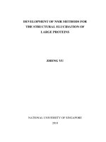
Fig. 2 Light and electron microscopy of mitochondria in the oocytes of Argulus bengalensis. (a) Light micrograph after mitochondria
specific vital staining with Janus green B showing distribution pattern of mitochondria within the cytoplasm of an early previtellogenic
oocyte (o); mitochondrial clouds (indicated by boxes) are differentiated beside the nucleus (n). Bar, 18 lm. (b) Transmission electron
micrograph of an early previtellogenic oocyte showing similar mitochondria rich zone around the nucleus (n). Numerous small vesicles or
granulofibrillar material (GFM) are distributed within this mitochondria rich zone. (c) Ultrastructure of mitochondrion of an early
previtellogenic oocyte exhibiting its tubular cristae with dilated terminal. The mitochondrion exhibits a loose association with an ER.
Numerous small but dense granules (g) are observed within the inner matrix. Bar, 0.34 lm. (d) A magnified view (2·) of the association of
mitochondria with ER – showing tethers (t) and ribosomes (r) in the upper pannel, and the lower panel is the schematic diagram of the
same. Bar, 0.2 lm.
Mitochondrial types in Argulus bengalensis
323
Fig. 3 Transmission electron micrograph of oocyte mitochondria at dynamic state in Argulus bengalensis. The upper left panel exhibits
out pocketing, an indication of fission of mitochondria. The upper right panel exhibits mitochondrial fusion indicated by diffused
membrane (arrow head) between mitochondria. The lower left panel shows two mitochondria immediately after completion of fission.
Bar, 0.54 lm. The graphic in the lower right panel represents number of mitochondria (Mt) in four previtellogenic oocytes (O1, O2, O3,
and O4); mitochondria undergoing fusion or fission are counted as a single unit. Mean of five readings with ±standard error is presented
in the graphics.
found at a right angle to the medially located mitochondrion.
It is pear shaped in cross section and spans about half of the
flagellum. In its course through the flagellum, the alignment
is changed with respect to the middle mitochondrion. The inner membrane of this mitochondrion is clearly distinguishable
but looses its connection with the transverse cristae. The transverse cristae are closely stacked into a cluster, and 20 numbers
of F1 particles are aligned at regular intervals at the outer
periphery of the cluster. Fifteen singlet microtubules, each
comprises of 12 protofilaments, are attached to the circumference of this mitochondrion through motor proteins (Fig. 5d).
tethering structures of endoplasmic reticulum (Fig. 6b) are also
being observed. A gap of 78–86 nm is maintained between the
mitochondria and the tethering vesicles where several ribosomes (Fig. 6b) are distributed.
In A. bengalensis other than classical type, six mitochondrial
variants are observed in different cell types to meet up the energy demands under varied physiological states of its parasitic
mode of life.
Striated muscle cell mitochondria
Proboscis gland cell and spinal gland cell mitochondria
The mitochondria of striated muscles from the thoracic
appendages are oval in shape and provided by orthodox cristae
(Fig. 6a and Table 1). Inner matrix of those is compartmentalized further by the extension of some cristae. In the sarcomeres, the mitochondria are distributed adjacent to the
myofibrils and are intimately associated with the ER through
tight junction (Fig. 6c). Their associations with the vesicular
Feeding apparatus of Argulus spp. is a secondary acquisition
and comprises of a proboscis and a preoral spine. A pair of
proboscis gland consisting two giant cells is associated with
the proboscis, and one spinal gland consisting four large cells
is located at the base of the spine. The spine is used to pierce
the host tissue, and the tissue fluid and blood ooze out are
ingested through the proboscis. The spinal gland produces
Discussion
324
A. Banerjee and S.K. Saha
Fig. 4 Transmission electron microscopy of mitochondria in the testicular cells of Argulus bengalensis. (a) Nebenkern (N) in the nurse
cell (Nc). Nurse cell is present in between the primary (Ps) and secondary spermatocytes (Ss). The nebenkern (N) is positioned at the base
of the cytoplasmic projection (P). Small classical mitochondria (mt) are also randomly distributed beside the nucleus (n) of the nurse cell.
(b) Primary spermatocyte also exhibits a large nebenkern (N) beside its nucleus (n) and small classical mitochondria (mt) are randomly
distributed around the nebenkern. Bar, 1 lm. (c) Magnified view (4 ·) of the nebenkern showing its stacked zigzag cristae. Bar, 0.2 lm. (d)
Highly magnified view (17·) of the nebenkern showing cristae with overlapping at regular interval. The right corner panel is a schematic
diagram shows the arrangement of cristae and variation in cristae diameter. Bar, 54 nm.
an anesthetic substance which is injected into the fish’s body
for effortless feeding activity, and the proboscis glands produce an anticoagulant that prevents ingested blood from clotting within the gut [10]. Condensed state of cristae in the
mitochondria found in these glandular cells correspond to
their high workload of ATP production [11] required for their
synthesis activities. An unusual type of closed membrane vesicles containing one or more mitochondria was observed in
the spinal gland cells. Similar type of mitochondrial concealment was also observed in aging wheat coleoptiles and in
neural tissue (Table 2) [12,13]. Concealment of the mitochondria within vesicles may preserve their structure and activity
[12] and thereby protect them from exposure to the glandular
synthesis (Table 1). In proboscis gland, cluster of small mitochondria is concealed in an area surrounding the nucleus.
Similar type of mitochondrial cluster was also observed in
human fetal and adult female germ cells (Table 2) [14]. Concealment of mitochondria within vesicles or in specialized
area definitely protect the mitochondria from the detrimental
effect of glandular synthesis and help to restore their structural and functional organization and thereby confers some
adaptive advantages to the organisms in their parasitic mode
of feeding or hematophagy.
Oocyte mitochondria
One of the most prominent features of the early previtellogenic
oocytes of A. bengalensis is the mitochondrial aggregation into
cluster known as Yolk nucleus and Balbiani body. The term
mitochondrial cloud is often more appropriate to these clustered organelles and has frequently been used [15]. Light
microscopy of the previtellogenic oocyte with Janus green B,
which stains mitochondria supravitally [16], able to detect such
clusters sometimes referred as the Balbiani body or Yolk nucleus in the oocyte of Xenopus sp. and human (Table 2)
[14,15,17] which plays an important role in germinal granule
localization in the vegetal pole [17]. The ultrastructure of the
mitochondrial aggregates reveals that they consist of large
numbers of discrete mitochondria, but the image is not quite
that expected from a simple aggregate of separate mitochondria. However, during our study of the mitochondrial clusters
of Argulus sp., we became impressed with the dynamicity
Mitochondrial types in Argulus bengalensis
325
Fig. 5 Transmission electron micrograph of mitochondria in the sperm flagellum of Argulus bengalensis. (a) Mitochondrial (mt)
alignment surrounding the axoneme (Ax). (b) Three vesicular filamentous mitochondria immediately adjacent to the axoneme with clearly
distinguishable outer membrane (om), inner membrane (im) and transverse cristae (tc). Cristae are uniting at the central meeting point
(mp) to compartmentalize the inner matrix. Bar, 74 nm. (c) An unusual association of mitochondrion with numerous microtubules (am).
The transverse cristae (tc) are originated from the inner membrane and extending up to the inner membrane of the opposite side. Twenty
F1 particles are arranged near the base of each cristae. Bar, 74 nm. The bottom left panel is a magnified (6·) view showing the
microtubular association with the outer membrane (om) by motor protein (mp). Bar, 13 nm. The bottom right panel is a diagrammatic
representation of the microfilament. The microfilament is a complete circle of twelve protofilament (pf). It remains attached with the outer
mitochondrial membrane (om) with a motor protein (mp).
accompanied with both fusion and fission leading profound
morphogenetic changes, reflecting changing metabolic requirements. The complexity of the mitochondrial profiles is further
validated by its association with other organelles. The endoplasmic reticulum composes a loose junction with the mitochondria which is occupied by granulo fibrillar material
(GFM). Mitochondrial association with the endoplasmic reticulum will be important to supply energy for translation. One
of the important features of an ectoparasite is its reproductive
investment which involves much energy production and utilization to meet up the needs for maturation of gametes (Table 1). Previtellogenic stage is the most active stage in the
maturation process when the oocytes become prepare for several synthetic activities including synthesis of Yolk to carry out
the embryonic development of the parasite.
Spermatocytes, nurse cell and spermatozoan mitochondria
In the spermatocytes and nurse cell, a mitochondrial variant,
nebenkern is observed like that of other insect spermatocytes.
Nebenkern is formed through a multistep process by which the
numerous mitochondria are clustered together and fused to
produce a large spherical body [18–20]. The cristae of the
‘‘nebenkern’’ in the spermatocytes of Argulus are longer and
more closely packed which indicates that the cells are in hyper-
active metabolic state. Such type of zigzag orientation of cristae is also observed in hyper metabolically active tissue like
cardiac muscle cells of the canary and other birds [21]. Dorogova et al. [22] explained that marlin protein in the nebenkern
plays important role in spermatogenesis of Drosophila. The
nebenkern also unfolds and extends along with growing axoneme in Drosophila sperm. Similar role of nebenkern in Argulus species can be apprehended.
One rare type of mitochondrion is found in the flagellum of
the spermatozoa where it is associated with microtubules (Table 1). Microtubular association is also found in vitro with different cell types of the vertebrate like fibroblasts, macrophages,
smooth muscle cells and in neuronal axons (Table 2) [23,24]
but in those cases, mitochondria are associated with fewer
microtubules, whereas in vivo argulid sperm is associated with
numerous as more as 15 microtubules in a definite pattern. The
physiological significance of such association is that mitochondrion uses these microtubular tracts for its movement [25] with
the aid of motor proteins like dynein. During copulation of
Argulus, sperm is donated as packets or spermatophores [26].
So, the individual sperm does not require active motility at this
stage, but sudden active and regulated motility is required
immediately after its release from the spermatophore just before fertilization. The movement of mitochondrion through
the microtubular tracts must be related to energy distribution
326
A. Banerjee and S.K. Saha
Fig. 6 Transmission electron microscopy of mitochondria in the striated muscle. (a) Mitochondrion (mt) in association with
sarcoplasmic reticulum (Sr) showing attached putative ER vesicle (ERv). A narrow space is present in between the outer membrane (om)
and the inner membrane (im). (b) Higher magnified (5·) view of the ER vesicle (ERv) showing tethers (t) and associated ribosome (r).
Right panel is a schematic diagram showing molecular bridges that regulate the close contacts between ER and mitochondria. Bar, 43 nm.
(c) Tight association of the ER with outer membrane (om) of a mitochondrion. Bar, 70 nm. Right panel is a magnified view of the same.
and utilization along the length of the flagellum resulting regulated sperm motility for successful fertilization in Argulus.
This type of mitochondrial association with microtubules is
also observed in the flagellum of tongue worm, Pentastomid
Raillietiella in one of the publications of Wingstrand [27] that
justify their phylogenetic relationship.
Striated muscle cell mitochondria
The mitochondria of the striated muscle cells of thoracic
appendages are very large in comparison with the other types
of mitochondria found in Argulus (Table 1). The physical association between the endoplasmic reticulum (ER) and mitochon-
dria, which is known as the mitochondria-associated ER
membrane (MAM), has important roles in various cellular
‘‘housekeeping’’ functions [28]. Close contacts between the
ER membrane and the mitochondrial outer membrane have
been visualized by various authors in rat liver tissue and in
the pseudobranch gland of teleost (Table 2) [29,30]. ER and
mitochondria are held together by different molecular chaperones as stated by Hayashi et al. [28] and Rizzuto et al. [31].
Other than the vertebrate system, mitochondrial association
with ER is first time evident in an invertebrate like the parasitic
Argulus. Mitochondria regulated efflux of endoplasmic Ca2+
and Ca2+ signaling thereof [28,31] helps in regulating muscle
contraction, lipid transport [32], and cellular survival [33,34].
Mitochondrial types in Argulus bengalensis
Table 2
327
Comparative account of mitochondrial variants of Argulus bengalensis with that of other referred organisms.
Mitochondrial variants
Source tissue of A.
bengalensis
Other plant, invertebrate and
vertebrate sources
References
Confined around nucleus
Proboscis gland
Motta et al. [14]
Vesicle enclosed
Spinal gland
Mitochondria with fission and
fusion and ER associated
Nebenkern
Microtubule associated
Previtellogenic oocyte
Human fetal and adult female
germ cells
Aging wheat coleoptiles, neural
tissue
Oocytes of vertebrates like
Xenopus laevis
Spermatocytes of various insects
In vitro culture of. rat kidney cell,
human fibroblasts, peritoneal
macrophages and smooth muscle
of mouse
Liver cell, neuron and various
other cell types of vertebrates
ER associated
Spermatocyte
Sperm flagellum (in vivo it is
reported for the first time)
Sarcomere
A continuous mitochondrial Ca2+ uptake occurs in the muscle
tissue which in turn could facilitate mitochondrial Ca2+ overloading and membrane permeabilization [35]. Such type of
ER-mitochondria tethering ensures the propagation of TP3Rlinked Ca2+ signals to the mitochondria to coordinate ATP
production with the stimulated state of the cell, and it protects
the cell from energy depletion and maintain mitochondrial
metabolism [34]. The respiratory structures of the parasite are
located on the ventrolateral thoracic carapace, which is ventilated by the continuous movement of the three pairs of thoracic
appendages when the parasite remains attached to the host
body with a pair of suckers. Continuous movement of appendages needs uninterrupted muscular function. The physical association between the endoplasmic reticulum (ER) and the
mitochondria must play important role in energy production
and utilization to confer the stimulated state of the cells to bestow the parasitic adaptive advantages.
Bakeeva et al. [12], Mishchenko
[13]
Billett and Adam [15], Motta
et al. [14], Wilk et al. [17]
Beams et al. [19], Tokuyasu [20].
Goldman and Follett [23],
Heggeness et al. [24]
Copeland and Dalton [29],
Morre et al. [30]
Funding
This work was supported by the University Grants Commission, Government of India through a Major Research Project
F.33-333/2007(SR) dt. 10th March 2008.
Conflict of interest
The authors have declared no conflict of interest.
Acknowledgements
We have pleasure to acknowledge STA-TEM section, SAIF–
North Eastern Hill University, Shillong for extending assistance in electron microscopy.
References
Conclusions
In A. bengalensis, mitochondria are highly dynamic structures
and appear in varied forms and numbers in different cell types
at varying physiological states. It readily undergoes fission and
fusion in cells like oocytes and even can move on cytoskeletal
track for efficient energy distribution and utilization in a specific cell type like argulid sperm. Muscle cells in continuous action can utilize the close association of mitochondria with the
endoplasmic reticulum not only for efficient energy production
and utilization but also for regulated contraction brought
about by regulated Ca2+ release from the endoplasmic reticulum. The mitochondria of glandular cells associated with the
feeding apparatus of Argulus are well protected within cytoplasmic vesicles. The tissue specific mitochondrial variability
of this parasitic organism has its implication on the biology
of the cell and hence on the biology of the organism which bestow several adaptive advantages to its parasitic mode of life.
The phylogenetic relationship of argulids with pentastomids
is a long pending issue; mitochondrial association with microtubules in the flagellum of the sperm adds further evidence in
support of it.
[1] Palade GE. An electron microscope study of the mitochondrial
structure. J Histochem Cytochem 1953;1:188.
[2] Bereiter-Hahn J, Voth M. Dynamics of mitochondria in living
cells: shape changes, dislocations, fusion, and fission of
mitochondria. Microsc Res Techniq 1994;27:198–219.
[3] Chan DC. Mitochondrial fusion and fission in mammals. Annu
Rev Cell Dev Biol 2006;22:79–99.
[4] Frey TG, Mannella CA. The internal structure of mitochondria.
Trends Biochem Sci 2000;25:319–24.
[5] Attardi G, Schatz G. Biogenesis of mitochondria. Annu Rev
Cell Biol 1988;4:289–333.
[6] Green DR, Reed JC. Mitochondria and apoptosis. Science
1998;281:1309–12.
[7] Saraste M. Oxidative phosphorylation at the fin de sie`cle.
Science 1999;283:1488–93.
[8] Bereiter-Hahn J. Behavior of mitochondria in the living cell. Int
Rev Cytol 1990;122:1–63.
[9] Ramakrishna G. Notes on the Indian species of the genus
Argulus Mu¨ller (Crustacea: Copepoda) parasitic on fishes. Rec
Indian Mus 1951;49:207–15.
[10] Saha SK, Guha A, Banerjee A. Feeding apparatus and
associated glands in the freshwater fish ectoparasite Argulus
siamensis
Wilson,
1926
(Branchiura).
Crustaceana
2011;84:1153–68.
328
[11] Perkins GA, Ellisman MH. Mitochondrial configurations in
peripheral nerve suggest differential ATP production. J Struct
Biol 2011;173:117–27.
[12] Bakeeva LE, Kirnos MD, Aleksandrushkina NI, Kazimirchyuk
SB, Shorning BY, Zamyatnina VA, et al. Subcellular
reorganization of mitochondria producing heavy DNA in
aging wheat coleoptiles. FEBS Lett 1999;457:122–5.
[13] Mishchenko Y. Automation of 3D reconstruction of neural
tissue from large volume of conventional serial section
transmission electron micrographs. J Neurosci Meth
2009;176:276–89.
[14] Motta PM, Nottola SA, Makabe S, Heyn R. Mitochondrial
morphology in human fetal and adult female germ cells. Hum
Reprod 2000;15:129–47.
[15] Billett FS, Adam E. The structure of the mitochondrial cloud of
Xenopus laevis oocytes. J Embryol Exp Morphol
1976;33:697–710.
[16] Cooperstein SJ, Dixit PK, Lazarow A. Studies on the
mechanism of Janus green B staining of mitochondria. IV.
Reduction of Janus green B by isolated cell fractions. Anat Rec
1960;138:49–66.
[17] Wilk K, Bilinski S, Dougherty MT, Kloc M. Delivery of
germinal granules and localized RNAs via the messenger
transport organizer pathway to the vegetal cortex of Xenopus
oocytes occurs through directional expansion of the
mitochondrial cloud. Int J Dev Biol 2004;49:17–21.
[18] Bowen RH. Studies on insect spermatogenesis. III. On the
structure of the nebenkern in the insect spermatid and the origin
of nebenkern patterns. Biol Bull 1922;42:53–82.
[19] Beams HW, Tahmisian TN, Devine RL, Roth LE. Phasecontrast and electron microscope studies on the nebenkern, a
mitochondrial body in the spermatids of the grasshopper. Biol
Bull 1954;107:47–56.
[20] Tokuyasu KT. Dynamics of spermiogenesis in Drosophila
melanogaster. VI. Significance of ‘‘onion’’ nebenkern
formation. J Ultrastruct Res 1975;53:93–112.
[21] Slautterback DB. Mitochondria in cardiac muscle cells of the
canary and some other birds. J Cell Biol 1965;4:1–21.
[22] Dorogova NV, Akhmametyeva EM, Kopyl SA, Gubanova NV,
Yudina OS, Omelyanchuk LV, et al. The role of Drosophila
Merlin in spermatogenesis. BMC Cell Biol 2008;9:1–15.
A. Banerjee and S.K. Saha
[23] Goldman RD, Follett EAC. The structure of the major cell
processes of isolated BHK21 fibroblasts. Exp Cell Res
1969;57:263–76.
[24] Heggeness MH, Simon M, Singer SJ. Association of
mitochondria with microtubules in cultured cells. Proc Natl
Acad Sci USA 1978;75:3863–6.
[25] Vos M, Lauwers E, Verstreken P. Synaptic mitochondria in
synaptic transmission and organization of vesicle pools in health
and disease. Front Synaptic Neurosci 2010;2:1–10.
[26] Avenant-Oldewage A, Everts L. Argulus japonicus: sperm
transfer by means of a spermatophore on Carassius auratus
(L). Exp Parasitol 2010;126:232–8.
[27] Wingstrand KG. Comparative spermatology of a pentastomid
Raillietiella hemiductyli and a branchiuran crustacean Argulus
foliaceus with a discussion of pentastomid relationships. Biol Skr
Dan Vid Selsk 1972;19:l–72.
[28] Hayashi T, Rizzuto R, Hajnoczky G, Su T. MAM: more than
just a housekeeper. Trends Cell Biol 2009;19:81–8.
[29] Copeland DE, Dalton AJ. An association between mitochondria
and the endoplasmic reticulum in cells of the pseudobranch
gland of a teleost. J Biophys Biochem Cytol 1959;5:393–5.
[30] Morre DJ, Merritt WD, Lembi CA. Connections between
mitochondria and endoplasmic reticulum in rat liver and onion
stem. Protoplasma 1971;73:43–9.
[31] Rizzuto R, Marchi S, Bonora M, Aguiari P, Bononi A, de
Stefani D, et al. Ca2+ transfer from the ER to mitochondria:
when, how and why. Biochim Biophys Acta 2009;1787:
1342–51.
[32] Voelker DR. Bridging gaps in phospholipid transport. Trends
Biochem Sci 2005;30:396–404.
[33] Hajno´czky G, Davies E, Madesh M. Calcium signaling and
apoptosis. Biochem Biophys Res Co 2003;304:445–54.
[34] Hayashi T, Su TP. Sigma-1 receptor chaperones at the ERmitochondrion interface regulate Ca2+ signaling and cell
survival. Cell 2007;131:1–15.
[35] Bernardi P. Mitochondrial transport of cations: channels,
exchangers, and permeability transition. Physiol Rev
1999;79:1127–55.