Isolation and morphological characterization of endophytic fungi isolated from ten different varieties of mango
Bạn đang xem bản rút gọn của tài liệu. Xem và tải ngay bản đầy đủ của tài liệu tại đây (820.1 KB, 10 trang )
Int.J.Curr.Microbiol.App.Sci (2019) 8(3): 717-726
International Journal of Current Microbiology and Applied Sciences
ISSN: 2319-7706 Volume 8 Number 03 (2019)
Journal homepage:
Original Research Article
/>
Isolation and Morphological Characterization of Endophytic Fungi Isolated
from Ten Different Varieties of Mango
Mahesh S. Dashyal1, C.G. Sangeetha1*, Vikram Appanna2,
G.K. Halesh3 and V. Devappa1
¹Department of Plant Pathology, College of Horticulture, University of Horticultural Sciences
Campus, GKVK, Bengaluru, 560 065, Karnataka, India
²Department of Agricultural Microbiology, College of Horticulture, University of
Horticultural Sciences Campus, Mysuru-571130, Karnataka, India
³Department of Biotechnology and Crop Improvement, College of Horticulture, University of
Horticultural Sciences Campus, GKVK, Bengaluru, 560 065, Karnataka, India
*Corresponding author
ABSTRACT
Keywords
Endophytes,
Mango,
Anthracnose, Fungi,
morphology
Article Info
Accepted:
07 February 2019
Available Online:
10 March 2019
Endophytes are the microbes that colonize the healthy tissues of plant. Endophytes
associated with Mangifera indica (mango) are less understood. In this study, endophytic
fungi were isolated fromten different mango varieties viz., Alphonso, Totapuri, Neelam,
Anfas, Willard, Badam model, Khaderi, Pancharasi, White Sari and KisanBhog from
Bengaluru, Karnataka, India. Endophytic fungi were isolated fromleaf and stem tissues.
Nine endophytic fungi were isolated from Alphonso out of which four were from leaf and
five were from stem tissues. Two endophytic fungi were isolated from the leaf tissue of
Totapuri, and five from the stem tissue. From Neelam, only four endophytic fungi were
isolated from stem tissue and no endophytic fungi grew from the leaf tissue. One
endophytic fungi was isolated from leaf and stem tissue each from Anfas, Willard, Badam
Model, Kadari, Pancharasi and White sari, whereas two fungi were isolated from leaf
tissue of KisanBogh and one from stem tissue. The isolates were studied for to their
morphological characters and naming was done based on the variety and the part isolated.
production of 185.27 lakh MT. In Karnataka,
it is grown in 1.78 lakh hectares with a
production of 1.78 metric tonnes and
productivity is 10 t/ha (Anon., 2014). Mango
is affected by a number of diseases at all the
stages of its development right from seedling
in nursery to the fruit in storage or transit
Introduction
Mango (Mangifera indica L.) is considered as
one of the choicest fruitcrops grown all
around the world (Shad et al., 2002). It is
grown in more than 100 nations, however
nowhere it is enormously esteemed as in India
where 40 per cent of the total fruits grown is
mango (Swamy, 2012). In India, mango is
grown in 21.63 lakh hectares with a
Endophytes are bacterial or fungal
microorganisms that colonize plant tissue
717
Int.J.Curr.Microbiol.App.Sci (2019) 8(3): 717-726
intercellularly as well as intracellularly
without causing any clear manifestations
(Wilson, 1995). Endophyte, by definition, is
one which lives in the tissues underneath the
epidermal cell layers and makes no evident
damage to the host (Stone et al., 2000). They
are present in all the plants parts, colonize all
plants, and have been found from all plants
analyzed till date (Nair and Padmavathy,
2014). To keep up consistent beneficial
interaction, endophytes deliver different
compounds that advance development of
plants and help plant to better environmental
adoption. Plants growing in areas of great
biodiversity also have the prospect of housing
endophytes with great biodiversity (Strobel et
al., 2003).Out of 1.5 million fungal species
evaluated to be available, only 97,861 fungal
species have been studied (Hawksworth,
1991). They deliver an immense range of
compounds which are valuable for plants for
their development, insurance to natural
circumstances and supportability (Nair and
Padmavathy, 2014). They additionally assume
an essential part in nutrient cycling,
biodegradation and bioremediation (Das and
Varma, 2009; Lee et al., 2004). Hence in this
study we aimed at studying the diversity of
endophytic fungi associated with ten different
varieties of mango in terms of presence and
their characteristics.
varieties. Healthy leaf and stem tissues of 10
mango varieties viz., Alphonso, Totapuri and
Neelam were collected from the orchards of
University
of
Horticultural
Sciences,
Bagalkot, Regional Horticulture Research
Station, Bengaluru. Similarly, healthy leaf
and stem tissues of Anfas, Willard, Khaderi,
Pancharasi, Kisanbhog, White Sari and
Badam model were collected from Indian
Institute of Horticultural Research Station,
Bengaluru. Samples were collected from three
randomly selected plants from each variety
and collected leafs are clubbed and collected
in a plastic bags, labeled, transported to lab
within 12 hours and used for isolation.
Although rapid changes in endophyte
colonization probably do not occur
immediately following collection, all samples
were handled carefully and processed as
quickly as possible. Samples were air-dried to
remove any surface moisture before transport
or storage. During transport, samples were
kept cool and dry. Samples of Alphonso,
Totapuri, Neelam were collected from six
year old trees and the samples from the other
remaining varieties viz., Anfas, Willard,
Khaderi, Pancharasi, KisanBhog, White Sari
and Badam model were collected from 40
years old trees.
Materials and Methods
Endophytic fungi were isolated from leaf and
stem tissues collected from the different
mango varieties like Anfas, Willard, Khaderi,
Pancharasi, KisanBhog, Alphanso, Totapuri,
Neelum, White Seri and Badam Model.
Isolation of fungal endophytes
The foregoing investigations were conducted
at the Department of Plant Pathology, College
of Horticulture, Bangalore, University of
Horticultural Sciences, Bagalkot.
All the leaf and stem samples were washed
under running water prior to surface
sterilization. Leaf discs were cut from leaf
using a sterile blade to isolate endophytes.
Stem segments were cut from internal tissues
of stems. Size of the sampling unit and
surface
sterilization
procedures
vary
according to the preferences of the
Isolation of endophytic fungi
Collection of sample
For isolation of endophytic fungi, mature,
healthy, green, asymptomatic leaves and stem
tissues were collected from ten mango
718
Int.J.Curr.Microbiol.App.Sci (2019) 8(3): 717-726
investigator, the species of host plant, and
host tissue type sampled. In our study we
followed the modified method of Michereff et
al., (2014) as discussed below:
varieties, out of which 14 isolates were from
leaf and 21 isolates were from stem tissues
(Table 1). The result showed that more
number of endophytic fungi was isolated from
stem tissue as compared to the leaf tissues.
The isolates were designated with first two
alphabets denoting endophytic fungi (EF),
next letter the first letter of the variety from
which the fungi was isolated (Eg.:AAlphonso), the fourth letter the tissue from
which it was isolated, wherein L stands for
leaf and S stands for stem. The endophytes
isolated are designated as A, B, C, D and so
forth.
Leaf segments were surface sterilized in 75
per cent ethanol for 1 minute and 2 per cent
sodium hypochlorite for 1 minute. The stem
fragments were surface sterilized in 75 per
cent ethanol for 1minute and 2 per cent
sodium hypochlorite for 2 minute. Sterilized
segments were rinsed in sterilized distilled
water and then dried on sterilized paper. The
effectiveness of the sterilization procedure
was tested using the imprint method (Schulz
et al., 1993). Three fragments were placed
evenly in petri dishes (9 cm dia.) containing
potato dextrose agar (PDA) medium amended
with streptomycin to suppress bacterial
growth and incubated at 28ºC. The fungi
growing out of the segments during the
incubation period were recorded as
endophytes and then those endophytes were
pure cultured.
Similar isolations from plants were done by
other workers like Nayak (2015) who isolated
17 fungal endophytic species from the
ornamental mango (Mangiferaindica) and
Michereffet al., (2014) isolated 169 fungal
endopytes from noncommercial mango plant;
Amin et al., 2014 isolated endophytes from
cultivars which were resistant and susceptible
to vascular streak disease.
Characterization of fungal endophytes
Morphological characterization of fungal
endophytes
Morphological study
Morphological characteristics of all the 35
isolates like mycelial characters and spore
production were examined using cultures
grown on PDA. There was a lot of variation
in the morphological characters of all the
endophytic fungi isolated from different
varieties (Table 2).
Cultures on PDA media were assessed
according to their morphology. Colony
character of the fungal growth, topography
was noted and characters of mycelial colour,
type and spore production were observed.
Results and Discussion
Isolation of fungal endophytes
different mango varieties
Out of nine fungal endophytes isolated from
Alphonso variety, two endophytes EFAL-A
and EFAS-E exhibited slow growth and two
isolated EFAL-B and EFAS-C exhibited
medium growth. Five endophyes isolated
from leaf as well as stem, EFAL-C, EFAL-D,
EFAS-A, EFAS-B and EFAS-D exhibited fast
growth. Five endophytes EFAL-A, EFAL-C,
EFAL-D, EFAS-C and EFAS-D showed
greyish mycelium. All the isolates were non-
from
Endophytic fungi were isolated from leaf and
stem of different mango varieties like
Alphanso, Totapuri, Neelum, Anfas, Willard,
Khaderi, Pancharasi, KisanBhog, White Sari
and Badam Model. Totally 35 fungal
endophytes were isolated from mango
719
Int.J.Curr.Microbiol.App.Sci (2019) 8(3): 717-726
sporulating except EFAS-E. Four endophytes
isolated from leaf EFAL-A, EFAL-B, EFALC and EFAL-D showed septate mycelium and
five endophytes isolated from stem EFAS-A,
EFAS-B, EFAS-C, EFAS-D and EFAS-E
showed aseptate mycelium.
The fungal isolates EFAL-A, EFAS-E,
EFATL-A, EFATL-B, EFTS-A, EFTS-B,
EFTS-C, EFWDL-A, EFBML-A, EFKAL-A
and EFKAS-B were slow growing and EFALB, EFAS-C, EFTS-D, EFTS-E, EFNS-B,
EFNS-C, EFNS-D, EFANS-A, EFBMS-A,
EFPL-A, EFPS-A, EFWSL-A, EFKBL-A and
EFKBL-B showed medium growth rate and
EFAL-C, EFAL-D, EFAS-A, EFAS-B,
EFAS-D, EFNS-A, EFWS-A, EFANFL-A,
EFWS-A and EFKBS-A were reported to be
fast growing.
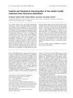
Two endophytes EFAL-D and EFAS-A
showed cottony type mycelium and three
endophytic fungi viz., EFAL-B, EFAL-C and
EFAS-D showed fluffy mycelium on PDA
(Figure 1).
The results revealed that, out of total 35
fungal isolates, 17 endophytes EFAL-A,
EFAL-B, EFAL-C, EFAL-D, EFTS-A,
EFTS-B, EFTS-D, EFTS-E, EFNS-A, EFNSB, EFNS-C, EFWL-A, EFWS-A, EFBML-A,
EFKAL-A, EFPL-A, EFWSL-A, EFKBL-B
and EFKBS-A showed septate mycelium
which was dark coloured and the remaining
18 endophytes showed aseptate mycelium and
were hyaline in colour.
Six isolates out of 35 showed fluffy cottony
growth of mycelia (EFAL-B, EFAL-C,
EFAS-D, EFNS-B, EFNS-C and EFKBS-A)
on PDA and 11 isolates out of 35 showed
cottony growth (EFAL-D, EFAS-A, EFATLA, EFATL-B, EFTS-A, EFTS-C, EFTS-D,
EFTS-E, EFNS-D, EFWS-A and EFKBL-B).
Nine endophytic fungi (EFAL-A, EFAL-C,
EFAL-D, EFAS-C, EFAS-D, EFNS-B,
EFWS-A, EFBML-A and EFKL-A) out of 35
isolates showed greyish colony on PDA
media, three (EFTL-A, EFTS-B and EFTS-E)
showed blackish growth on PDA. Six (EFASB, EFTL-B, EFTS-C, EFNS-C, EFWDL-A
and EFKBS-A) endophytes showed mixed
colony colour on PDA media.
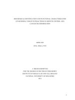
Majority of them did not produce spores and
only two endophytes EFTS-E and EFKBL-A
isolated from the stem tissue of totapuri and
leaf tissue of Kisanbhog respectively
produced hyaline spores (Figure 2).
Table.1 The endophytic fungi isolated from ten different varieties of mango
Sl. No
Varieties
1
2
3
4
5
6
7
8
9
10
Alphonso
Totapuri
Neelam
Anfas
Willard
Badam model
Khaderi
Pancharasi
White Sari
KisanBogh
Total
Fungal strains isolated
Leaf
Stem
4
5
2
5
0
4
1
1
1
1
1
1
1
1
1
1
1
1
2
1
14
21
720
Int.J.Curr.Microbiol.App.Sci (2019) 8(3): 717-726
Table.2 Morphological characteristics of endophytic fungi isolated from ten different varieties of mango
Sl. No
Variety
Isolate
Spore
production
Mycelium
characters
Topography of the
fungal growth
Color of the colony
Growth
rate
AFAL-A
EFAL-B
EFAL-C
EFAL-D
EFAS-A
EFAS-B
EFAS-C
EFAS-D
EFAS-E
No
No
No
No
No
No
No
No
Yes
Septatecoloured
Septatecoloured
Septatecoloured
Septatecoloured
Aseptate hyaline
Aseptate hyaline
Aseptate hyaline
Aseptate hyaline
Aseptate hyaline
Flat uniform
Fluffy
Fluffy uniform
Cottony type mycelium
Cottony type mycelium
Flat Silky
Flat spongy
Fluffy uniform
Flat milky growth of
mycelium
Greyish
Dull whitish
Greyish
Greyish
White and yellow
White and yellow
Greyish
Greyish black
Yellowish white
Slow
Medium
Fast
Fast
Fast
Fast
Medium
Fast
Slow
EFTL-A
EFTL –B
No
No
Aseptate hyaline
Aseptate hyaline
Cottony flat
Cottony flat
Slow
Slow
EFTS –A
No
Septatecoloured
13
EFTS –B
No
Septatecoloured
Blackish grey
Slow
14
EFTS –C
No
Aseptate hyaline
Cottony and slightly
raised
Corky growth with wavy
margin slightly
undulated
Cottony flat
Black
Centrally brown,
whitish margin
Reddish brown
Slow
15
EFTS –D
No
Septatecoloured
Cottony flat type
Medium
16
17
EFTS –E
No
Septatecoloured
Cottony flat
Reddish center with
white margin
Dark brownish centre
with brown margin
Black
EFNS-A
No
Septatecoloured
Flat uniform
Brownish yellow
Fast
Alphonso
1
2
3
4
5
6
7
8
9
10
11
12
Leaf
Stem
Totapuri
Leaf
Stem
Neelum
Stem
721
Slow
Medium
Int.J.Curr.Microbiol.App.Sci (2019) 8(3): 717-726
18
19
EFNS –B
EFNS –C
No
No
Septatecoloured
Septatecoloured
20
EFNS –D
No
21
22
23
24
25
26
27
28
Anfas
Leaf
EFANL-A
Stem
EFANS-A
Willard
Leaf
EFWL-A
Stem
EFWS-A
Badam Modal
Leaf
EFBML-A
Stem
EFBMS-A
Kadari
Leaf
EFKL-A
Stem
EFKS-A
31
32
Pancharasi
Leaf
EFPL-A
Stem
EFPS-A
White Seri
Leaf
EFWSL-A
Stem
EFWSS-A
33
34
35
KisanBogh
Leaf
EFKBL-A
EFKBL-B
Stem
EFKBS-A
29
30
Greyish
White and greyish
Medium
Medium
Aseptate hyaline
Fluffy uniform
Fluffy cottony growth
raised at center
Cottony uniform
Whitish purple
Medium
No
No
Aseptate hyaline
Aseptate hyaline
Flat uniform
Flat uniform growth
Dull brown
Whitish yellow
Fast
Medium
No
No
Septatecoloured
Septatecoloured
Undulated
flat cottony growth
Yellow and grey
Grey
Slow
Fast
No
No
Septatecoloured
Aseptate hyaline
Raised colony
Uniform undulated
Grey
Whitish yellow
Slow
Medium
No
No
Septatecoloured
Aseptatehyaline
Raised undulated
Flat wavy
Grey
Red releasing blackish
brown
Slow
Slow
No
No
Septatecoloured
Aseptatehyaline
Flat uniform
Flat undulated
Brownish black
Yellowish white
Medium
Medium
No
No
Septatecoloured
Aseptate hyaline
Flat wavy
Flat undulated wavy
growth
Dark brown
Yellow
Medium
Medium
Yes
No
No
Aseptate hyaline
Septatecoloured
Septatecoloured
Flat milky growth
Flat cottony growth
Uniform raised fluffy
growth
Dull yellow
Brownish black
Grey and dull white
Medium
Medium
Fast
722
Int.J.Curr.Microbiol.App.Sci (2019) 8(3): 717-726
Fig.1 Colony morphology of fungal endophytes isolated from Alphonso variety
723
Int.J.Curr.Microbiol.App.Sci (2019) 8(3): 717-726
Fig.2 Microscopic characters of some fungal endophytes isolated from different mango tissues
EFKL-A
EFKBL-A
EFBML-A
EFBMS-A
EFKBS-A
EFAS-E
724
Int.J.Curr.Microbiol.App.Sci (2019) 8(3): 717-726
Bisby’s Dictionary of the fungi. VI.
Edn. Commonwe. mycol. Inst., Kew,
Surrey, England, pp. 663.
Amin, N., Salam, M., Junaid, M., Asman and
Baco, M. S. 2014. Isolation and
identification of endophytic fungi from
cocoa plant resistant VSD M.05 and
cocoa plant Susceptible VSD M.01 in
South Sulawesi, Indonesia. Int. J. Curr.
Microbiol. App. Sci., 3(2):459-467.
Anonymous. 2014. Indian Horticulture
Database,
National
Horticultural
Board,Ministry
of
Agriculture,
Government of India, Gurgaon.
Barnett, H.L., And B. B. Hunter, 1998, The
Illustrated Genera of Imperfect Fungi.
Fourth Edition Aps Press, American
Phytopathol.
society,
St.
Paul,
Minnesota. pp.218.
Carmichael, J.W., Kendrick, W.B., Conners,
I.L. and Sigler, L., 1980, Genera of
Hyphomycetes. The University of
Alberta Press, Edmonton, Alberta,
Canada.pp.386.
Correa, R. C. G., Rhoden, S. A., Mota, T. R.,
Azevedo, J. L., Pamphile, J. A., Souza,
C. G. M., Polizeli, M. L. T., 2014,
Endophytic fungi: expanding the arsenal
of industrial enzyme producers. J. Ind.
Microbiol. Biotechnol., 41: 1467–1478.
Das, A. and Varma, A., 2009, Symbiosis: the
art of living, In Symbiotic Fungi
Principles and Practice. Ed. Varma, A.
and Kharkwal, A. C., Springer
Publication, pp. 1–28.
Hawksworth, D. L., 1991, The fungal
dimension of biodiversity: magnitude,
significance, and conservation, Mycol.
Res., 95(6): 641-655.
Huang W.Y, CAI Y. Z., Surveswaran, S.,
Hyde K. D., Corke, H., Sun, M., 2009,
Molecular phylogenetic identification of
endophytic fungi isolated from three
Artemisia species. Fungal Diversity 36:
69–88.
Lee, S., Flores-Encarnacion, M., Contreras-
Similar morphological studies in fungi done
by Huang et al., 2009 isolated 108 fungal
isolates from three medicinal Artemisia
species was first carried out according to
colony or hyphal morphology of the fungal
culture, characteristics of the spores;
Carmichael et al., (1980), Barnett and Hunter,
(1998), Ainsworth et al., (1973), Shekhawat
et al., (2010) studied morphological studies
from endophytic fungi isolated from leaves of
Melia azedarach L. (Meliaceae); Correa et
al., 2014 studied morphological studies on
endophytic
fungi
from
Actinidia
macrosperma.
In conclusion, endophytic miccrobes are
known to exist in unique ecological niches
and influence the distribution, ecology,
physiology and characteristic of the plants.
The present investigation was undertaken to
know the diversity and distribution of the
endophytic fungi in ten different varieties of
mango. The study revealed that there exists
diversity in the number and morphological
characteristics of the endophytic fungi
isolated from the various tissue of the host.
There was also variations in the
mycelialcolour, sepatations and the colony
characters similar to the variation in the host
characters.
Further
studies
on
the
identification of the fungi, their role in the
growth and development of the plant and their
ability to suppress the pathogens will play a
significant role in horticulture.
Acknowledgment
I would like to acknowledge Department of
Plant Pathology, College of Horticulture,
Bengaluru, UHS, Bagalkot for smooth
conduct of experiment during my M.Sc.
research work.
References
Ainsworth, G. C., 1973, Ainsworth and
725
Int.J.Curr.Microbiol.App.Sci (2019) 8(3): 717-726
Zentella, M., Garcia-Flores, L.,
Escamilla, J. E. and Kennedy, C., 2004,
Indole-3-aceticacid biosynthesis is
deficient
in
Gluconacetobacterdiazotrophicus
strains with mutations in Cytochrome c
biogenesis genes, J. Bacteriol., 186(16):
5384–5391.
Michereff, S., Morais, M. A., Hyde, K. D. and
Marcos, P. S. C., 2014, Endophytic
species of Colletotrichum associated
with mango in north eastern Brazil.
Fungal Diversity, 67(1): 181–202.
Nair, D. and Padmavathy, S., 2014, Impact of
endophytic microorganisms on plants,
environment and humans. Sci.world j.,
2014: 250693.
Nayak, B. K., 2015, Studies on endophytic
fungal diversity from different leaf
samples of Pongamia pinnata. Int. J.
Medi. Pharm. Res., 1:134-138.
Schulz, B., Wanke, U., Drager, S. and Aust,
H. J., 1993. Endophytes from
herbaceous
plants
and
shrubs:
effectiveness of surface sterilization
methods. Mycol. Res., 97: 1447-1450
Shad, M.A., Ansari, T.M., Pervez, H., Rubab,
M. and Mahmood, T., 2002,
Department
of
Chemistry,
BahauddinZakariya University, Multan60800, Pakistan Mango Research
Station,
Shujabad,
Punjab,
Pakistan.Online Journal of Biological
Science, 2(10): 694-696.
Shekhawat KK, Rai DV, Batra A., 2010,
Morphological study of endophytic
fungi
inhabiting
leaves
of
Meliaazedarach
L.
Int.
J.
Pharmaceutical
Sci.
Review
Research5(3), 177-180.
Stone JK, Bacon CW, White JF, 2000, An
overview of endophytic microbes:
endophytism defined. In: Bacon CW,
White JF (eds) Microbial endophytes.
Dekker, New York, pp.3–30.
Strobel,
G.
and
Bryn
D.,
2003,
Bioprospecting
for
microbial
endophytes and their natural products,
Microbiol. Mol. Biol., 67(4): 491–502.
Swamy, J. S., 2012, Anthracnose- A
devastating pre and post-harvest disease
in mango, Int. J. Pl. Protec., 5(2): 429437.
Wilson, D., 1995, Endophyte: the evolution of
a term, and clarification of its use and
definition, Oikos, 73(2): 274–276.
How to cite this article:
Mahesh S. Dashyal, C.G. Sangeetha, Vikram Appanna, G.K. Halesh and Devappa, V. 2019.
Isolation and Morphological Characterization of Endophytic Fungi Isolated from Ten Different
Varieties of Mango. Int.J.Curr.Microbiol.App.Sci. 8(03): 717-726.
doi: />
726