Therapeutic effect of green tea extract on alcohol induced hepatic mitochondrial DNA damage in albino wistar rats
Bạn đang xem bản rút gọn của tài liệu. Xem và tải ngay bản đầy đủ của tài liệu tại đây (1.6 MB, 7 trang )
Journal of Advanced Research (2017) 8, 289–295
Cairo University
Journal of Advanced Research
ORIGINAL ARTICLE
Therapeutic effect of green tea extract on alcohol
induced hepatic mitochondrial DNA damage in
albino wistar rats
Hymavathi Reddyvari a,1, Suresh Govatati a,1, Sumanth Kumar Matha b,
Swapna Vahini Korla c, Sravanthi Malempati d, Sreenivasa Rao Pasupuleti e,
Manjula Bhanoori f, Varadacharyulu Nallanchakravarthula a,*
a
Department of Biochemistry, Sri Krishnadevaraya University, Anantapur 515 003, India
Department of Environmental Sciences, Andhra University, Visakhapatnam 530 003, India
c
Department of Biotechnology, Dr BR Ambedkar University, Srikakulam 532 410, India
d
Department of Biochemistry, Krishna University Dr. MRAR PG Center, Nuzvid 521 201, India
e
Department of Advanced Research Centre, Narayana Medical College and Hospital, Nellore 524 003, India
f
Department of Biochemistry, Osmania University, Hyderabad 500 007, India
b
G R A P H I C A L A B S T R A C T
* Corresponding author. Fax: +91 8554 255244.
E-mail address: (V. Nallanchakravarthula).
1
These authors contributed equally to this work.
Peer review under responsibility of Cairo University.
Production and hosting by Elsevier
/>2090-1232 Ó 2017 Production and hosting by Elsevier B.V. on behalf of Cairo University.
This is an open access article under the CC BY-NC-ND license ( />
290
A R T I C L E
H. Reddyvari et al.
I N F O
Article history:
Received 22 December 2016
Received in revised form 12 February
2017
Accepted 16 February 2017
Available online 24 February 2017
Keywords:
Alcohol
Green tea extract
Antioxidant
ROS
Mitochondrial DNA
D-loop
A B S T R A C T
The present study principally sought to investigate the effect of green tea extract (GTE) supplementation on hepatic mitochondrial DNA (mtDNA) damage in alcohol receiving rats. MtDNA
was isolated from hepatic tissues of albino wistar rats after alcohol treatment with and without
GTE supplementation. Entire displacement loop (D-loop) of mtDNA was screened by PCRSanger’s sequencing method. In addition, mtDNA deletions and antioxidant activity were measured in hepatic tissue of all rats. Results showed increased frequency of D-loop mutations in
alcoholic rats (ALC). DNA mfold analysis predicted higher free energy for 15507C and
16116C alleles compared to their corresponding wild alleles which represents less stable secondary structures with negative impact on overall mtDNA function. Interestingly, D-loop mutations observed in ALC rats were successfully restored on GTE supplementation. MtDNA
deletions were observed in ALC rats, but intact native mtDNA was found in ALC + GTE group
suggesting alcohol induced oxidative damage of mtDNA and ameliorative effect of GTE. Furthermore, markedly decreased activities of glutathione peroxidise, superoxide dismutase, catalase and glutathione content were identified in ALC rats; however, GTE supplementation
significantly (P < 0.05) restored these levels close to normal. In conclusion, green tea could be
used as an effective nutraceutical against alcohol induced mitochondrial DNA damage.
Ó 2017 Production and hosting by Elsevier B.V. on behalf of Cairo University. This is an open
access article under the CC BY-NC-ND license ( />4.0/).
Introduction
Alcohol (ethanol) is a commonly abused psychoactive drug
affecting diverse cellular and molecular processes in the liver
and other organs of the body with no exception [1] As per
the reports of World health organization (2014) there are
nearly three billion alcoholics worldwide now and chronic
excessive alcohol consumption is the third leading cause of global deaths accounting for 6% of the total deaths. Harmful use
of alcohol is an important cause of mortality and morbidity
associated with a number of diseases with multiple pathologies, such as malnutrition, gastritis, chronic pancreatitis, cardiomyopathy, alcoholic liver disease (ALD) and cancers of
all organs leading to death [2,3]. Elevated oxidative stress
due to the excessive liberation of reactive oxygen species
(ROS) in ethanol metabolism affects the antioxidant defense
system leading to various diseases including cancer [4,5].
Mitochondria are highly dynamic and energy transducing
cell organelles playing a key role in cellular ATP generation
via oxidative phosphorylation [6]. In addition, mitochondria
involved in antioxidant defense system, fat oxidation, intermediary metabolic processes which includes alcohol metabolism
and bioenergetics of the hepatocytes [7]. Ethanol induced hepatotoxicity often exhibits mitochondrial dysfunction associated with mitochondrial DNA (mtDNA) damage [8].
Hepatic mitochondria are more susceptible for alcoholic damage as 90% of ingested alcohol is metabolized here [9] producing its metabolites and free radicals which in turn lead to
damage of several biomolecules including mtDNA.
Mitochondrial genome is a double-stranded, closed-circular
DNA molecule of 16.5 kb in size (16.313 kb in rats) and
encodes for 13 essential subunits of the respiratory chain complexes along with 2 ribosomal and 22 transfer rRNAs [10]. The
mutation rate of mtDNA is higher than nuclear DNA due to
the presence of limited DNA repair mechanisms and lack of
associated histones. Displacement loop (D-loop), the only regulatory site of mitochondrial genome, is a hot spot for mtDNA
mutations providing a unique opportunity to investigate the
ethanol-induced hepatic mtDNA damage for which therapeutic strategy is sought [11].
Polyphenols exert a broad spectrum of therapeutic health
effects against various chronic pathological conditions and diseases associated with oxidative stress such as ALD, cancer,
neurodegenerative diseases, diabetes, and cardiovascular diseases [12]. Green tea (Camellia sinensis L.), a widely used beverage is rich in polyphenols. As compared to conventional
pharmaceutical drugs, the ‘biosafety’ of green tea constituents,
in particular, catechins are considerably higher and can more
easily be incorporated into lifestyle changes [12]. Hence,
polyphenols of green tea have become a nucleus of scientific
interest targeted for developing novel therapeutic agents. Earlier studies suggested the protective effect of green tea catechins as effective scavengers of ROS, a key factor of
mtDNA damage [13,14]. So far no information is available
on the protective effect of green tea on alcohol induced mitochondrial DNA damage. The present study is an attempt to
investigate the effect of green tea supplementation on hepatic
mtDNA damage in alcohol receiving rats with a view to recommend the same for therapeutic purpose.
Material and methods
All the chemicals and reagents used in the current study were
purchased from Sigma-Aldrich chemical Co. (St. Louis, MO,
USA) and SRL chemicals (Mumbai, India). Aqueous green
tea leaf extract dry powder (extract contains 75% catechins
with 50% EGCG) was obtained from Guardian Biosciences,
Phoenix, Arizona, USA.
Animals
Albino wistar rats weighing 120–140 g procured from Sri Venkateswara Agencies, Bangalore, India, were maintained on a
standard pellet diet (M/s. Hindustan Lever Ltd., Mumbai,
Green Tea Extract and Mitochondrial DNA Damage
India) and water ad libitum with 24 h light-dark cycle in the
university animal house. After acclimatization for a week, animals were divided into four groups (n = 8) viz., group-I control (C), group-II alcohol (ALC), group-III green tea extract
supplemented (GTE) and group-IV alcoholic rats with green
tea extract supplementation (ALC + GTE). Alcohol (20%)
was administered at a dose of 5 g/kg b.wt/day and GTE was
administered at a dose of 300 mg/kg b.wt/day for 60 days.
Experimentation and animal maintenance were done with prior
approval of institutional animal ethical committee (Registered
No:
1889/GO/Re/S/16/CPCSEA;
F.No:
25/30/2015CPCSEA, dated 30-05-2016). Animals of all experimental
groups were fasted overnight and sacrificed by cervical dislocation at the end of 60 days period. Livers were collected and
used for experimentation.
Isolation of total DNA
291
Comprehensive screening of mtDNA D-loop
The entire mitochondrial D-loop region (np15416-16313) was
screened by PCR-Sanger’s sequencing analysis using specific
primers (Table 1) as described earlier [16]. PCR amplicons of
432 bp (primer set 1) and 519 bp were subjected to gelpurification and sequences were obtained by direct sequencing
technique using an automated DNA-sequencer (Applied
BioSystems, USA).
For mutational analysis, the mtDNA sequence of all experimental animals was compared with the reference mtDNA
sequence (wistar rat strain BBDP/Rhw; Acc. No. FJ919760).
Sequences were aligned using CLUSTAL-X software and
mutations were scored as described earlier [17]. Impact of identified mutations on D-loop secondary structures was assessed
by DNA mfold web server.
Determination of mtDNA deletions
Total DNA was extracted from frozen liver tissues by using
proteinase K and sodium dodecyl sulfate (SDS) as per the
methods described previously [15]. DNA was quantified by
Biophotometer (Eppendorf) using absorbance at 260 nm.
The extract containing both nuclear DNA and mtDNA, was
used for PCR and sequencing analysis without further
purification.
Table 1
MtDNA deletions were analyzed by PCR method as described
earlier [18] using specific primers (Table 2). Whole mtDNA
genome was amplified by long extension PCR using Expand
Long Template PCR system (Roche). Whole mitochondrial
genome was amplified using 25 cycles of primary PCR followed by nested PCR. The 1st primers set (primary PCR)
Primers used for PCR-Sanger’s sequencing analysis of mtDNA D-loop.
S. no.
Primer sequences
NT location
Amplicon (bp)
1
F: 50 -CACCATCAACACCCAAAGC-30
R: 50 -GGCCCTGAAGTAAGAACCA-30
15358-15376
15771-15789
432 bp
2
F: 50 -GGTTCTTACTTCAGGGCCATC-30
R: 50 -GTGGAATTTTCTGAGGGTAGGC-30
15772-15792
16269-16290
519 bp
Table 2
Primers and PCR conditions used for mtDNA deletion analysis.
Primer set
Primer sequences
NT location
Amplicon
size (bp)
PCR conditions
1
F: 50 -CCATCCTCCGTGAAATCAACAACCCG-30
R: 50 -CTTTGGGTGTTGATGGTGGGGAGGTAG-30
F: 50 -AAGACATCTCGATGGTAACGGGTC-30
R: 50 -CCAGAGATTGGTATGAGAATGAGG-30
15671-15696
15377-15350
15826-15849
15233-15209
16,007 bp
93 °C for 15 s, 62 °C for 30 s,
68 °C for 15 min, 25 cycles
2
Table 3
15,708 bp
Mitochondrial DNA D-loop mutations observed in the present study.
Locus (position in D-loop)
Nucleotide position
Ref sequence
Base change
IUPAC code
ETAS1 (15446-15503)
TAS-D (15497-15511)
TAS-C (15520-15531)
TAS-A (15571-15584)
CB (15673-15979)
MT-CSB3(16116-16133)
15483
15507
15529
15572
15779
16116
A
T
T
A
G
T
G
C
C
G
A
C
R
Y
Y
R
R
Y
Status
C
AL
GT
AG
U
Â
Â
U
Â
Â
U
U
U
U
U
U
U
Â
Â
U
Â
Â
U
Â
Â
U
Â
Â
ETAS: Extended Termination-associated sequence; TAS: Termination associated sequence; CB: Central Block; MT-CSB: Conserved sequence
block; IUPAC: International Union of Pure and Applied Chemistry; C: Control rats; AL: Alcoholic rats; GT: Green Tea Extract supplemented
rats; and AG: Alcoholic rats with Green Tea Extract supplementation.
292
H. Reddyvari et al.
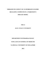
Fig. 1 Mitochondrial DNA D-loop mutations identified in the present study: Chromatogram of sequence analysis and consequent
secondary structure alterations are shown. (A) ETAS1 15483 A/G; (B) TAS-D 15507 T/C; (C) TAS-C 15529 T/C; (D) TAS-A 15572 A/G;
(E) CB 15779 G/A; and (F) MT-CSB3 16116 T/C.
Green Tea Extract and Mitochondrial DNA Damage
293
Effect of mutations on secondary structure of D-loop
To find out the impact of D-loop mutations on its secondary
structure conformation, in silico analysis was performed using
DNA mfold web server (Fig. 1). Results showed lesser free
energy for 15483G (ETAS1), 15572G (TAS-A) alleles and
higher free energy for 15507C (TAS-D), 16116C (MT-CSB3)
alleles when compared to their corresponding wild alleles
(Fig. 1). However, for 15529 T/C (TAS-C) and 15779 G/A
(CB) variants no considerable difference was observed in free
energy levels between wild and mutant alleles.
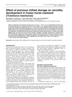
Fig. 2 Long-extension PCR analysis of mtDNA deletions in
hepatic tissue of experimental rats: M: DNA size marker; C:
Controls; ALC: Alcohol; and GTE: Green tea extract.
amplifies mtDNA fragment of 16,007 bp size while the 2nd primers set (nested PCR) amplifies a 15,708 bp fragment. The
quality of PCR amplification products was analyzed by agarose gel electrophoresis.
Mitochondrial DNA deletions
Whole mitochondrial genome from all the investigated groups
was analyzed by Long-extension PCR technique. Large scale
mtDNA deletions were observed only in alcoholic (ALC) rats
while intact wild type mtDNA was observed in rats of C, GTE
and ALC + GTE groups (Fig 2).
Activity of liver antioxidants
Activity of liver antioxidants
Liver tissue was homogenized (10% w/v) in ice cold 0.1 M Tris
buffer (pH 7.4), and supernatant was collected by centrifugation (10,000g for 20 min at 4 °C) and used to assess the activities of enzymatic and non-enzymatic antioxidants. Total
glutathione (GSH) content was measured by Ellman’s method
[19] and the activities of glutathione peroxidise (GPx) [20],
catalase [21] and superoxide dismutase (SOD) [22] were determined. Protein concentration was estimated by standard protocols [23].
Results
Mitochondrial DNA D-loop mutations
A total of 6 mutations were identified in the D-loop region of
investigated groups (Table 3; Fig. 1). All the identified mutations were transition substitutions of purines (Y) or pyrimidines (R). Among them, 4 were present in alcoholic rats (ALC)
while remaining 2 were present in all experimental groups viz.,
C, ALC, GTE and ALC + GTE groups. In overall, 4 mutations were present in the termination associated sequences
(TAS, ETAS), 1 was in the central block (CB) and 1 was
located in conserved sequence block 3 (MT-CSB3).
Table 4
Parameter
GSH
GPx
SOD
CAT
The data on the effects of green tea extract on liver antioxidants in alcohol administered rats are summarized in Table 4.
The activities of antioxidant enzymes viz., GPx, SOD, catalase
and the content of GSH were markedly decreased in alcohol
administered rats in comparison with the other experimental
groups. Treatment of green tea extract to alcohol administered
rats significantly (P < 0.05) restored these levels close to normal levels.
Discussion
Green tea has many bioactive components, chiefly catechins
viz., epigallocatechingallate (EGCG), epigallocatechin
(EGC), epicatechingallate (ECG), and epicatechin (EC) along
with other constituents such as caffeine, theobromine,
theophylline, organic acids, free amino acids, carbohydrates,
alkaloids and minerals [24]. The antioxidant activity of green
tea polyphenols was primarily attributed to catechins.
However, polyphenols are highly target specific with different
efficacies and bio-availabilities. Earlier studies have shown that
green tea catechins are effective scavengers of ROS including
superoxide anions [14]. Thus, by lowering the levels of ROS
and oxidative stress, green tea catechins may ameliorate
mtDNA damage, and at the same time, the possibility of
Effect of green tea extract on antioxidant enzymes and glutathione content of liver in alcohol administered rats.
C
ALC
a
6.2 ± 0.29
9.2 ± 0.24a
34 ± 3.8a
5.4 ± 0.19a
GTE
b
3.4 ± 0.16
5.6 ± 1.3b
23 ± 3.3b
3.6 ± 0.38b
ALC + GTE
a
6.7 ± 0.43
9.9 ± 0.4a
36 ± 2.1a
5.8 ± 0.21a
5.9 ± 0.33a
8.6 ± 0.7a
31 ± 4.5b
5.1 ± 0.15a
GSH is expressed as mg/mg protein and remaining values as mmole/min/mg protein. Values are mean ± SD of eight rats in each group. a,
b
Within a row, means not sharing a common superscript letter are significantly different at P < 0.05 (Tukey HSD method post hoc analysis for
all groups, P < 0.01). C: Control rats; ALC: Alcohol fed rats; GTE: Green tea extract fed rats; and ALC + GTE: Alcohol and green tea extract
fed rats.
294
involvement of several other mechanisms related to beneficiary
actions of catechins cannot be ruled out.
Mitochondrial DNA D-loop, the key regulating site of
mtDNA function, is highly vulnerable to oxidative damage
[25]. Thus, D-loop mutations might affect the overall mitochondrial function by altering mitochondrial replication, transcription and/or biogenesis. Numerous studies have reported
association between D-loop mutations and risk of developing
various complex diseases [26–28]. The present study reports
increased frequency of D-loop mutations in ALC group rats
(Table 3). Alcohol metabolism linked production of ROS
might be responsible for this enhancement. However, alcoholic
rats supplemented with green tea extract (ALC + GTE)
showed no D-loop mutations that were observed in ALC
group (Table 3). This could be due to the effective ROS scavenging nature of green tea catechins.
It is evident that DNA secondary structures can influence
the molecular mechanisms of replication, transcription and
recombination [29,30]. In general, hairpin or cruciform structures serve as binding sites for several transacting elements
[31,32]. Hence, local intra-strand DNA secondary structures
have a key role in replication and transcription processes. As
key regulatory site of mtDNA replication and transcription,
D-loop mutations can influence overall mtDNA stability.
Therefore, impact of identified mutations on D-loop secondary
structure was analyzed. Results showed higher free energy for
15507C (TAS-D) and 16116C (MT-CSB3) alleles compared to
their corresponding wild alleles (Fig. 1). Higher free energy
represents less stable secondary structures which may have
negative impact on overall mtDNA function. The 15507C
(TAS-D) and 16116C (MT-CSB3) variants observed in alcoholic rats were not present in alcoholic rats supplemented with
GTE, indicating ameliorative effect of green tea. However, further studies are warranted to clarify the underlying molecular
mechanisms involved in these findings.
DNA mfold analysis predicted lesser free energy for 15483G
(ETAS1) and 15572G (TAS-A) alleles when compared to their
corresponding wild alleles (Fig. 1). Lesser free energy represents more stable secondary structures. Interestingly, both of
these variants were present in all groups of rats; hence, they
can be considered as single nucleotide polymorphisms rather
than mutations. The remaining 2 variants observed in alcoholic rats [15529 T/C (TAS-C) and 15779 G/A (CB)] showed
no much difference in free energy levels (Fig. 1) and were
restored by GTE treatment.
Oxidative stress can lead to the accumulation of mtDNA
deletions [33,34]. Large scale deletions of mitochondrial genome have been reported in several complex diseases including
diabetes [26,27]. Altered mtDNA replication and/or repair
system could lead deletions in mtDNA [35,36]. The present
study identified mtDNA deletions in alcoholic rats while
ALC + GTE group rats showed no detectable mtDNA
deletions (Fig. 2). This could be attributed to elevated oxidative stress by alcohol induced ROS in ALC group and ameliorative effect of green tea catechins on mtDNA damage by ROS
scavenging nature in ALC + GTE group. Although this is an
interesting finding, further studies are warranted to clarify the
underlying molecular mechanisms.
SOD, CAT and GPx are the major antioxidant enzymes
that stand in the first-line of defense against oxidative damage
[37]. These antioxidants play a key role in scavenging ROS,
H. Reddyvari et al.
reduction in hydrogen peroxide and maintaining redox balances in biological system. GSH, an important nonenzymatic antioxidant biomolecule in tissues, is the substrate
for GPx and GST. It plays a central role in the maintenance
of membrane protein thiols and elimination of free oxygen species, such superoxide anions, alkoxy radicals including H2O2
[38]. The present study showed diminished activities of SOD,
CAT and GPx and reduced GSH content in alcohol administered rats (Table 4). The lowered GSH content might be
responsible for the reduced GPx activity. Decreased catalase
activity accounts for less hydrogen peroxide decomposition,
consequently the possible overproduction of hydroxyl radicals
via fenton reaction. Decreased GSH content and lowered
activity of catalase, SOD and GPx favor the environment for
oxidative stress, which leads to mtDNA damage. Amelioration
of mtDNA damage and restoration of antioxidant status in
terms of GSH content and activities of defense enzymes to normal level in alcoholic rats receiving GTE supplementation are
evident from the results of the study. This finding confirms the
reports of Lodhi et al. [39] and others who reported such GTE
induced restorative effect in antioxidant status in alcohol
receiving rats.
Conclusions
The present study reports therapeutic effect of green tea
extract against alcohol induced hepatic mitochondrial DNA
damage in rats. To the best of our knowledge, this is the first
report demonstrating the ameliorative effect of green tea
extract on alcohol mediated mtDNA damage. However, further investigation is warranted to explore the molecular mechanisms involved in the reported findings.
Conflict of interest
The authors have declared no conflict of interest.
Acknowledgments
Dr. Suresh Govatati acknowledges the financial support from
the University Grants Commission, New Delhi, under its Dr.
D.S. Kothari postdoctoral scheme [No. F.4-2/2006 (BSR)/131014/2013 (BSR)].
References
[1] Wang Z, Su B, Fan S, Fei H, Zhao W. Protective effect of
oligomeric proanthocyanidins against alcohol-induced liver
steatosis and injury in mice. Biochem Biophys Res Commun
2015;458:757–62.
[2] Adjemian MK, Volpe RJ, Adjemian J. relationships between
diet, alcohol preference, and heart disease and type 2 diabetes
among Americans. Plos One 2015;11:e0124351.
[3] Louvet A, Mathurin P. Alcoholic liver disease: mechanisms of
injury and targeted treatment. Nat Rev Gastroenterol Hepatol
2015;4:231–42.
[4] Pyun CW, Mandal PK, Hong GE, Lee CH. Effect of chronic
alcohol consumption on phosphatidylcholine hydroperoxide
content of rat liver and brain. Trop J Pharm Res
2015;7:1225–30.
Green Tea Extract and Mitochondrial DNA Damage
[5] Chuang SC, Lee YC, Wu GJ, Straif K, Hashibe M. Alcohol
consumption and liver cancer risk: a meta-analysis. Cancer
Causes Control 2015;26:1205–31.
[6] Yin F, Cadenas E. Mitochondria: the cellular hub of the
dynamic coordinated network. Antioxid Redox Signal
2015;12:961–4.
[7] Song BJ, Akbar M, Abdelmegeed MA, Byun K, Lee B, Yoon
SK, et al. Mitochondrial dysfunction and tissue injury by
alcohol, high fat, nonalcoholic substances and pathological
conditions through post-translational protein modifications.
Redox Biol 2014;3:109–23.
[8] Zelickson BR, Benavides GA, Johnson MS, Chacko BK,
Venkatraman A, Landar A, et al. Nitric oxide and hypoxia
exacerbate alcohol-induced mitochondrial dysfunction in
hepatocytes. Biochim Biophys Acta 2011;12:1573–82.
[9] Han D, Ybanez MD, Johnson HS, McDonald JN, Mesropyan
L, Sancheti H, et al. Dynamic adaptation of liver mitochondria
to chronic alcohol feeding in mice biogenesis, remodeling, and
functional alterations. J Biol Chem 2012;50:42165–79.
[10] Anderson S, Bankier AT, Barrell BG, De Bruijn MH, Coulson
AR, Drouin J, et al. Sequence and organization of the human
mitochondrial genome. Nature 1981;290:457–65.
[11] Nassir F, Ibdah JA. Role of mitochondria in alcoholic liver
disease. World J Gastroenterol 2014;9:2136–42.
[12] Kim HS, Quon MJ, Kim JA. New insights into the mechanisms
of polyphenols beyond antioxidant properties; lessons from the
green tea polyphenol, epigallocatechin 3-gallate. Redox Biol
2014;2:187–95.
[13] Guo Q, Zhao B, Shen S, Hou J, Hu J, Xin W. ESR study on the
structure–antioxidant activity relationship of tea catechins and
their epimers. Biochim Biophys Acta 1999;1427:13–23.
[14] Velayutham P, Babu A, Liu D. Green tea catechins and
cardiovascular health: an update. Curr Med Chem
2008;15:1840–50.
[15] Govatati S, Singamsetty GK, Nallabelli N, Malempati S, Rao
PS, Madamchetty VKK, et al. Contribution of cyclin D1
(CCND1) and E-cadherin (CDH1) alterations to colorectal
cancer susceptibility: a case–control study. Tumor Biol
2014;35:12059–67.
[16] Govatati S, Challa K, Reddy SB, Pramod K, Deenadayal M,
Chakravarty B, et al. BRCA1 alterations are associated with
endometriosis, but BRCA2 alterations show no detectable
endometriosis risk: a study in Indian population. J Assist
Reprod Genet 2015;2:277–85.
[17] Govatati S, Tipirisetti NR, Perugu S, Kodati VL, Deenadayal
M, Satti V, et al. Mitochondrial genome variations in advanced
stage endometriosis: a study in South Indian population. Plos
One 2012;7:e40668.
[18] Govatati S, Malempati S, Saradamma B, Divyamaanasa D,
Naidu BP, Bramhachari PV, et al. Manganese-superoxide
dismutase (Mn-SOD) overexpression is a common event in
colorectal cancers with mitochondrial microsatellite instability.
Tumor Biol 2016;37:10357–64.
[19] Ellman’s. Tissue sulfhydryl groups. Arch Biochem Biophys
1959;82:70–7.
[20] Rotruck JT, Pope AL, Ganther HE, Swanson AB, Hafeman
DG, Hoekstra WG. Selenium: biochemical role as a component
of glutathione peroxidase. Science 1973;179:588–90.
[21] Aebi H. Catalase in vitro. Meth Enzymol 1984;105:21–6.
[22] Marklund S, Marklund G. Involvement of the superoxide
anion radical in the autoxidation of pyrogallol and a
295
[23]
[24]
[25]
[26]
[27]
[28]
[29]
[30]
[31]
[32]
[33]
[34]
[35]
[36]
[37]
[38]
[39]
convenient assay for superoxide dismutase. Eur J Biochem
1974;47:469–74.
Lowry OH, Nira JR, Farr L, Rose JR. Protein measurement
with the Follin phenol reagent. J Biol Chem 1951;193:265–75.
Chaturvedula VS, Prakash I. The aroma, taste, color and
bioactive constituents of tea. J Med Plants Res 2011;5:2110–24.
Tan X, Lei Z, Jiang Y, Yang Y, Zhang W, Li Y, et al. Post
conditioning ameliorates mitochondrial DNA damage and
deletion after renal ischemic injury. Nephrol Dial Transplant
2013;28:2754–65.
Tipirisetti NR, Govatati S, Pullari P, Malempati S, Thupurani
MK, Perugu S, et al. Mitochondrial control region alterations
and breast cancer risk: a study in South Indian population. Plos
One 2014;1:e85363.
Govatati S, Saradamma B, Malempati S, Dasi D, Thupurani
MK, Nagesh N, et al. Association of mitochondrial
displacement loop polymorphisms with risk of colorectal
cancer in south Indian population. Mitochond DNA A DNA
Mapp
Seq
Anal
2016.
/>24701394.2016.1160076.
Govatati S, Deenadayal M, Shivaji S, Bhanoori M.
Mitochondrial displacement loop alterations are associated
with endometriosis. Fertil Steril 2013;7:1980–6.
Seffens W, Digby D. MRNAs have greater negative folding free
energies than shuffled or codon choice randomized sequences.
Nucl Acids Res 1999;27:1578–84.
Katz L, Burge CB. Widespread selection for local RNA
secondary structure in coding regions of bacterial genes.
Genome Res 2003;13:2042–51.
Wright BE, Reschke DK, Schmidt KH, Reimers JM, Knight W.
Predicting mutation frequencies in stem-loop structures of
derepressed genes: implications for evolution. Mol Microbiol
2003;48:429–41.
Hoede C, Denamur E, Tenaillon O. Selection acts on DNA
secondary structures to decrease transcriptional mutagenesis.
Plos Genet 2006;2:e176.
Ikushima T, Andoh T, Kaikawa T, Hashiguchi K. Induction of
a large deletion in mitochondrial genome of mouse cells induced
by X-ray irradiation. Int Congr 2002;1236:331–4.
Murphy J, Nugent S, Seymoura C, Mothersill C. Mitochondrial
DNA point mutations and a novel deletion induced by direct
low-LET radiation and by medium from irradiated cells. Mutat
Res 2005;12:127–36.
Krishnan KJ, Reeve AK, Samuels DC, Chinnery PF,
Blackwood JK, Taylor RW, et al. What causes mitochondrial
DNA deletions in human cells? Nat Genet 2008;3:275–9.
Sadikovic B, Wang J, El-Hattab A, Landsverk M, Douglas G,
Brundage EK, et al. Sequence homology at the breakpoint and
clinical phenotype of mitochondrial DNA deletion syndromes.
Plos One 2010;12:e15687.
Maturu P, Reddy VD, Padmavathi P, Varadacharyulu N.
Ethanol induced adaptive changes in blood for the pathological
and toxicological effects of chronic ethanol consumption in
humans. Exp Toxicol Pathol 2012;64:697–703.
Padmavathi P, Reddy VD, Varadacharyulu N. Influence of
chronic cigarette smoking on serum biochemical profile in male
human volunteers. J Health Sci 2009;55:265–70.
Lodhi P, Tandan N, Singh N, Kumar D, Kumar M. Camellia
sinensis (L.) Kuntze Extract Ameliorates Chronic EthanolInduced Hepatotoxicity in Albino Rats. Evid Based
Complement Alternat Med 2014, ID:787153.