Báo cáo y học: "The effect of open lung ventilation on right ventricular and left ventricular function in lung-lavaged pigs" pps
Bạn đang xem bản rút gọn của tài liệu. Xem và tải ngay bản đầy đủ của tài liệu tại đây (546.35 KB, 9 trang )
Open Access
Available online />Page 1 of 9
(page number not for citation purposes)
Vol 10 No 3
Research
The effect of open lung ventilation on right ventricular and left
ventricular function in lung-lavaged pigs
Dinis Reis Miranda
1
, Lennart Klompe
2
, Filippo Cademartiri
3
, Jack J Haitsma
1
,
Alessandro Palumbo
3
, Johanna JM Takkenberg
2
, Burkhard Lachmann
1
, Ad JJC Bogers
2
and
Diederik Gommers
1
1
Department of Anesthesiology, Erasmus MC, Rotterdam, The Netherlands
2
Department of Cardio-Thoracic Surgery, Erasmus MC, Rotterdam, The Netherlands
3
Department of Radiology, Erasmus MC, Rotterdam, The Netherlands
Corresponding author: Dinis Reis Miranda,
Received: 1 Jan 2006 Revisions requested: 15 Feb 2006 Revisions received: 18 Apr 2006 Accepted: 11 May 2006 Published: 8 Jun 2006
Critical Care 2006, 10:R86 (doi:10.1186/cc4944)
This article is online at: />© 2006 Miranda et al.; licensee BioMed Central Ltd.
This is an open access article distributed under the terms of the Creative Commons Attribution License ( />),
which permits unrestricted use, distribution, and reproduction in any medium, provided the original work is properly cited.
Abstract
Introduction Ventilation according to the open lung concept
(OLC) consists of recruitment maneuvers, followed by low tidal
volume and high positive end-expiratory pressure, aiming at
minimizing atelectasis. The minimization of atelectasis reduces
the right ventricular (RV) afterload, but the increased
intrathoracic pressures used by OLC ventilation could increase
the RV afterload. We hypothesize that when atelectasis is
minimized by OLC ventilation, cardiac function is not affected
despite the higher mean airway pressure.
Methods After repeated lung lavage, each pig (n = 10) was
conventionally ventilated and was ventilated according to OLC
in a randomized cross-over setting. Conventional mechanical
ventilation (CMV) consisted of volume-controlled ventilation with
5 cmH
2
O positive end-expiratory pressure and a tidal volume of
8–10 ml/kg. No recruitment maneuvers were performed. During
OLC ventilation, recruitment maneuvers were applied until
PaO
2
/FiO
2
> 60 kPa. The peak inspiratory pressure was set to
obtain a tidal volume of 6–8 ml/kg. The cardiac output (CO), the
RV preload, the contractility and the afterload were measured
with a volumetric pulmonary artery catheter. A high-resolution
computed tomography scan measured the whole lung density
and left ventricular (LV) volumes.
Results The RV end-systolic pressure–volume relationship,
representing RV afterload, during steady-state OLC ventilation
(2.7 ± 1.2 mmHg/ml) was not significantly different compared
with CMV (3.6 ± 2.5 mmHg/ml). Pulmonary vascular resistance
(OLC, 137 ± 49 dynes/s/cm
5
versus CMV, 130 ± 34 dynes/s/
cm
5
) was comparable between groups. OLC led to a
significantly lower amount of atelectasis (13 ± 2% of the lung
area) compared with CMV (52 ± 3% of the lung area).
Atelectasis was not correlated with pulmonary vascular
resistance or end-systolic pressure–volume relationship.
The LV contractility and afterload during OLC was not
significantly different compared with CMV. Compared with
baseline, the LV end-diastolic volume (66 ± 4 ml) decreased
significantly during OLC (56 ± 5 ml) ventilation and not during
CMV (61 ± 3 ml). Also, CO was significantly lower during OLC
ventilation (OLC, 4.1 ± 0.3 l/minute versus CMV, 4.9 ± 0.3 l/
minute).
Conclusion In this experimental study, OLC resulted in
significantly improved lung aeration. Despite the use of elevated
airway pressures, no evidence was found for a negative effect of
OLC on RV afterload or LV afterload, which might be associated
with a loss of hypoxic pulmonary vasoconstriction due to alveolar
recruitment. The reductions in the CO and in the mean
pulmonary artery pressure were consequences of a reduced
preload.
CMV = conventional mechanical ventilation; CO = cardiac output; CT = computed tomography; ECG = electrocardiogram; ESPVR = end-systolic
pressure–volume relationship; FiO
2
= Inspired oxygen fraction;HU = Houndsfield units; LV = left ventricular; OLC = open lung ventilation; PaO
2
=
partial arterial oxygen pressure; PCWP = pulmonary capillary wedge pressure; PEEP = positive end-expiratory pressure; PVR = pulmonary vascular
resistance; REDV = right ventricular end-diastolic volume; RV = right ventricular.
Critical Care Vol 10 No 3 Miranda et al.
Page 2 of 9
(page number not for citation purposes)
Introduction
The open lung concept (OLC) is a ventilation strategy
intended to avoid atelectasis causing shear forces during
repeated opening and closing of atelectatic lung areas [1,2].
This is achieved with a recruitment maneuver and an applica-
tion of sufficient positive end-expiratory pressure (PEEP) to
counterbalance retractive forces. This strategy increases
intrathoracic pressure, however, which could increase the
right ventricular (RV) afterload [3-7] and could reduce safety.
Many studies (without recruiting the lung) show that elevated
airway pressures increase the RV afterload in patients with
respiratory failure [3,6-8]. One reason for this increase in RV
afterload is alveolar overdistention of aerated lung areas in the
presence of atelectasis; another reason is the occurrence of
hypoxic vasoconstriction in atelectatic lung areas, as shown in
experimental studies by Duggan and colleagues [9] and
Cramer and colleagues [10]. We have shown that avoiding
atelectasis by application of OLC ventilation did not lead to an
increased RV afterload in cardiac surgery patients, despite the
use of increased airway pressures [11,12]. Data on RV after-
load in these latter studies were obtained by means of a pul-
monary artery catheter or use of echocardiography. These
methods are often used for measuring RV afterload, but they
have not yet been validated. In addition, in these latter studies
we were not able to assess atelectasis and therefore could not
demonstrate a relationship between RV afterload and
atelectasis.
We therefore designed an experimental study, investigating
RV afterload during OLC ventilation compared with a low air-
way pressure ventilation strategy allowing atelectasis. RV
afterload is assessed by the load-independent [13] afterload
marker end-systolic pressure–volume relationship (ESPVR)
[14-16]. The amount of atelectasis was assessed with a multi-
slice whole lung computed tomography (CT) scan. As the
influence of OLC during steady-state ventilation on left ven-
tricular (LV) afterload is unknown, LV volumes were also meas-
ured during the whole cardiac cycle using this multi-slice CT
scan.
We hypothesized that when atelectasis is minimized by OLC
ventilation, the RV afterload and LV afterload are not affected
despite the use of higher mean airway pressures in an experi-
mental lung injury model.
Methods
The study was approved by the institutional animal investiga-
tion committee, and the care and handling of the animals were
in accordance with the European Community guidelines. In 10
pigs weighing 32 ± 1.3 kg, anesthesia was induced with ket-
amine hydrochloride (35 mg/kg, intramuscularly) and mida-
zolam (0.5 mg/kg, intramuscularly). The animals were
tracheotomized, connected to a Servo ventilator 300 (Sie-
mens-Elema, Solna, Sweden) and were ventilated in a volume-
controlled mode, with pure oxygen, at a rate of 20 breaths/
minute, a tidal volume of 8 ml/kg, a PEEP of 5 cmH
2
O and an
inspiratory/expiratory ratio of 1:2. Neuromuscular block was
induced with pancuronium bromide (0.5 mg/kg intravenously),
and anesthesia was maintained with a continuous infusion of
fentanyl (20 μg/kg/hour), midazolam (0.3 mg/kg/hour) and
pancuronium bromide (0.3 mg/kg/hour).
After induction, an indwelling ParaTrend 7+ blood gas ana-
lyzer probe (Philips, Boblingen, Germany) was inserted in the
carotid artery for continuous blood gas analyses. An 8-Ch
Foley catheter was inserted into the femoral vein. A correct
position in the inferior caval vein was assured by CT scan of
the abdomen. To reduce the cardiac preload, the Foley balloon
was inflated with 5 ml water. One CCO 774HF75 series pul-
monary artery catheter (Edwards, Irvine, CA, USA) was
inserted through the right internal jugular vein with the tip in the
pulmonary artery (measuring pulmonary artery pressures), and
another catheter was also inserted through the jugular vein
with the tip in the right ventricle (measuring RV pressures).
Hemodynamic measurements consisted of the right atrial
pressure, the right ventricular pressure, the pulmonary arterial
pressure, and the pulmonary capillary wedge pressure
(PCWP). The cardiac output (CO), the RV end-diastolic vol-
ume (REDV) and the RV ejection fraction were calculated
using a Vigilence cardiac output computer (Edwards), con-
nected with the pulmonary artery catheter. From these values,
the pulmonary vascular resistance (PVR = (mean pulmonary
artery pressure - PCWP)/CO × 79.9) and the RV end-systolic
volume were calculated.
The ESPVR was considered in each animal. During each ven-
tilation strategy, the ESPVR was measured by calculating the
slope of the end-systolic pressure and volume obtained with
and without inflation of the balloon on the Foley catheter in the
inferior caval vein. RV stroke work was calculated by the fol-
lowing equation: 0.0136 × (mean pulmonary artery pressure -
right atrial pressure) × stroke volume [14]. The preload recruit-
able stroke work was considered in each animal during each
ventilation strategy as the slope of RV stroke work and REDV
obtained with and without inflation of the balloon on the Foley
catheter in the inferior caval vein. Systemic vascular resistance
was calculated as: (mean arterial pressure – right atrial pres-
sure)/CO × 79.9.
After instrumentation, respiratory failure was induced by
repeated saline lavage (50 ml/kg; 37°C) as described by
Lachmann and colleagues [17]. Lavages were repeated at 3-
minute intervals until the PaO
2
was below 13 kPa.
To minimize the effect of confounding variables, conventional
ventilation and OLC ventilation were applied in a cross-over
design. The order of the applied ventilation strategies was ran-
domized by sealed envelopes. Ten minutes after the last lung
Available online />Page 3 of 9
(page number not for citation purposes)
lavage, the first ventilation strategy was started. Before each
ventilation strategy, the ventilation was disconnected for 15
seconds, which has been shown to result in an immediate lung
collapse [18] and was substantiated by the CT measure-
ments. Conventional mechanical ventilation (CMV) was
started with volume control ventilation at the following set-
tings: tidal volume, 8–10 ml/kg; PEEP, 5 cmH
2
O; inspiratory/
expiratory ratio, 1:2; FiO
2
, 1.0; and respiratory rate adjusted to
achieve PaCO
2
, 4.5–5.5 kPa.
Ventilation according to the OLC was started by switching the
ventilator to a pressure-controlled mode with a respiratory fre-
quency of 40/minute. The FiO
2
was set at 1.0, the PEEP was
10 cmH
2
O, the inspiratory/expiratory ratio was 1:1, and a driv-
ing pressure suitable to obtain a tidal volume of 6–8 ml/kg aim-
ing at a PaCO
2
of 4.5 and 5.5 kPa was used. A lung
recruitment maneuver was performed by increasing the peak
inspiratory pressure to 40 cmH
2
O during 10 seconds in order
to increase the PaO
2
/FiO
2
ratio to a value greater than 60 kPa.
If this value was not reached, a recruitment maneuver was
repeated by adding 5 cmH
2
O to the previous peak inspiratory
airway pressure, up to a maximum peak inspiratory airway
pressure of 60 cmH
2
O. If the PaO
2
/FiO
2
ratio decreased
slowly below 60 kPa after recruitment, indicating renewed
lung collapse, the PEEP was increased with 2 cmH
2
O and the
recruitment maneuver (again beginning at 40 cmH
2
O) was
repeated. If the PaO
2
/FiO
2
ratio decreased below 60 kPa dur-
ing the study period, the PEEP was not increased but a new
recruitment maneuver was performed.
All measurements were performed once before lung lavage (at
baseline) and twice after lung lavage during each ventilation
strategy. Following lung lavage, one CT scan of the thorax was
made to confirm lung collapse. During both ventilation strate-
gies, measurements were performed once without balloon
inflation of the Foley catheter in the inferior caval vein and once
with inflation (5 ml saline) of the balloon of the Foley catheter.
Fluid management during the study was based on the REDV
provided by the pulmonary artery catheter. The REDV before
lung lavage was considered the optimal REDV. After lung lav-
age (and a REDV below the optimal value), the REDV was
treated with starch colloids (Voluven
®
, Fresenius Kabi, Bad
Homburg, Germany). A decrease of REDV during inflation of
the Foley balloon was not treated.
The CT-scan protocol was performed using a state-of-the-art
64-slice Sensation 64 CT scanner (Siemens Medical Solu-
tions, Forchheim, Germany) with a 0.4 mm voxel size and a
330 ms gantry rotation time. Each scan was performed twice:
first with a standard protocol for thoracic imaging (standard
scan), and then with dedicated software able to synchronize
the reconstructed image with the cardiac phase (electrocardi-
ogram (ECG) gated scan) [19]. The scan parameters were as
follows: number of slices, 64/rotation; individual detector
width, 0.6 mm; effective spatial resolution, 0.4 mm
3
; 120 kV,
120 mA/s (900 mA/s for the ECG gated scan); feed:rotation,
58 mm/pitch:1 (11.52 mm/pitch:0.2 for the ECG gated scan);
effective reconstructed slice thickness, 0.6 mm; and recon-
struction increment, 0.4 mm. The standard scan was recon-
structed as a volumetric dataset, and a slice was selected
Table 1
Hemodynamic data at baseline and during conventional ventilation and open lung ventilation
Baseline Conventional mechanical ventilation Open lung ventilation
Heart rate (beats/min) 105 ± 5 86 ± 5
**
94 ± 5
Mean arterial pressure (mmHg) 93 ± 4 104 ± 4
**
80 ± 4
*,**
Right atrial pressure (mmHg) 4.2 ± 1 5.9 ± 1
**
8.1 ± 2
**
Cardiac output (l/min) 5.3 ± 0.3 4.9 ± 0.3 4.1 ± 0.3
*,**
Right ventricular end-diastolic volume (ml) 165 ± 11 173 ± 13 148 ± 13
Right ventricular end-systolic volume (ml) 112 ± 10 119 ± 11 103 ± 11
Systolic pulmonary pressure (mmHg) 30 ± 3 31 ± 3 28 ± 2
Mean pulmonary arterial pressure (mmHg) 17 ± 3 20 ± 2 17 ± 2
*
Pulmonary capillary wedge pressure (mmHg) 9.3 ± 2 12.1 ± 3 12.5 ± 2
**
Right ventricular ejection fraction (%) 33.1 ± 1.7 33.1 ± 1.7 31.1 ± 1.9
Pulmonary vascular resistance (dynes/s/cm
5
) 126 ± 38 130 ± 34 137 ± 49
Left ventricular end-diastolic volume (ml) 66 ± 4 61 ± 3 56 ± 5
**
Systemic vascular resistance (dynes sec cm
5
) 1379 ± 120 1693 ± 139
**
1508 ± 124
Left ventricular ejection fraction (ml) 49.5 ± 1.6 53.2 ± 2.1 43.2 ± 5.6
*
P < 0.05 open lung ventilation versus conventional mechanical ventilation,
**
P < 0.05 versus baseline.
Critical Care Vol 10 No 3 Miranda et al.
Page 4 of 9
(page number not for citation purposes)
every 20 mm starting at the apex of the thorax for the analysis.
For the assessment of the left ventricle a short-axis multiphasic
reconstruction was performed, dividing the cardiac cycle
(using as two R waves reference points) into 10 phases and
the left ventricle into eight levels [20]. The standard thoracic
scan was used to analyze the lung parenchyma by means of
dedicated PulmoCT software (Siemens Medical Solutions).
The ECG gated scan and the left ventricle were analyzed with
a dedicated ARGUS software platform (Siemens Medical
Solutions).
CT data analysis was performed in all cases by an experienced
radiologist. For the lung parenchyma evaluation we used three
main ranges of attenuation, measured in Houndsfield Units
(HU), representing the usual location of tissues in the HU
spectrum: -1000 HU to -600 HU as good aerated lung tissue
(voxels with a prevalent content of air); -600 HU to -200 HU
as poorly aerated lung tissue (mostly voxels with air and with
some soft tissues or fluid); and -200 HU to +200 HU as non-
aerated lung tissue (mostly voxels with a mixture of fat, fluid
and soft tissues).
The operator segmented in a semi-automatic mode the lung
parenchyma of the right lung slice by slice (usually between 10
and 14 slices depending on the phase of the experiment and
on the size of the animal's lung). The results were expressed
as percentages of each subrange of attenuation as compared
with the total lung area.
For evaluation of the left ventricle, the endocardial contours
were semi-automatically detected by the operator on the
images reconstructed on the short axis. The eight levels
throughout the left ventricle allowed a volumetric interpolation
of the whole left myocardium, allowing calculation of the end-
systolic volume and of the end-diastolic volume.
Statistics
Between-group differences for hemodynamic parameters
were tested with a paired, two-sided Student t test. Results
are presented as the mean ± standard error of the mean. A
relationship between the end-systolic pressure and volume
was calculated for each pig and these regression coefficients
were then averaged. The relationship between RV afterload
and lung aeration was calculated by the Pearson's correlation
coefficient.
Results
Hemodynamic data are presented in Table 1. In summary, the
mean pulmonary artery pressure, the CO, and the mean
arterial pressure were higher during CMV compared with OLC
ventilation.
As indicators of RV afterload, the regression coefficients
between systolic pulmonary pressure and RV end-systolic vol-
ume were comparable between the two ventilation strategies
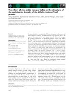
(Table 1). Within the applied fluid management the dynamic
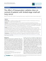
pressure–flow diagram (Figure 1) showed a significantly lower
CO during OLC (Table 1), but the pressure drop through the
pulmonary circulation (pulmonary artery mean pressure -
PCWP pressure) was not significantly higher during OLC ven-
tilation (OLC, 6.0 ± 2.3 mmHg versus CMV, 7.4 ± 2.5 mmHg).
The PVR was comparable between the two groups (Table 1).
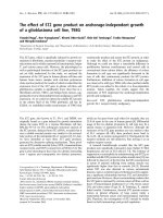
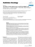
Contractility in the right ventricle during OLC was not signifi-
cantly different compared with during CMV. The regression
coefficient of the ESPVR was comparable between the
groups (OLC, 2.7 ± 1.2 mmHg/ml versus CMV, 3.6 ± 2.5; Fig-
ure 2). The regression coefficient of the preload recruitable
stroke work was also no different between groups (OLC, 0.07
± 0.07 g.m/beat.m
2
.ml versus CMV, 0.24 ± 0.16 g.m/
beat.m
2
.ml; P = 0.36). The RV ejection fraction was also no
different between the two groups (Table 1).
Contractility in the left ventricle during OLC was not signifi-
cantly different compared with during CMV. The regression
coefficient of the ESPVR was comparable between the
groups (OLC, 43 ± 26 mmHg/ml versus CMV, 61 ± 30
mmHg/ml). The LV ejection fraction was also no different
between the two groups (Table 1). The systemic vascular
resistance, reflecting the LV afterload, tended to be lower dur-
ing OLC ventilation compared with during CMV (P = 0.056)
(Table 1).
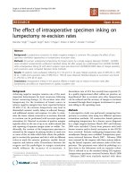
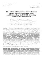
Considering the aeration of the lungs (Figure 3), 13 ± 2% of
the lung was atelectatic during OLC whereas significantly
more lung tissue was atelectatic in the CMV group (52 ± 3%,
with a HU density between -200 and +200) (Table 2). The
Figure 1
Dynamic pressure–flow plotDynamic pressure-flow plot. The effect of open lung ventilation (OLC)
on flow and pressure drop through the pulmonary circulation is dis-
played compared with conventional mechanical ventilation (CMV). On
the vertical axis, change of pressure drop through the pulmonary circu-
lation is displayed: mean pulmonary artery pressure (PAmean)–pulmo-
nary capillary wedge pressure during OLC – PAmean–pulmonary
capillary wedge pressure during CMV. On the horizontal axis, the
change of cardiac output (CO) is displayed.
Available online />Page 5 of 9
(page number not for citation purposes)
amount of poorly aerated lung tissue (HU density -600 to -
200) was also significantly higher in the OLC group compared
with the CMV group (Table 2). The amount of good aerated
lung tissue (HU -1000 to -600) was also higher in the OLC
group compared with the CMV group (Table 2). OLC ventila-
tion could not, however, restore the area of good aerated lung
tissue to baseline values (Table 2).
There was no significant correlation between the PVR, the CO
and the pressure drop through the pulmonary circulation with
the amount of lung aeration (Table 3).
Discussion
In this experimental study, the amount of atelectatic lung area
was not correlated with the parameters of RV afterload. OLC
ventilation significantly increased the PaO
2
/FiO
2
ratio and sig-
nificantly reduced atelectasis compared with CMV. Indicators
of RV afterload or contractility were not affected by the chosen
ventilation strategy. Indicators of LV afterload and contractility
were also no different between the different ventilation
strategies.
This study showed that ventilation according to the OLC effec-
tively reduced atelectasis. These findings are in agreement
with results of Tusman and colleagues [21] and Amato and
colleagues [22], who found that atelectasis is greatly reduced
during OLC ventilation in children and in patients suffering
from acute respiratory distress syndrome patients. The
present study, however, also shows that there is still a small
portion of nonaerated lung tissue during OLC ventilation. This
is probably explained by the impossibility to exclude all (small)
lung vasculature from lung density measurements. This falsely
increases the amount of nonaerated lung tissue since lung
Table 2
Ventilatory measurements at baseline and during conventional ventilation and open lung ventilation
Baseline Conventional mechanical ventilation Open lung ventilation
Intrinsic + extrinsic positive end-expiratory pressure (cmH
2
O) 5 ± 0.4 6 ± 0.3 14 ± 0.6
*,**
Peak inspiratory airway pressure (cmH
2
O) 20 ± 0.5 28 ± 1
**
26 ± 0.4
**
Tidal volume (ml) 271 ± 5 270 ± 6 240 ± 11
*,**
PaO
2
/FiO
2
(kPa) 60 ± 5 13 ± 2 72 ± 2
*
-1000 HU to -600 HU (%) 51 ± 3 10 ± 2
**
29 ± 3
*,**
-600 HU to -200 HU (%) 29 ± 2 36 ± 2 57 ± 3
*,**
-200 HU to +200 HU (%) 20 ± 2 52 ± 3
**
13 ± 2
*
HU, Houndsfield units, expressed as the percentage of the lung area.
*
P < 0.05 versus conventional mechanical ventilation,
**
P < 0.05 versus
baseline.
Table 3
Correlation between lung aeration and indicators of right ventricular afterload
Correlation coefficient Good aeration (-1000 HU to -600 HU) Poor aeration (-600 HU to -200 HU) Nonaeration (-200 HU to +200 HU)
Pulmonary vascular resistance 0.7 -0.1 -0.7
Cardiac output -0.2 -0.2 -0.2
Mean pulmonary artery pressure–
pulmonary capillary wedge pressure
a
0.2 -0.1 -0.7
HU, Houndsfield units, expressed as the percentage of the lung area. None of the correlations was significant.
a
Pressure drop through the
pulmonary circulation.
Figure 2
End-systolic pressure–volume relationshipEnd-systolic pressure–volume relationship. The right ventricular
end-systolic pressure (RV) versus the right ventricular end-systolic vol-
ume. The end-systolic pressure, and the volume with and without bal-
loon inflation, is connected with a straight line for conventional
mechanical ventilation and with the interrupted line for open lung
ventilation.
Critical Care Vol 10 No 3 Miranda et al.
Page 6 of 9
(page number not for citation purposes)
vasculature has the same density as nonaerated lung tissue.
This effect could be pronounced with this very high-resolution
CT technique, also measuring very small pulmonary vessels.
We therefore think that the amount of nonaerated lung tissue
is negligible when considering the effect of OLC ventilation on
RV contractility and RV afterload.
It is unlikely that OLC ventilation caused alveolar overdisten-
tion. The lung was less aerated during OLC compared with
baseline. In some studies [23-25] overdistention (or emphy-
sema) is characterized by the HU density ranging from -1000
HU to -900 HU. The limit between air and tissue in the lungs
is arbitrary, however, because the spatial resolution dramati-
cally affects the capability of the scanner to distinguish a voxel
Figure 3
Computed tomography scan examples of basal lung areasduring expirationComputed tomography scan examples of basal lung areasduring expiration. Upper two scans, during baseline, before lung lavage. Middle two
scans, conventional mechanical ventilation after lung lavage. Lower two scans, during open lung ventilation after lung lavage. Good aerated lung
areas (-1000 Houndsfield units (HU) to -600 HU) are coded red in the right-hand scans, poorly aerated areas (-600 HU to -200 HU) are coded
green, and non-aerated lung areas (-200 HU to +200 HU) are coded blue.
Available online />Page 7 of 9
(page number not for citation purposes)
with air from a fluid/solid voxel on axial slices. Even with very
high spatial resolution, as is the case in the present study (0.4
mm
3
is the highest available resolution for volumetric CT scan-
ning), the distal part of the airways are too thin for this imaging
modality. The borders of aerated lungs have recently been
described as lower than -500 HU [25], while the limit for soft
tissues is higher than -380 HU [26]. We therefore decided to
have three homogeneous ranges of 400 HU, each starting at
-1000 HU and ending at +200 HU.
In the present study, ESPVR, indicating RV afterload, was not
correlated with atelectasis. This relationship was described by
Duggan and colleagues [9] and Creamer and colleagues [10],
who showed experimentally that atelectasis causes a signifi-
cant increase in RV afterload. This effect of atelectasis on RV
afterload during mechanical ventilation could be explained by
two mechanisms: overdistention in aerated lung areas [27,28],
and local hypoxic pulmonary vasoconstriction in nonaerated
lung areas [29]. In the present study, we found no correlation
between atelectasis and indicators of RV afterload. The effect
of avoiding atelectasis (and thereby reducing hypoxic pulmo-
nary vasoconstriction) by means of OLC ventilation on RV
afterload is probably counterbalanced by the effect of a high
intrathoracic pressure.
The RV afterload was not increased by the application of OLC
ventilation. The mean arterial pulmonary pressure was even
significantly decreased during OLC ventilation, suggesting a
decreased RV afterload. This was not, however, consistent
with other parameters of RV afterload. This decreased pulmo-
nary artery mean pressure might be explained by a decreased
preload during OLC ventilation. During OLC ventilation the
CO decreased together with a decreased LV end diastolic vol-
ume, indicating a decreased preload. ESPVR is a load-inde-
pendent afterload marker, and did not suggest a decreased
RV afterload during OLC ventilation. We therefore think it is
more prudent to state that RV afterload is unchanged during
OLC ventilation.
The PVR is one of the parameters that indicate the RV after-
load is unchanged during OLC ventilation. Using the PVR as
an indicator of RV afterload, however, is heavily criticized [30].
Naeije [31] therefore proposed using a pressure–flow dia-
gram; on the vertical axis the pressure drop through the pulmo-
nary circulation (pulmonary artery mean pressure – PCWP) is
displayed, and on the horizontal axis CO is displayed.
Changes in pulmonary artery mean pressure – PCWP and
changes in CO (the latter is also preload and contractility
dependent) are compared with baseline values, indicating pul-
monary vasoconstriction or dilatation. Despite the reduction of
CO during OLC ventilation, the pulmonary artery mean pres-
sure – PCWP value did not change, suggesting that RV after-
load was not changed during OLC ventilation.
Another parameter reflecting ventricular afterload was pro-
posed by Pinsky using the ESPVR [14]. When afterload varies
while contractility is unaltered, as shown by the ESPVR, then
the end-systolic pressure and volume varies – but along the
line described by the ESPVR. The end-systolic pressure and
volume did not differ significantly between the two ventilation
strategies. In the case that RV contractility is not changed,
therefore, the RV afterload is not affected by OLC ventilation.
RV contractility was comparable between both ventilation
strategies. Ventricular contractility was assessed by the slope
of the ESPVR and by the slope of the preload recruitable
stroke work [32-34]. Both parameters adequately reflect con-
tractility [13,32-34] and seem generally to be considered
preload independent [13-16]. In addition, the ESPVR even
correlated with myocardial oxygen consumption [35]. The
slopes of both parameters were comparable, indicating an
unchanged RV contractility during OLC ventilation. As the RV
contractility did not change, the parameters for RV afterload
were not affected by RV contractility – we therefore conclude
that the RV afterload was not increased by application of OLC.
OLC also did not affect LV contractility and did tend to
decrease LV afterload. The ESPVR, representing LV contrac-
tility, was not influenced by the applied ventilation strategy.
The systemic vascular resistance, representing LV afterload,
even tended to decrease during OLC ventilation. The CO and
subsequently the mean arterial pressure, however, did
decrease during OLC ventilation. The CO is preload, contrac-
tility and afterload dependent [14]. Indicators of LV preload,
LV end-diastolic volume, LV contractility and LV afterload did
not change significantly during OLC ventilation compared with
during CMV. The LV end-diastolic volume, however, was sig-
nificantly lower during OLC ventilation compared with base-
line, whereas the LV end-diastolic volume during CMV was
comparable with baseline. We therefore assume that a
decrease of CO during OLC ventilation is primarily attributable
to a preload effect. This hypothesis is supported by Wise and
colleagues [36] and Fellahi and colleagues [37], who found no
change of LV contractility during PEEP increment in patients
with normal LV function [37].
Conclusion
In this experimental study, OLC resulted in significantly
improved lung aeration. Despite the use of elevated airway
pressures, no evidence was found for a negative effect of OLC
on RV or LV afterload that might be associated with a loss of
hypoxic pulmonary vasoconstriction due to alveolar recruit-
ment. The reduction in the CO and mean pulmonary artery
pressure were consequences of a reduced preload.
Key messages
• OLC improves lung aeration.
• OLC does not increase RV afterload.
Critical Care Vol 10 No 3 Miranda et al.
Page 8 of 9
(page number not for citation purposes)
Competing interests
This study was supported by a grant from Edwards
LifeSciences.
Authors' contributions
DRM participated in the design of the study, data acquisition
and preparing the manuscript. LK participated in data acquisi-
tion and preparing the manuscript, JJH participated in the
design of the manuscript, data acquisition and revising the
manuscript, JJMT helped in the statistical analysis and revising
the manuscript. BL and AJJCB helped in the design of the
study and revising the manuscript. FC and AP participated in
the design of the study, data acquisition and revising the man-
uscript. DG participated in the design of the study, fund acqui-
sition, data acquisition and revising the manuscript.
Financial disclosure
This study was sponsored by a grant from Edwards Life sci-
ences. The authors have no financial disclosure.
Acknowledgements
The authors thank Stefan and Ilona Krabbendam for animal handling,
and Laraine Visser-Isles (Department of Anesthesiology) for correcting
the English in this paper.
References
1. Hartog A, Vazquez de Anda GF, Gommers D, Kaisers U, Lach-
mann B: At surfactant deficiency, application of 'the open lung
concept' prevents protein leakage and attenuates changes in
lung mechanics. Crit Care Med 2000, 28:1450-1454.
2. Tusman G, Bohm SH, Suarez-Sipmann F, Turchetto E: Alveolar
recruitment improves ventilatory efficiency of the lungs during
anesthesia. Can J Anaesth 2004, 51:723-727.
3. Biondi JW, Schulman DS, Soufer R, Matthay RA, Hines RL, Kay
HR, Barash PG: The effect of incremental positive end-expira-
tory pressure on right ventricular hemodynamics and ejection
fraction. Anesth Analg 1988, 67:144-151.
4. Schmitt JM, Vieillard-Baron A, Augarde R, Prin S, Page B, Jardin F:
Positive end-expiratory pressure titration in acute respiratory
distress syndrome patients: impact on right ventricular out-
flow impedance evaluated by pulmonary artery Doppler flow
velocity measurements. Crit Care Med 2001, 29:1154-1158.
5. Dambrosio M, Fiore G, Brienza N, Cinnella G, Marucci M, Ranieri
VM, Greco M, Brienza A: Right ventricular myocardial function
in ARF patients. PEEP as a challenge for the right heart. Inten-
sive Care Med 1996, 22:772-780.
6. Poelaert JI, Reichert CL, Koolen JJ, Everaert JA, Visser CA: Trans-
esophageal Echo-doppler evaluation of the hemodynamic
effects of positive-pressure ventilation after coronary artery
surgery. J Cardiothorac Vasc Anesth 1992, 6:438-443.
7. Spackman DR, Kellow N, White SA, Seed PT, Feneck RO: High
frequency jet ventilation and gas trapping. Br J Anaesth 1999,
83:708-714.
8. Poelaert JI, Visser CA, Everaert JA, De Deyne CS, Decruyenaere J,
Colardyn FA: Doppler evaluation of right ventricular outflow
impedance during positive-pressure ventilation. J Cardiotho-
rac Vasc Anesth 1994, 8:392-397.
9. Duggan M, McCaul CL, McNamara PJ, Engelberts D, Ackerley C,
Kavanagh BP: Atelectasis causes vascular leak and lethal right
ventricular failure in uninjured rat lungs. Am J Respir Crit Care
Med 2003, 167:1633-1640.
10. Creamer KM, McCloud LL, Fisher LE, Ehrhart IC: Ventilation
above closing volume reduces pulmonary vascular resistance
hysteresis. Am J Respir Crit Care Med 1998, 158:1114-1119.
11. Reis Miranda D, Gommers D, Struijs A, Meeder H, Schepp R, Hop
W, Bogers A, Klein J, Lachmann B: The open lung concept:
effects on right ventricular afterload after cardiac surgery. Br
J Anaesth 2004, 93:327-332.
12. Reis Miranda D, Klompe L, Mekel J, Bommel vJ, Struijs A, Bogers
A, Gommers D: High PEEP during open lung ventilation does
not increase right ventricular afterload. Intensive Care Med
2005, 31:s21.
13. Carabello BA: Evolution of the study of left ventricular function:
everything old is new again. Circulation 2002, 105:2701-2703.
14. Pinsky MR: The hemodynamic consequences of mechanical
ventilation: an evolving story. Intensive Care Med 1997,
23:493-503.
15. Cho PW, Levin HR, Curtis WE, Tsitlik JE, DiNatale JM, Kass DA,
Gardner TJ, Kunel RW, Acker MA: Pressure–volume analysis of
changes in cardiac function in chronic cardiomyoplasty. Ann
Thorac Surg 1993, 56:38-45.
16. Kass DA: Clinical evaluation of left heart function by conduct-
ance catheter technique. Eur Heart J 1992, 13(Suppl E):57-64.
17. Lachmann B, Robertson B, Vogel J: In vivo lung lavage as an
experimental model of the respiratory distress syndrome.
Acta Anaesthesiol Scand 1980, 24:231-236.
18. Dyhr T, Bonde J, Larsson A: Lung recruitment manoeuvres are
effective in regaining lung volume and oxygenation after open
endotracheal suctioning in acute respiratory distress
syndrome. Crit Care 2003, 7:55-62.
19. Flohr TG, Schaller S, Stierstorfer K, Bruder H, Ohnesorge BM,
Schoepf UJ: Multi-detector row CT systems and image-recon-
struction techniques. Radiology 2005, 235:756-773.
20. Juergens KU, Grude M, Maintz D, Fallenberg EM, Wichter T, Hein-
del W, Fischbach R: Multi-detector row CT of left ventricular
function with dedicated analysis software versus MR imaging:
initial experience. Radiology 2004, 230:403-410.
21. Tusman G, Bohm SH, Tempra A, Melkun F, Garcia E, Turchetto E,
Mulder PG, Lachmann B: Effects of recruitment maneuver on
atelectasis in anesthetized children. Anesthesiology 2003,
98:14-22.
22. De Matos GFJ, Borges JBS, Stanzani F, Correa AG, Caserta CR,
Rodrigues M, Santos C, Funari MBG, Hoelz C, Amato MB, et al.:
Tidal recruitment decreases after stepwise recruitment
maneuver: multislice thoracic CT analysis. Am J Respir Crit
Care Med 2004, 169:A720.
23. Grasso S, Terragni P, Mascia L, Fanelli V, Quintel M, Herrmann P,
Hedenstierna G, Slutsky AS, Ranieri VM: Airway pressure-time
curve profile (stress index) detects tidal recruitment/hyperin-
flation in experimental acute lung injury. Crit Care Med 2004,
32:1018-1027.
24. Henzler D, Pelosi P, Dembinski R, Ullmann A, Mahnken AH, Ros-
saint R, Kuhlen R: Respiratory compliance but not gas
exchange correlates with changes in lung aeration after a
recruitment maneuver: an experimental study in pigs with
saline lavage lung injury. Crit Care 2005, 9:R471-R482.
25. van der Lee I, van Es HW, Noordmans HJ, van den Bosch JM,
Zanen P: Alveolar volume determined by single-breath helium
dilution correlates with the high-resolution computed tomog-
raphy-derived nonemphysematous lung volume. Respiration
2005. DOI: 10.1159/000088711
26. Shaker SB, Dirksen A, Laursen LC, Maltbaek N, Christensen L,
Sander U, Seersholm N, Skovgaard LT, Nielsen L, Kok-Jensen A:
Short-term reproducibility of computed tomography-based
lung density measurements in alpha-1 antitrypsin deficiency
and smokers with emphysema. Acta Radiol 2004, 45:424-430.
27. Yamaki S, Abe A, Sato K, Takahashi T: Microatelectasis in
patients with secundum atrial septal defect and its relation to
pulmonary hypertension. Jpn Circ J 1997, 61:384-389.
28. Pirlo AF, Benumof JL, Trousdale FR: Atelectatic lobe blood flow:
open vs. closed chest, positive pressure vs. spontaneous
ventilation. J Appl Physiol 1981, 50:1022-1026.
29. Barer GR, Howard P, McCurrie JR, Shaw JW: Changes in the
pulmonary circulation after bronchial occlusion in anesthe-
tized dogs and cats. Circ Res 1969, 25:747-764.
30. Versprille A: Pulmonary vascular resistance. A meaningless
variable. Intensive Care Med
1984, 10:51-53.
31. Naeije R: Pulmonary vascular resistance. A meaningless
variable? Intensive Care Med 2003, 29:526-529.
32. Glower DD, Spratt JA, Snow ND, Kabas JS, Davis JW, Olsen CO,
Tyson GS, Sabiston DC Jr, Rankin JS: Linearity of the Frank–
Starling relationship in the intact heart: the concept of preload
recruitable stroke work. Circulation 1985, 71:994-1009.
Available online />Page 9 of 9
(page number not for citation purposes)
33. Kass DA, Maughan WL: From 'Emax' to pressure–volume rela-
tions: a broader view. Circulation 1988, 77:1203-1212.
34. Haney MF, Johansson G, Haggmark S, Biber B: Analysis of left
ventricular systolic function during elevated external cardiac
pressures: an examination of measured transmural left ven-
tricular pressure during pressurevvolume analysis. Acta
Anaesthesiol Scand 2001, 45:868-874.
35. Suga H, Hayashi T, Shirahata M: Ventricular systolic pressure–
volume area as predictor of cardiac oxygen consumption. Am
J Physiol 1981, 240:H39-H44.
36. Wise RA, Robotham JL, Bromberger-Barnea B, Permutt S: Effect
of PEEP on left ventricular function in right-heart-bypassed
dogs. J Appl Physiol 1981, 51:541-546.
37. Fellahi JL, Valtier B, Beauchet A, Bourdarias JP, Jardin F: Does
positive end-expiratory pressure ventilation improve left ven-
tricular function? A comparative study by transesophageal
echocardiography in cardiac and noncardiac patients. Chest
1998, 114:556-562.