A review on the effects of bisphenol a and its derivatives on skeletal health
Bạn đang xem bản rút gọn của tài liệu. Xem và tải ngay bản đầy đủ của tài liệu tại đây (597.94 KB, 8 trang )
Int. J. Med. Sci. 2018, Vol. 15
Ivyspring
International Publisher
1043
International Journal of Medical Sciences
2018; 15(10): 1043-1050. doi: 10.7150/ijms.25634
Review
A Review on the Effects of Bisphenol A and Its
Derivatives on Skeletal Health
Kok-Yong Chin1, Kok-Lun Pang2, Wun Fui Mark-Lee3
1.
2.
3.
Department of Pharmacology, Faculty of Medicine, Universiti Kebangsaan Malaysia
Biomedical Science Programme, School of Diagnostic and Applied Health Sciences, Faculty of Health Sciences, Universiti Kebangsaan Malaysia
School of Chemical Sciences and Food Technology, Faculty of Science and Technology, Universiti Kebangsaan Malaysia
Corresponding author: Kok-Yong Chin, Level 17, Preclinical Building, Department of Pharmacology, Universiti Kebangsaan Malaysia Medical Centre, Jalan
Yaacob Latif, Bandar Tun Razak, 56000 Cheras, Kuala Lumpur, Malaysia. Email: ; Tel: +603-9145-9573
© Ivyspring International Publisher. This is an open access article distributed under the terms of the Creative Commons Attribution (CC BY-NC) license
( See for full terms and conditions.
Received: 2018.02.20; Accepted: 2018.06.06; Published: 2018.06.22
Abstract
Bisphenol A (BPA) is an endocrine disruptor which can bind to the oestrogen receptor. It also
possesses oestrogenic, antiandrogenic, inflammatory and oxidative properties. Since bone responds
to changes in sex hormones, inflammatory and oxidative status, BPA exposure could influence bone
health in humans. This review aimed to summarize the current evidence on the relationship
between BPA and bone health derived from cellular, animal and human studies. Exposure to BPA
(0.5-12.5 µM) decreased the proliferation of osteoblast and osteoclast precursor cells and induce
their apoptosis. Bisphenol AF (10 nM) enhanced transforming growth factor beta signalling but
bisphenol S (10 nM) inhibited Wnt signalling involved in osteoblast differentiation in vitro. In animals,
BPA and its derivatives demonstrated distinct effects in different models. In prenatal/postnatal
exposure, BPA increased femoral bone mineral content in male rats (at 25 ug/kg/day) but decreased
femoral mechanical strength in female mice (at 10 µg/kg/day). In oestrogen deficiency models, BPA
improved bone mineral density and microstructures in aromatase knockout mice (at very high dose,
0.1% or 1.0% w/w diet) but decreased trabecular density in ovariectomized rats (at 37 or 370
ug/kg/day). In contrast, bisphenol A diglycidyl ether (30 mg/kg/day i.p.) improved bone health in
normal male and female rodents and decreased trabecular separation in ovariectomized rodents.
Two cross-sectional studies have been performed to examine the relationship between BPA level
and bone mineral density in humans but they yielded negligible association. As a conclusion, BPA and
its derivatives could influence bone health and a possible gender effect was observed in animal
studies. However, its effects in humans await verification from more comprehensive longitudinal
studies in the future.
Key words: Bone; Endocrine discruptor; Oestrogen; Osteoporosis; Xenoestrogen
Introduction
Bisphenol A (BPA) is a raw material in the
production of epoxy resins and polycarbonate plastics
used in various household appliances, such as
electronic devices/media, children toys, kitchen
utensils, water pipes, reusable bottles and food
storage containers [1, 2]. Humans are exposed to BPA
directly through oral and topical routes, and
indirectly via environmental pollution and food chain
[3-6].
The biological effects of BPA are exerted via its
bindings to various receptors in the body, including
the bone. Due to its structural similarity with the
endogenous 17β-oestradiol (E2), it can exert
oestrogenic activities via binding with both oestrogen
receptor (ER) α and β [7]. However, its affinity is
approximately 2000 to 10000-fold weaker compared to
E2 [7, 8]. Exposure to BPA has been associated with
reduced testosterone level, suggesting the possibly
antiandrogenic activity of BPA [9]. Furthermore, BPA
also possesses the antiandrogenic activity indirectly
via upregulation of aromatase enzyme to convert
androgens to oestrogens [10, 11]. The complex
Int. J. Med. Sci. 2018, Vol. 15
interactions between BPA and sex hormones could
bear significant biological implications to the bone, a
target organ of sex hormones.
Besides, BPA also possesses inflammatory
activities
by
stimulating
production
of
pro-inflammatory cytokines, such as tumor necrosis
factor-α (TNF-α) and interleukin (IL)-6, but inhibiting
the production of anti-inflammatory cytokines, such
as IL-10 and transforming growth factor-β (TGF-β), in
cellular studies via ER/nuclear factor-κB (NF-κB)
signaling pathway [12]. On the other hand, BPA has
been shown to produce reactive oxygen species (ROS)
via mitochondrial dysfunction, downregulation of
antioxidant enzymes, and alteration of cellular
signalling [13, 14]. Bisphenol A-mediated ROS
production subsequently causes oxidative DNA
damage and cell death [8, 15]. Cross-sectional studies
also revealed that BPA exposure was linked with
inflammation and oxidative stress in men and
postmenopausal women [16, 17]. Since both
inflammation and oxidative stress are associated with
decreased bone health [18, 19], exposure to BPA might
have degenerative effects on the bone.
Since BPA influences several biological
processes associated with skeletal health, it may have
an impact on skeletal development and pathogenesis
of osteoporosis. A number of studies have been
performed to investigate the skeletal action of BPA
and its derivatives but the results are inconsistent [20,
21]. The current review aimed to summarize the
evidence on the effects of BPA exposure on bone.
Evidence derived from cellular, animal and human
studies were considered to provide a comprehensive
overview of the subject matter.
Evidence from in vitro studies
Bone remodelling is a dynamic process
orchestrated by three main skeletal cells, i.e.
osteoclasts from haematopoietic lineage responsible
for bone resorption, osteoblasts from mesenchymal
lineage responsible for bone formation, and
osteocytes formed from terminally differentiated
osteoblasts permanently entombed in the bone
matrix. Osteocytes are mediators of the bone
remodelling process [22]. The modelling and
remodelling of bone can be influenced by endogenous
and exogenous factors, including chemical pollutants
like BPA, through various receptors present on the
cell membrane [23]. When bone remodelling is
skewed to bone resorption, bone loss occurs
ultimately resulting in osteoporosis. In this section,
the effects of BPA on two major cell types, osteoblasts
and osteoclasts, are presented. Currently, the
evidence on osteocytes is largely absent.
1044
Osteoblasts synthesize the bone matrix and
mineralize it. The formation of mature functional
osteoblasts involves the expression of transcriptional
factors, such as runt-related factor-2 (RUNX2) and
osterix by osteoprogenitor cells [24]. Their bone
formation activities can be estimated by the secretion
of bone matrix protein (type 1 collagen, alkaline
phosphatase, osteocalcin, osteopontin etc.) and
calcium nodules formed in culture plate [25].
Treatment of BPA (2.5-12.5 µM) reduced the
osteoblast and bone formation by MC3T3-E1
preosteoblasts, indicated by alkaline phosphatase
activities and formation of calcium nodules in the
culture plate [26]. Coincidentally, gene expressions of
RUNX2, osterix and beta-catenin critical in osteoblast
formation were decreased [26]. Apoptosis of
MC3T3-E1 associated with increased BCL-2 gene
expression (proapoptotic gene) and caspase 9
(initiator of apoptosis) was also found [26].
Comparison of the effects of BPA, p-n-nonylphenol
(NP) and bis(2-ethylohexyl)phthalate (DEHP) on
M3T3-E1 preosteoblasts were performed by Kanno et
al. (2004). All three compounds reduced the
proliferation of preosteoblasts but only BPA (1 µM to
10 µM) alone increased the activity of alkaline
phosphatase and cellular calcium content [27]. This
might indicate that BPA promoted early osteoblast
differentiation in this study. The results of Kanno et
al. (2004) were significantly different from Hwang et
al. (2013), possibly due to use of stripped foetal blood
serum (FBS) and the range of concentrations used.
Stripped FBS avoids the interference of endogenous
stimulants for growth but it is not similar with the in
vivo condition. Mika et al. (2016) showed that BPA
might exert its effects on osteoblasts through steroid
and xenobiotic receptor (SXR). This receptor was only
detected in osteoblasts but not osteoclasts of adult and
foetal bone tissues. Treatment with BPA increased
SXR responsive genes in human foetal preosteoblast
cell line (hFOB transfected with SXR) and
osteoblast-like cells, MG-63. The proliferation and
collagen productions of hFOB transfected with SXR
were increased at lower concentrations of BPA
compared to control cells [28].
The effects of long-term exposure to BPA and its
analogues, bisphenol AF (BPAF) and bisphenol S
(BPS) (10 nM) on human osteosarcoma cells were
compared [29]. After three months of exposure, BPAF
and BPS significantly enriched 5 and 11 skeletal
biological processes according to the genome-wide
gene expression assay, but BPA exposure was not
associated with changes in any skeletal genes [29].
Some of the processes enhanced by BPAF and BPS
included development of embryonic skeletal system,
osteoclast differentiation and hedgehog signalling
Int. J. Med. Sci. 2018, Vol. 15
pathway [29]. Bisphenol AF by itself enriched
TGF-beta signalling pathway whereas BPS reduced
expression of genes related to Wnt signalling pathway
(low-density lipoprotein receptor-related protein 5
and Wnt5A) and specific osteoblast markers
(RUNX-2, osteoprotegerin, collagen type 1 alpha 1)
[29]. The differential effects of BPA analogues on
skeletal process might be related to their affinity
towards cell receptors. For instance, BPAF was shown
to have a higher affinity towards oestrogen receptor
and thus higher oestrogenic activities [30]. A
derivative of BPA, bisphenol A diglycidyl ether
(BADGE), is a potent antagonist of peroxisome
proliferator-activated receptor gamma (PPARγ). Yu et
al. (2012) showed that human bone mesenchymal
stem cells incubated with BADGE demonstrated
lower adipogenesis but not higher osteogenesis [31].
Osteoclasts reabsorb damaged bone and make
way for new bone formation. However, excessive
reabsorption can damage bone health. In cellular
studies,
osteoclasts
are
differentiated
from
macrophages using specific factors [32]. Formation of
tartrate resistance acid phosphatase (TRAP) positive
cells (osteoclast-like cells) from RAW 264.7
macrophages were dose-dependently reduced by
1045
BPA (0.5-12.5 µM) [26]. This was associated with
suppressed expression of osteoclastic genes, receptor
activator of nuclear factor-κB (RANK) and nuclear
factor of activated T cells (NFATc1) triggered by
inhibition of JNK, p38, ERK and Akt phosphorylation
[26]. The viability of RAW 264.7 macrophages was
also decreased by BPA. This was induced by decreasing the expression of BCL2 and upregulation of
caspases 3 and 8 (initiator of apoptosis) [26]. Overall,
in vitro studies of BPA on osteoclasts are limited.
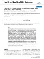
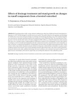
The effects of BPA and its derivatives on bone
cells are summarized in Figure 1.
Evidence from animal studies
Many studies have been conducted on the effects
of BPA on skeletal health in rodents, ranging from the
foetal/neonate skeletal development model, the
diabetic bone loss to the classic oestrogen deficiency
(knockout or castrated) osteoporosis model. This
appropriately encompasses the skeletal health from
early development to old age similar in humans.
Developmental programming or changes in
metabolic environmental during the prenatal and
postnatal period can influence disease development
in the later stage of life [33]. To investigate the effects
Figure 1. The effects of BPA and its derivatives on bone cells. The effect varies according to the derivatives, probably depending on the affinity towards
different receptors on bone cells. Abbreviations: BADGE=bisphenol A diglycidyl ether; BPAF=bisphenol AF; BPA= bisphenol A; BPS=bisphenol S; MAPK=
mitogen-activated protein kinase; RUNX-2=runt related factor-2; OSX=osterix; TGFβ=transforming growth factor beta.
Int. J. Med. Sci. 2018, Vol. 15
of xenoestrogens on skeletal programming, Pelch et
al. (2012) compared the skeletal effects of BPA,
diethylstilbestrol
(DES,
used
in
hormone
replacement), and ethinyl oestradiol (EE, used in oral
contraceptives) exposure to mice nine days prenatal
and 12 days postnatal [34]. The skeletal health of these
mice was assessed during adulthood when they had
reached peak bone mass. The study revealed that
exposure to 10 µg/kg/day BPA significantly
increased femoral length in male mice but decrease
biomechanical strength (energy to failure) in female
mice [34]. In contrast, DES and EE increased the femur
length in female mice but decreased biomechanical
strength in mice [34]. In addition, male mice exposed
to 0.1 µg/kg/day DES had significantly lower
marrow cavity diameter, higher cortical bone width,
lower endosteal to periosteal medio-lateral diameter
ratio [34]. This was not seen in other treatment
groups. None of the treatment affected circulating
bone remodelling markers [34]. The stronger effects of
DES on bone compared to BPA might arise due to a
stronger oestrogen receptor binding affinity. This
study showed that early exposure of BPA, EE or DES
could lead to reduced bone strength and low-trauma
fractures.
In a similar study, Lejonklou et al. supplemented
Wistar rats with 25-50,000 µg/kg BPA from
gestational day 7 until 22 days postnatal. Their bone
health was examined at three months old. The results
showed that femoral length of the rats exposed to all
doses of BPA was significantly higher than controls
[20]. The femoral diaphyseal bone mineral content
(BMC) of the female rats exposed to BPA at 250 µg/kg
was significantly lower compared to rats exposed to
50,000 µg/kg BPA [20]. Male rats exposed to 25 µg/kg
BPA had significant thicker diaphyseal cortex, total
and cortical BMC, as well as cortical cross-sectional
area compared to rats exposed to 250 µg/kg BPA [20].
Bone biomechanical strength and metaphyseal
geometry of the femur was not affected by BPA
exposure [20]. This did not necessarily indicate the
skeletal geometrical changes were insufficient to
produce a specific effect. Since the biomechanical test
(three-point-bending) only applied stress to a certain
part of the bone, it might not reflect the weakest bone
segment. This study highlighted the gender difference
in the skeletal response of the rats towards moderate
exposure of BPA in their early stage of life. Bone
mineral content deteriorated in female rats but
increased in the male rats. The exact reason is not
known at the moment.
Female aromatase-knockout (ArKO) mice are a
model of oestrogen deficiency because they lack
aromatase enzymes essential in the production of
oestrogen [35]. Toda et al. (2002) supplemented
1046
five-week-old female ArKO mice with 0.1% or 1.0%
(w/w) BPA in the diet for five months. They found
that BPA exhibited strong oestrogenic effects by
preventing the degeneration of uteri and ovaries,
normalizing the gene expression of progesterone
receptor and vascular endothelial growth factor in the
uteri and insulin-like growth factor-1 receptor, bone
morphogenetic protein-15 and growth differentiation
factor-9 in the ovaries of ArKO mice [36]. With
regards to their bone health, total BMD of the ArKO
mice was improved in a dose-dependent manner by
BPA. Peripheral quantitative computed tomography
demonstrated that degenerative changes in the
femoral trabecular bone of the ArKO mice were
reversed by BPA [36]. In contrast, BPA did not
improve BMD and bone structure of the wildtype
mice in this study [36]. This might be due to the
relatively lower binding affinity of BPA to oestrogen
receptors compared with oestrogens (2,000-10,000
fold lower compared to 17β-oestradiol) [37]. It should
be noted that the dose of BPA used in this study (1%
in diet) was 1x10⁵ higher than the environmental
exposure.
Seidlova-Wuttke et al. (2004) compared the
oestrogenic effects of BPA (37 or 370 ug/kg),
dibutylphtalate (DBP, 92.5 or 462.5 mg/kg) and
benzophenone-2 (BP2, 185 or 925 mg/kg) in
ovariectomized rats for three months. The affinity of
BPA to ER-β was high but to ER-α was low in
oestrogen-binding assay [38]. However, oestrogenic
activities of BPA on oestrogenic responsive tissues,
such as uterine epithelium, endometrium and
myometrium were not significant [38]. With respect to
skeletal health, BPA reduced the trabecular density at
the tibial metaphyseal of the ovariectomized rats by
5%. Osteocalcin (bone formation marker) level was
increased in BPA-treated rats but C-terminal of
collagen crosslinks (bone resorption marker) level was
not affected [38]. In contrast, BP2 exhibits strong
oestrogenic activities on uterine tissues and increased
tibial metaphyseal trabecular bone density, while DBP
had the least effects on uterine and bone tissues [38].
The researchers suggested that the oestrogenic
activities of BPA were overcome by its antiandrogenic
and aryl hydrocarbon receptor binding activities,
which were associated with reduced bone health.
A derivative of BPA, BADGE, is a component of
epoxy resin coatings for cans, tanks and concrete vats
[39]. The skeletal effects of BADGE on bone have also
been studied. Botolin and McCabe (2006)
administered BADGE at 30 mg/kg daily (i.p.) to
15-week-old male mice with insulin-deficient induced
by streptozotocin and normal mice. These mice
suffered from bone loss, bone marrow adiposity,
hyperglycaemia and hyperlipidaemia induced by
Int. J. Med. Sci. 2018, Vol. 15
diabetes [40]. Treatment with BADGE inhibited the
development of hyperlipidaemia and bone marrow
adiposity but not bone loss and suppression of bone
formation genes (runt-related factor 2 and osteocalcin)
[40]. By itself, BADGE did not suppress osteoblastrelated gene expression or decrease the bone mineral
density of the rats [40].
In a subsequent study by Duque et al. (2012),
nine-month-old male mice were treated with BADGE
alone (30 mg/kg i.p. daily) or in combination with
1,25-dihydroxyvitamin D (the biologically active form
of vitamin D, delivered using a subcutaneous osmotic
pump, 18 mp/day) for six weeks. Mice receiving
BADGE alone or in combination with 1,25dihydroxyvitamin D showed increased bone volume,
trabecular number, thickness and unmineralized
osteoid at the distal femoral metaphysis [41]. This
might be contributed by increased bone formation,
indicated by higher levels of osteocalcin (bone
formation marker), osteoblast number and mineral
apposition rate (at both cortical and trabecular bone)
at the femur of the stated groups [41]. The treatment
also reduced bone marrow adiposity concurrently
with the downregulation of genes related to
adipogenesis (PPARγ and CCAAT/enhancer binding
protein α (C/EBPα)) [41]. The extracted bone marrow
cells from mice treated with BADGE and
1,25-dihydroxyvitamin D showed more colony
forming units and higher protein expression of
osteocalcin and runt-related factor-2, but a lower
expression of osteopontin [41]. Osteopontin expression is critical in bone mineralization. Therefore, the
researchers suggested that the lower osteopontin
expression was related to the unmineralized osteoid,
demonstrating high bone matrix synthesis exceeding
its capability to mineralized.
1047
Li et al. investigated the effects of BADGE (30
mg/kg daily for 12 weeks, i.p.) on five-month-old
ovariectomized or normal female rats. Bone structural
indices were improved in normal female rats
receiving BADGE, demonstrated by increased bone
density and volume, increased trabecular thickness,
number and lower separation. This was contributed
by increased bone formation, indicated by higher
mineral apposition rate, bone formation rate,
osteoblast number and N-terminal propeptide of type
I collagen (a bone formation marker) [21]. Bone
marrow adiposity was lowered in the treated group
[21]. These physical changes were reflected in the
gene expression level, whereby the expression of
adipogenesis gene (PPARγ and C/EBPα) was
lowered while expression of osteogenesis genes
(osteocalcin and RUNX2) was increased with
treatment [21]. The beneficial skeletal effects of
BADGE were attenuated by ovariectomy. Apart from
a reduced trabecular separation and bone marrow
adiposity, no other changes including gene expression
were detected in BADGE treated ovariectomized rats
[21].
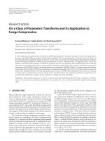
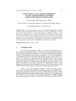
Figure 2 shows the effects of BPA and its
derivatives on rodents in various models, ranging
from prenatal exposure to disease states.
Evidence from human studies
Two cross-sectional studies have examined the
relationship between BPA and bone health in
humans. A small-scale study among 51 Korean
post-menopausal women aged between 50-82 years
(mean age 64.5 years) receiving osteoporotic
treatment in a hospital found that serum BPA did not
correlate significantly with bone mineral density
(lumbar spine, total femur and femoral neck), body
mass index, 25-hydroxyvitamin D and bone
remodelling markers [42]. Since the
sample size was small, this study
might be underpowered. The
subjects were restricted to women on
osteoporosis treatment, which might
mask the effects of BPA on bone [42].
In a population of 246 pre- and
post-menopausal Chinese women
from Shanghai China aged 35.2±0.6
years, positive relationships between
urinary BPA level, fat mass and
leptin level were found [43].
However, the associations between
urinary BPA and bone mineral
density (lumbar spine and femoral
neck), bone remodelling markers,
Figure 2. The skeletal effects of BPA and BADGE in animal models. Abbreviation:
serum oestradiol level were not
ArKO=aromatase knock-out; BMC= bone mineral content; BMD=bone mineral density;
BADGE=bisphenol A diglycidyl ether; BPA=bisphenol A; OVX=ovariectomized.
significant after the adjustment with
Int. J. Med. Sci. 2018, Vol. 15
body mass index [43]. In the final multivariate model,
fat-free mass was a strong predictor of bone mineral
density in these subjects instead of fat mass [43]. This
might explain the absence of mediating effects by BPA
in the relationship between body anthropometry and
bone mineral density [43]. It should be noted that
these women were healthy, with regular menses and
normal body mass index (21.2±0.2 kg/m2), so they
were not at risk for osteoporosis [43]. To date, no
study has been performed to investigate the
relationship between BPA level and risk of fragility
fracture.
Discussion
The pharmacokinetics studies on BPA revealed
that it undergoes rapid and extensive metabolism in
the body [44]. Most of the BPA undergoes
glucuronidation and sulfation by the liver to form
hydrophilic products and a very small amount is
excreted unchanged via the biliary route or urine [45].
More than 90% of the BPA is eliminated 24 hours after
ingestion [44]. Typical BPA exposure through food
ingestion would produce picomolar or subpicomolar
circulating concentration in the body [46]. Deposition
of BPA in body tissues is not well characterised but
some studies showed that it can be found in the
adipose tissue and breast milk of humans [47, 48]. The
BPA level in skeletal tissue is relatively unknown.
Therefore, it is difficult to judge whether the
concentrations in cellular studies or dosage in animal
studies are realistic.
There is evidence reporting that the
dose-response of BPA is biphasic non-monotonic [49,
50]. One study summarised that 20% of the published
dose-response studies on BPA demonstrated this
characteristic [50]. This suggests that the
dose-response curve of BPA could be inverted-U
shaped. It might be beneficial at lower doses, and
harmful to the bone at higher doses. This would
explain some of the heterogeneous results seen in
bone cell studies. However, this property of BPA on
bone is not scrutinized more closely in any study and
remains speculative at best at this moment.
Studies on the relationship between BPA level
and bone health are scarce at this moment. The two
available studies that examined the association
between BPA level and bone mineral density
demonstrated a non-significant relationship [42, 43].
Besides, the populations studied are small. The
subjects in the study by Zhao et al. (2012) were
relatively young and they were not at risk of
developing osteoporosis [43]. Meanwhile, Kim et al.
(2012) studied an osteoporotic population receiving
osteoporosis treatment [42]. Thus, the relationship
between BPA and bone health in humans is not
1048
conclusive and awaits larger studies. Longitudinal
cohort studies are necessary to investigate the risk of
fragility fracture and BPA exposure.
Ultimately, it is hard to quantify the effects of a
single xenoestrogen on bone health in humans as we
are constantly exposed to a myriad of pollutants with
potential
skeletal
effects.
Phthalates,
1,1
-dichloro-2,2-bis(p-chloropheny1)-ethylene
(DDE),
dioxin and cadmium are some of the pollutants
exhibiting skeletal activities in humans [51-54]. This
was confounded by the presence of dietary and
endogenous factors that regulate bone metabolism
[55-59]. Hence, it is impossible to delineate the skeletal
action of each pollutant in humans.
Conclusion
Bisphenol A is an endocrine disruptor which
could affect bone. However, due to the cellular and
animal model used in the investigations, the skeletal
effects of BPA and its derivatives are heterogeneous
(Table 1), whereby both positive and negative effects
have been reported. A possible gender effect of BPA
on bone has been revealed in animal studies
(beneficial in males, deleterious in females) but this
awaits further examinations. There is a paucity of
epidemiological studies on the effects of BPA
exposure and bone health in humans. The current
evidence from cross-sectional studies revealed a
negligible relationship between BPA level and bone
mineral density but this is not conclusive. A more
comprehensive longitudinal study is needed to verify
the relationship BPA and bone health in humans,
especially in fracture risk assessment.
Table 1. The skeletal effects of bisphenol A and its derivatives
Compounds
Bisphenol A
Bisphenol AF
Bisphenol S
Bisphenol A
diglycidyl ether
In vitro actions
Inhibit osteoblast
formation.
Induce apoptosis of
osteoblasts and
osteoclasts.
In vivo actions
Decrease bone strength and bone
mineral content in female rodents
but increase bone strength and
bone mineral content in male
rodents prenatally.
Further induce bone loss in
ovariectomized rats.
Encourage osteoblast Not tested.
formation.
Inhibit osteoblast
Not tested.
formation.
Inhibit adipocyte
Promote bone formation in normal
formation.
rats.
Decrease bone loss in
ovariectomized rats.
Acknowledgement
We thank Universiti Kebangsaan Malaysia for
funding the researchers through GUP-2017-060 and
AP-2017-009/1.
Int. J. Med. Sci. 2018, Vol. 15
Competing Interests
The authors have declared that no competing
interest exists.
References
1.
2.
3.
4.
5.
6.
7.
8.
9.
10.
11.
12.
13.
14.
15.
16.
17.
18.
19.
20.
21.
22.
23.
24.
25.
Staples Ca, Dom PB, Klecka GM, Sandra TO, Harris LR. A review of the
environmental fate, effects, and exposures of Bisphenol A. Chemosphere.
1998; 36: 2149-73.
Huang YQ, Wong CK, Zheng JS, Bouwman H, Barra R, Wahlstrom B, et al.
Bisphenol A (BPA) in China: a review of sources, environmental levels, and
potential human health impacts. Environ Int. 2012; 42: 91-9.
Morgan MK, Nash M, Barr DB, Starr JM, Scott Clifton M, Sobus JR.
Distribution, variability, and predictors of urinary bisphenol A levels in 50
North Carolina adults over a six-week monitoring period. Environment
International. 2018; 112: 85-99.
Toner F, Allan G, Dimond SS, Waechter JM, Beyer D. In vitro percutaneous
absorption and metabolism of Bisphenol A (BPA) through fresh human skin.
Toxicology in Vitro. 2018; 47: 147-55.
Björnsdotter MK, de Boer J, Ballesteros-Gómez A. Bisphenol A and
replacements in thermal paper: A review. Chemosphere. 2017; 182: 691-706.
Jalal N, Surendranath AR, Pathak JL, Yu S, Chung CY. Bisphenol A (BPA) the
mighty and the mutagenic. Toxicology Reports. 2018; 5: 76-84.
Bolli A, Galluzzo P, Ascenzi P, Del Pozzo G, Manco I, Vietri MT, et al. Laccase
treatment impairs bisphenol A-induced cancer cell proliferation affecting
estrogen receptor alpha-dependent rapid signals. IUBMB Life. 2008; 60: 843-52.
Bolli A, Bulzomi P, Galluzzo P, Acconcia F, Marino M. Bisphenol A impairs
estradiol-induced protective effects against DLD-1 colon cancer cell growth.
IUBMB Life. 2010; 62: 684-7.
Akingbemi BT, Sottas CM, Koulova AI, Klinefelter GR, Hardy MP. Inhibition
of testicular steroidogenesis by the xenoestrogen bisphenol A is associated
with reduced pituitary luteinizing hormone secretion and decreased
steroidogenic enzyme gene expression in rat Leydig cells. Endocrinology.
2004; 145: 592-603.
Castro B, Sanchez P, Torres JM, Preda O, del Moral RG, Ortega E. Bisphenol A
Exposure during Adulthood Alters Expression of Aromatase and
5a-Reductase Isozymes in Rat Prostate. PLoS One. 2013; 8: e55905.
Kim JY, Han EH, Kim HG, Oh KN, Kim SK, Lee KY, et al. Bisphenol
A-induced aromatase activation is mediated by cyclooxygenase-2
up-regulation in rat testicular Leydig cells. Toxicol Lett. 2010; 193: 200-8.
Liu Y, Mei C, Liu H, Wang H, Zeng G, Lin J, et al. Modulation of cytokine
expression in human macrophages by endocrine-disrupting chemical
Bisphenol-A. Biochem Biophys Res Commun. 2014; 451: 592-8.
Ooe H, Taira T, Iguchi-Ariga SM, Ariga H. Induction of reactive oxygen
species by bisphenol A and abrogation of bisphenol A-induced cell injury by
DJ-1. Toxicol Sci. 2005; 88: 114-26.
Lin Y, Sun X, Qiu L, Wei J, Huang Q, Fang C, et al. Exposure to bisphenol A
induces dysfunction of insulin secretion and apoptosis through the damage of
mitochondria in rat insulinoma (INS-1) cells. Cell Death Dis. 2013; 4: e460.
Xin F, Jiang L, Liu X, Geng C, Wang W, Zhong L, et al. Bisphenol A induces
oxidative stress-associated DNA damage in INS-1 cells. Mutat Res Genet
Toxicol Environ Mutagen. 2014; 769: 29-33.
Yang YJ, Hong YC, Oh SY, Park MS, Kim H, Leem JH, et al. Bisphenol A
exposure is associated with oxidative stress and inflammation in
postmenopausal women. Environ Res. 2009; 109: 797-801.
Savastano S, Tarantino G, D'Esposito V, Passaretti F, Cabaro S, Liotti A, et al.
Bisphenol-A plasma levels are related to inflammatory markers, visceral
obesity and insulin-resistance: a cross-sectional study on adult male
population. J Transl Med. 2015; 13: 169.
Wauquier F, Leotoing L, Coxam V, Guicheux J, Wittrant Y. Oxidative stress in
bone remodelling and disease. Trends in Molecular Medicine. 2009; 15: 468-77.
Mundy GR. Osteoporosis and Inflammation. Nutrition Reviews. 2007; 65:
S147-S51.
Lejonklou MH, Christiansen S, Orberg J, Shen L, Larsson S, Boberg J, et al.
Low-dose developmental exposure to bisphenol A alters the femoral bone
geometry in wistar rats. Chemosphere. 2016; 164: 339-46.
Li G, Xu Z, Hou L, Li X, Li X, Yuan W, et al. Differential effects of bisphenol A
diglicydyl ether on bone quality and marrow adiposity in ovary-intact and
ovariectomized rats. Am J Physiol Endocrinol Metab. 2016; 311: E922-e7.
Florencio-Silva R, Sasso GR, Sasso-Cerri E, Simoes MJ, Cerri PS. Biology of
Bone Tissue: Structure, Function, and Factors That Influence Bone Cells.
Biomed Res Int. 2015; 2015: 421746.
McGowan JA. Bone: target and source of environmental pollutant exposure.
Otolaryngol Head Neck Surg. 1996; 114: 220-3.
Xiao W, Wang Y, Pacios S, Li S, Graves DT. Cellular and Molecular Aspects of
Bone Remodeling. Front Oral Biol. 2016; 18: 9-16.
Mechiche Alami S, Gangloff SC, Laurent-Maquin D, Wang Y, Kerdjoudj H.
Concise Review: In Vitro Formation of Bone-Like Nodules Sheds Light on the
Application of Stem Cells for Bone Regeneration. Stem Cells Transl Med. 2016;
5: 1587-93.
1049
26. Hwang JK, Min KH, Choi KH, Hwang YC, Jeong IK, Ahn KJ, et al. Bisphenol A
reduces differentiation and stimulates apoptosis of osteoclasts and osteoblasts.
Life Sci. 2013; 93: 367-72.
27. Kanno S, Hirano S, Kayama F. Effects of phytoestrogens and environmental
estrogens on osteoblastic differentiation in MC3T3-E1 cells. Toxicology. 2004;
196: 137-45.
28. Miki Y, Hata S, Nagasaki S, Suzuki T, Ito K, Kumamoto H, et al. Steroid and
xenobiotic receptor-mediated effects of bisphenol A on human osteoblasts.
Life Sci. 2016; 155: 29-35.
29. Fic A, Mlakar SJ, Juvan P, Mlakar V, Marc J, Dolenc MS, et al. Genome-wide
gene expression profiling of low-dose, long-term exposure of human
osteosarcoma cells to bisphenol A and its analogs bisphenols AF and S.
Toxicol In Vitro. 2015; 29: 1060-9.
30. Cao LY, Ren XM, Li CH, Zhang J, Qin WP, Yang Y, et al. Bisphenol AF and
Bisphenol B Exert Higher Estrogenic Effects than Bisphenol A via G
Protein-Coupled Estrogen Receptor Pathway. Environ Sci Technol. 2017; 51:
11423-30.
31. Yu WH, Li FG, Chen XY, Li JT, Wu YH, Huang LH, et al. PPARgamma
suppression inhibits adipogenesis but does not promote osteogenesis of
human mesenchymal stem cells. Int J Biochem Cell Biol. 2012; 44: 377-84.
32. Takahashi N, Udagawa N, Kobayashi Y, Suda T. Generation of osteoclasts in
vitro, and assay of osteoclast activity. Methods Mol Med. 2007; 135: 285-301.
33. Nesterenko TH, Aly H. Fetal and neonatal programming: evidence and
clinical implications. Am J Perinatol. 2009; 26: 191-8.
34. Pelch KE, Carleton SM, Phillips CL, Nagel SC. Developmental exposure to
xenoestrogens at low doses alters femur length and tensile strength in adult
mice. Biology of Reproduction. 2012; 86.
35. Fisher CR, Graves KH, Parlow AF, Simpson ER. Characterization of mice
deficient in aromatase (ArKO) because of targeted disruption of the cyp19
gene. Proc Natl Acad Sci U S A. 1998; 95: 6965-70.
36. Toda K, Miyaura C, Okada T, Shizuta Y. Dietary bisphenol A prevents ovarian
degeneration and bone loss in female mice lacking the aromatase gene (Cyp19
). Eur J Biochem. 2002; 269: 2214-22.
37. Acconcia F, Pallottini V, Marino M. Molecular Mechanisms of Action of BPA.
Dose-Response. 2015; 13: 1559325815610582.
38. Seidlova-Wuttke D, Jarry H, Wuttke W. Pure estrogenic effect of
benzophenone-2 (BP2) but not of bisphenol A (BPA) and dibutylphtalate
(DBP) in uterus, vagina and bone. Toxicology. 2004; 205: 103-12.
39. [Internet] National Center for Biotechnology Information. Bisphenol A
diglycidyl ether Last
Update: 13/1/2018. Accessed: 16/1/2018
40. Botolin S, McCabe LR. Inhibition of PPARgamma prevents type I diabetic
bone marrow adiposity but not bone loss. J Cell Physiol. 2006; 209: 967-76.
41. Duque G, Li W, Vidal C, Bermeo S, Rivas D, Henderson J. Pharmacological
inhibition of PPARgamma increases osteoblastogenesis and bone mass in male
C57BL/6 mice. J Bone Miner Res. 2013; 28: 639-48.
42. Kim DH, Oh CH, Hwang YC, Jeong IK, Ahn KJ, Chung HY, et al. Serum
bisphenol a concentration in postmenopausal women with osteoporosis. J
Bone Metab. 2012; 19: 87-93.
43. Zhao HY, Bi YF, Ma LY, Zhao L, Wang TG, Zhang LZ, et al. The effects of
bisphenol A (BPA) exposure on fat mass and serum leptin concentrations have
no impact on bone mineral densities in non-obese premenopausal women.
Clin Biochem. 2012; 45: 1602-6.
44. Thayer KA, Doerge DR, Hunt D, Schurman SH, Twaddle NC, Churchwell MI,
et al. Pharmacokinetics of bisphenol A in humans following a single oral
administration. Environ Int. 2015; 83: 107-15.
45. Kurebayashi H, Betsui H, Ohno Y. Disposition of a low dose of 14C-bisphenol
A in male rats and its main biliary excretion as BPA glucuronide. Toxicol Sci.
2003; 73: 17-25.
46. Teeguarden JG, Twaddle NC, Churchwell MI, Yang X, Fisher JW, Seryak LM,
et al. 24-hour human urine and serum profiles of bisphenol A: Evidence
against sublingual absorption following ingestion in soup. Toxicol Appl
Pharmacol. 2015; 288: 131-42.
47. Fernandez MF, Arrebola JP, Taoufiki J, Navalon A, Ballesteros O, Pulgar R, et
al. Bisphenol-A and chlorinated derivatives in adipose tissue of women.
Reprod Toxicol. 2007; 24: 259-64.
48. Kuruto-Niwa R, Tateoka Y, Usuki Y, Nozawa R. Measurement of bisphenol A
concentrations in human colostrum. Chemosphere. 2007; 66: 1160-4.
49. vom Saal FS, Hughes C. An Extensive New Literature Concerning Low-Dose
Effects of Bisphenol A Shows the Need for a New Risk Assessment.
Environmental Health Perspectives. 2005; 113: 926-33.
50. Vandenberg LN. Non-monotonic dose responses in studies of endocrine
disrupting chemicals: bisphenol a as a case study. Dose Response. 2014; 12:
259-76.
51. DeFlorio-Barker SA, Turyk ME. Associations between bone mineral density
and urinary phthalate metabolites among post-menopausal women: a
cross-sectional study of NHANES data 2005-2010. Int J Environ Health Res.
2016; 26: 326-45.
52. Eskenazi B, Warner M, Sirtori M, Fuerst T, Rauch SA, Brambilla P, et al. Serum
dioxin concentrations and bone density and structure in the Seveso Women's
Health Study. Environ Health Perspect. 2014; 122: 51-7.
53. Beard J, Marshall S, Jong K, Newton R, Triplett-McBride T, Humphries B, et al.
1,1,1-trichloro-2,2-bis (p-chlorophenyl)-ethane (DDT) and reduced bone
mineral density. Arch Environ Health. 2000; 55: 177-80.
Int. J. Med. Sci. 2018, Vol. 15
1050
54. James KA, Meliker JR. Environmental cadmium exposure and osteoporosis: a
review. Int J Public Health. 2013; 58: 737-45.
55. Chin KY, Ima-Nirwana S. Sex steroids and bone health status in men. Int J
Endocrinol. 2012; 2012: 208719.
56. Chin KY, Ima-Nirwana S. The effects of alpha-tocopherol on bone: a
double-edged sword? Nutrients. 2014; 6: 1424-41.
57. Chin KY, Ima-Nirwana S. Vitamin C and Bone Health: Evidence from Cell,
Animal and Human Studies. Curr Drug Targets. 2018; 19: 439-50.
58. Jolly J, Chin K-Y, Alias E, Chua K, Soelaiman I. Protective Effects of Selected
Botanical Agents on Bone. Int J Environ Res Public Health. 2018; 15: 963.
59. Mohamad NV, Soelaiman IN, Chin KY. A concise review of testosterone and
bone health. Clin Interv Aging. 2016; 11: 1317-24.