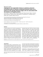
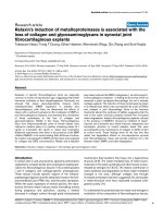
Locus 5p13.1 may be associated with the selection of cancer-related HBV core promoter mutations
Bạn đang xem bản rút gọn của tài liệu. Xem và tải ngay bản đầy đủ của tài liệu tại đây (727.71 KB, 8 trang )
Int. J. Med. Sci. 2019, Vol. 16
Ivyspring
International Publisher
990
International Journal of Medical Sciences
2019; 16(7): 990-997. doi: 10.7150/ijms.34297
Research Paper
Locus 5p13.1 may be associated with the selection of
cancer-related HBV core promoter mutations
Qin-Yan Chen1,#, Yan-Ling Hu2,#, Xue-Yan Wang1, Tim J. Harrison3, Chao Wang1, Li-Ping Hu1, Qing-Li
Yang1, Chuang-Chuang Ren1,4, Hui-Hua Jia1,4, and Zhong-Liao Fang1
1.
2.
3.
4.
Guangxi Zhuang Autonomous Region Center for Disease Prevention and Control, Guangxi Key Laboratory for the Prevention and Control of Viral
Hepatitis, Nanning, Guangxi 530028, China.
Center for Genomic and Personalized Medicine, Guangxi Medical University, 22 ShuangYong Road, Nanning, Guangxi 530021, China.
Division of Medicine, UCL Medical School, London, UK.
School of Preclinical Medicine, Guangxi Medical University, 22 ShuangYong Road, Nanning, Guangxi 530021, China.
# These authors contributed equally to this work.
Corresponding author: Zhong-Liao Fang, Guangxi Zhuang Autonomous Region Center for Disease Prevention and Control, 18 Jin Zhou Road, Nanning,
Guangxi, China, 530028. Tel: 0086 771 2518306; Fax: 0086 771 2518768; Email:
© Ivyspring International Publisher. This is an open access article distributed under the terms of the Creative Commons Attribution (CC BY-NC) license
( See for full terms and conditions.
Received: 2019.02.20; Accepted: 2019.05.21; Published: 2019.06.10
Abstract
Background: The basal core promoter (BCP) double mutations (A1762T and G1764A) of hepatitis
B virus (HBV) have been reported to be an aetiological factor of hepatocellular carcinoma (HCC).
What distinguishes the subset of HBV carriers in whom these mutations are selected?
Methods: A genome-wide association study (GWAS) was carried out on 218 asymptomatic HBsAg
carriers infected with HBV with BCP double mutations and 191 controls infected with HBV with the
wild type BCP. The highest ranking nucleotide polymorphisms (SNPs) were validated with other
study subjects, 203 cases and 181 controls. The expression of the gene nearest a SNP found to be
significant was examined using RT-PCR.
Results: Forty-five candidate SNPs were identified in the GWAS. Three SNPs were found to be
associated with the selection of HBV BCP double mutations in the replication stage, including
rs7717457 at 5p13.1, rs670011 at 17q21.2, rs2071611 at 6p22.2. Especially, rs7717457 (P=
4.57×10⁻⁵ combined P) reached the potential GWAS significance level. The expression of gene
complement component 7 (C7), nearest to SNP rs7717457, differed significantly between the case
and control groups (t=2.045, P=0.04), suggesting that SNP rs7717457 was associated with the
expression of its nearest gene.
Conclusions: SNP rs7717457 is associated with the selection of HBV BCP double mutations,
providing an important clue to understanding the mechanisms of oncogenesis of HBV BCP double
mutations.
Key words: Genome-wide association study (GWAS); hepatitis B virus (HBV); basal core promoter (BCP);
mutations; single nucleotide polymorphisms (SNPs).
Introduction
Worldwide, hepatocellular carcinoma (HCC) is
the fifth most common cancer in males and the
seventh in females and is the third most common
cause of cancer death [1]. The incidence of HCC varies
greatly according to the geographic area; the highest
incidence of HCC in the world is reported by
registries in Asia and Africa. Approximately 85% of
all liver cancers occur in these areas, with Chinese
registries alone reporting over 50% [2]. HCC in China
ranks as the second most common cause of cancer
death in males and the third in females. The mortality
rate from HCC is higher in males (37.4/100,000) than
in females (14.3/100,000) [3]. The major risk factors for
HCC in Asia and Africa are chronic hepatitis B virus
Int. J. Med. Sci. 2019, Vol. 16
(HBV) infection and aflatoxin B1 (AFB1) exposure.
HBV is responsible for 75 to 80% of virus-associated
HCC [4].
However, the mechanisms of the oncogenesis of
HBV remain obscure. Nonetheless, mutations in the
viral genome associated with tumour development
recently have become a major focus of research. The
precore mutation (G1896A), mutations in enhancer II
(C1653T) and the BCP (T1753V and the double
mutations, A1762T, G1764A), and deletions in the pre-S
region have been reported to be associated with the
development of HCC [5-11]. Perhaps the most
convincing association is with HBV with the double
mutations in the BCP; this has been confirmed by
several cohort studies, suggesting that the double
mutations are an aetiological factor of HCC [8, 12-13].
In addition to HBV and AFB1 exposure, host
factors may play a role in the development of HCC.
There have been a few genome-wide association
studies (GWAS) conducted on the genetic
susceptibility to HBV-related HCC. Various single
nucleotide polymorphisms (SNPs), such as rs7574865
at STAT4, rs9275319 at HLA-DQ and rs12682266,
rs7821974, rs2275959, rs1573266 at chromosome 8p12,
have been found to be associated with the
development of HBV-related HCC [14-15]. Combined
analyses of copy number variation (CNV), individual
SNPs, and pathways suggests that HCC susceptibility
is mediated by germline factors affecting the immune
response and differences in T-cell receptor processing
[16].
When we established the Long An cohort in
2004, we found that about half of the HBV-infected
individuals have BCP double mutations (A1762T,
G1764A) in the viral genome and more than 93% of
HCC cases occurred in those with BCP double
mutations[8]. Why are BCP double mutations selected
in a subset of HBV carriers? The answers may be
helpful in understanding the pathogenesis of HCC. It
has been reported from candidate-gene studies that
host genetic polymorphisms are associated with the
immune selection of HBV mutations [17]. This
phenomenon may also be seen in other viruses, such
as HIV-1 [18]. Therefore, we carried out a
genome-wide association study (GWAS), based on the
Long An cohort, to search for a genetic basis of the
selection of HCC-related, HBV BCP mutations and
which may potentially identify novel related SNPs.
Materials and Methods
Study subjects
The study subjects were recruited from the Long
An cohort, which was described previously [8]. The
cohort was recruited in early 2004 from agricultural
991
workers aged 30-55 living in the rural area of Long An
county, Guangxi, China, using stratified sampling.
This cohort comprises 2258 HBsAg-positive study
subjects, including a group (1261) with BCP double
mutations and a wild type BCP group (997). They
were further stratified into the male mutant (702) and
wild type (561) groups and female mutant (559) and
wild type (436) groups. When we recruited study
subjects for this study, we retested BCP sequence of
HBV of each subject in 2014. The selection criterion is
that they were infected with HBV with the same BCP
sequence as at baseline.
Informed consent in writing was obtained from
each individual. The study protocol conforms to the
ethical guidelines of the 1975 Declaration of Helsinki
and has been approved by the Guangxi Institutional
Review Board.
Serological Testing
Sera were tested for HBV serological markers
using enzyme immunoassays and AFP using a
Diagnostic Kit for the Quantitative Determination of
Alpha-feto-protein (ELISA) (Beijing Wantai Biological
Pharmacy Enterprise Co., Ltd., Beijing, China)
according to the manufacturer’s instructions. The
cut-off value of AFP for HCC was set at 20 ng/mL.
Alanine aminotransferase (ALT) concentrations were
determined using a kinetic method (Zhejiang Elikan
Biological Technology Company, Limited, Wenzhou,
Zhejiang, China).
Nested PCR for HBV DNA and nucleotide
sequencing
HBV DNA was extracted from 85 μl serum by
pronase digestion followed by phenol/chloroform
extraction. The method for amplification and
sequencing of the BCP region has been reported
previously [8].
Genotyping in GWAS
Peripheral blood mononuclear cell (PBMC) DNA
was extracted from 200 μl blood using a
QIAamp®DNA Mini Kit. PBMC DNA was sent to the
CapitalBio Corporation (Beijing 102206, China) for
genotyping. The Infinium® HumanCore BeadChips
(Illumina Inc.) was used for genotyping 306670 SNPs
in the GWAS stage. For the genotyping reactions, 250
ng of genomic DNA was analyzed using the
Infinium® Human Core Bead Chips according to the
manufacturer’s recommendations and using their
reagents [19]. Infinium® HumanCore BeadChips
Genotype data were generated using GenomeStudio
Genotyping Module v1.0. The genotyping was
performed by laboratory personnel blinded to the
study subjects.
Int. J. Med. Sci. 2019, Vol. 16
SNP selection and genotyping in the
replication study
If a locus had a SNP with a P value <1.0 × 10⁻⁴ in
the GWAS stage, it was chosen for replication. If
several SNPs were in the linkage disequilibrium with
R2>0.6, the SNP with the lowest P value was selected.
The iPLEX MassARRAY platform (Sequenom Inc.)
was used in the replication stage. 50 ng of genomic
DNA was analyzed using the iPLEX MassARRAY
platform
according
to
the
manufacturer’s
recommendations and using their reagents [19].
iPLEX MassARRAY platform Genotype data were
generated using MassARRAY® Typer 4.0 software.
The genotyping was performed by laboratory
personnel blinded to the study subjects.
Functional annotation and differential
expression analysis
Whole blood was collected in EDTA tubes and
RNALock Reagent (TIANGEN, China) was added
immediately. Total RNA was extracted from the
PBMC using RNAprep Pure Blood Kit (TIANGEN,
China) according to manufacturer’s instructions. The
RNA was reverse transcribed as PCR template using a
PrimeScriptTM II 1st Strand cDNA Synthesis Kit
(TaKaRa, China), followed by PCR with SYBR Premix
Ex TaqTM II (TaKaRa, China). The expression of
mRNA was detected by quantitative real-time reverse
transcriptase PCR (qRT-PCR) on CFX96 (BioRad). The
primers used for GAPDH, CARD6, PTGER4 and C7
were GAPDH-2F (5’ GAAGGTGAAGGTCGGAGTC
3’) and GAPDH-2R (5’ GAAGATGGTGATGGGATT
TC 3’), CARD6-F (5’ CCCACTGTGCTTGTATCTGC
3’) and CARD6-R (5’ CGGTAGCCATTGTTCCTGT
3’), PTGER4-F (5’ CGCAAGGAGCAGAAGGAGAC
3’) and PTGER4-R (5’CAGGCTGAAGAAGAGCAG
AATGAA 3’), C7-2F (5’ AACGGCAAGGAGCAGA
CG 3’) and C7-2R (5’ TGTCCAGTGCCCAGTTGTG
3’), respectively. GAPDH was chosen as an
endogenous control to normalize the relative mRNA
expression levels. Experiments were performed in
duplicate for each sample and fold changes were
calculated by the equation 2-ΔΔCt.
Statistical analysis
The PLINK package [20] was used to carry out
the quality control procedures and association
analyses. Quality control (QC) procedures were
carried out using GenomeStudio Genotyping Module
v1.0. The exclusion criteria were minor allele
frequency (MAF) <0.04, SNP call rate <90%, and
deviation from Hardy-Weinberg Equilibrium (p
<0.05). Genetic association tests were carried out by
multivariate analysis using logistic regression by
entering variables in the PLINK software. Statistical
992
comparisons of gene expression between cases and
controls were performed carried out using a
non-parametric paired t test (SPSS v.16.0). All P values
were two-tailed and P<0.05 was considered to be
significant.
Results
Genome-wide association analysis
In the initial discovery stage, we conducted a
GWAS using samples from 218 asymptomatic HBsAg
carriers with BCP double mutations (cases) and 191
asymptomatic HBsAg carriers with the wild type BCP
(controls). There are 122 males in the case group
(56.0%) and 102 males in control group (53.4%). The
average ages of the case and control groups are
50.8±6.3 and 51.0±6.5, respectively. There was no
difference between the two groups in terms of sex (χ2
=0.274, P>0.05) and age (F =1.974, P>0.05) (Table 1).
Table 1. General characteristics of the study subjects in the
GWAS
Variables
Number
Male
Female
Age, Years
Abnormal ALT, %
AFP (+), %
Total
409
224
185
50.9±6.4
9.3 (38/409)
4.9 (20/409)
Cases
218
122
96
50.8±6.3
10.1 (22/218)
5.5 (12/218)
Control
191
102
89
51.0±6.6
8.4 (16/191)
4.1 (8/191)
P value
P=0.604
P=0.782
P=0.551
P=0.538
Abnormal ALT: ≥40 IU/ml, AFP (+): >20 μg/L.
Quality control (QC) procedures were first
applied to 409 individuals. All 409 study subjects
passed the call rate of 90% and were used in the final
statistical analysis. The exclusion criteria were minor
allele frequency (MAF) <0.05, SNP call rate <90%, and
deviation from Hardy-Weinberg Equilibrium (p
<0.05). Based on these criteria, 25296 SNPs were
retained. The quantile-quantile plot for the cases and
controls is shown (Figure 1). The genomic inflation
factor for the cases and controls was 1.147, indicating
adequate control of population stratification and
systematic bias in our GWAS population. In the
GWAS stage, we assessed genome-wide associations
for the cases and controls using multivariate linear
models adjusted with age and sex. We found that
there are two regions with trends of significant
difference. They located in Chromosomes 5 and
10.The strongest association signals was SNP
rs2910830 in phosphodiesterase 4D (PDE4D), located
on chromosome 5q12 (P=1.136 × 10⁻⁵) (Figure 2).
SNP selection and genotyping in the
replication study
A P value <10-4 was considered to be statistically
significant and selected for the replication stage.
When several SNPs were in linkage disequilibrium
Int. J. Med. Sci. 2019, Vol. 16
993
with R2 >0.6, the SNP with the lowest P value was
selected. These parameters led to the identification of
45 candidate SNPs which were taken forward to the
replication stage. The study subjects were 203
asymptomatic HBsAg carriers infected with HBV
with BCP double mutations (case group) and 181
asymptomatic HBsAg carriers infected with HBV
with wild-type BCP (control group). There are 102
males in the case group (50.2%) and 100 males in the
control group (55.2%). The average ages of the case
and control groups are 50.4±7.0 and 47.5±7.0,
respectively. There are no significant differences
between the two groups in terms of sex and age (Table
2). Genotyping was carried out in the replication stage
using the iPLEX MassARRAY platform (Sequenom
Inc.). The primers and probes are available upon
request. The laboratory technicians who performed
the genotyping experiments were blinded to the
status of case and control. Three SNPs were found to
be differ significantly, rs7717457 (P=0.01387), rs670011
(P=0.04085) and rs2071611 (P=0.04627) (Table 3).
Table 2. General characteristics of the study subjects in the
replication study
Variables
Number
Male
Female
Age, Years
Abnormal ALT, %
AFP (+), %
HBeAg (+)
Total
384
202
182
49.0±7.1
2.3 (9/384)
3.4 (13/384)
6.0 (23/384)
Cases
203
102
101
50.4±7.0
1.5 (3/203)
4.9 (10/203)
0
Control
181
100
81
47.5±7.0
3.3 (6/181)
1.7 (3/181)
12.7 (23/181)
P value
P=0.327
P=0.001
P=0.235
P=0.08
P=0.001
Abnormal ALT: ≥40 IU/ml, AFP (+): >20 μg/L.
Table 3. Results of replication study for forty-five significant SNPs.
CHR
5
5
5
5
5
5
5
5
5
5
5
5
5
5
5
5
5
5
5
5
5
5
5
5
5
5
6
6
6
6
6
6
6
6
6
6
10
10
10
10
10
10
10
10
17
SNP
rs2935623
rs7717457
rs16887016
rs7703245
rs10940659
rs1588265
rs4700365
rs1544791
rs983280
rs2910830
rs2910829
rs4235479
rs35247
rs10057967
rs27135
rs253061
rs9293505
rs17085231
rs11741590
rs7707391
rs246430
rs3756309
rs13166904
rs17066036
rs1445844
rs1136377
rs4712415
rs2743582
rs1165159
rs670011
rs1150658
rs707898
rs198828
rs6457736
rs9365246
rs2981977
rs11253241
rs11005046
rs7908845
rs1245907
rs7916801
rs9422853
rs10781564
rs7098827
rs2071611
BP
2767442
40887679
57501466
60034662
60064514
60073967
60131915
60143255
60149310
60171370
60174072
60248826
68715157
75701931
76717018
76718175
88890652
95886452
95905423
103613967
143602616
150126061
160940790
165873965
179080269
179086140
19524844
19525822
25864397
25887731
26098527
26116992
26119231
33596635
161056632
167336662
5579665
55827304
55887821
109768722
109872687
126909164
131944418
131984924
41439409
OR(GWAS)
0.3544
1.725
1.829
0.482
0.49
0.482
0.5
0.5108
0.4767
0.4365
0.4606
0.5541
2.161
1.701
0.5228
0.5718
1.96
1.662
1.627
0.5803
0.2891
1.93
0.5477
0.5401
2.914
2.892
0.6175
0.5086
2.093
0.6324
0.5641
0.5634
0.559
2.135
1.571
0.582
0.5802
1.786
1.817
2.036
1.941
1.649
0.5955
0.4858
0.482
OR
1.448
1.466
1.095
0.8695
0.8695
1.018
0.8132
0.87
0.9854
0.9605
0.8797
1.186
1.252
1.105
1.064
1.006
1.268
1.023
0.8981
1.219
1.321
1.102
0.9373
1.061
1.129
1.135
0.9775
0.853
1.322
1.378
0.8705
0.8873
1.042
1.042
0.9879
1.045
0.9532
0.7658
0.8838
1.08
1.088
1.073
0.9054
0.8068
0.6775
P(GWAS)
0.0006732
0.000469
0.0009644
0.00006463
0.0001595
0.00006463
0.0007128
0.0004244
0.00005486
0.00001136
0.00007343
0.0009699
0.0003169
0.0005652
0.00008475
0.0008246
0.0006696
0.0004618
0.0006059
0.000974
0.0004727
0.0008312
0.0003813
0.0001008
0.0003181
0.000349
0.0006521
0.0002772
0.00004903
0.0002081
0.0001346
0.0001342
0.000004639
0.000546
0.0003458
0.00102
0.0007793
0.0001032
0.0001392
0.00001174
0.00001864
0.0006043
0.0005059
0.0007101
0.00006463
P value
0.2235
0.01387
0.6009
0.4721
0.4721
0.9259
0.3381
0.5163
0.9397
0.8298
0.5162
0.3299
0.3384
0.492
0.7675
0.973
0.2523
0.8803
0.4685
0.2356
0.3539
0.6101
0.7185
0.7235
0.5991
0.5798
0.8843
0.4193
0.3529
0.04085
0.4634
0.5196
0.7854
0.9035
0.938
0.82
0.7643
0.08212
0.4034
0.6255
0.59
0.6393
0.5081
0.3734
0.04627
P-hwe
1
0.305
0.04854
0.8108
0.8108
1
0.577
0.5777
1
0.6397
1
0.4829
0.6982
0.7575
4.36E-07
0.4289
0.7528
0.5238
0.8801
0.007409
1
0.2895
0.4022
0.452
0.4727
0.4727
0.2958
1
1
0.4132
0.03195
0.08859
0.7536
0.335
0.5261
1
0.3643
0.8746
0.5379
0.4795
0.8661
0.2301
0.8805
0.7227
0.8168
A1
G
G
C
T
A
A
A
C
T
A
A
C
T
T
G
C
T
T
T
T
A
C
T
A
A
C
T
T
G
A
T
C
A
C
G
G
T
C
C
A
A
G
G
T
T
A2
A
A
T
C
G
G
C
T
C
G
G
T
C
C
A
A
G
C
C
G
C
T
C
C
G
T
C
C
A
C
C
T
G
T
A
A
C
A
T
G
G
A
A
G
A
CHR: Chromosome; SNP: single nucleotide polymorphism; BP: base-pair position; OR: odds ratio; P-hwe: P value for Hardy-Weinberg equilibrium; A1 and A2 are Allele, A1
is mutant and A2 is wild type. MAF: minor allele frequency. GWAS: OR and P from genome-wide association study.
Int. J. Med. Sci. 2019, Vol. 16
994
Figure 1. Quantile-Quantile plot of genome-wide quantitative trait loci mapping for log-transformation.
Figure 2. Manhattan plot of genome-wide association analysis, adjusted with sex and age. The X-axis shows chromosomal positions. The Y-axis shows –log10
P-values from the linear regression.
We also carried out a combined analysis of data
of the GWAS and replication studies, using
multivariate linear models adjusted with age and sex.
We found that rs7717457 (P= 4.57×10⁻⁵) reached the
potential GWAS significance level. However,
rs2910830 (P= 1.136×10⁻⁵), which had the most
significant association at the GWAS stage, showed a P
value of 6.53×10⁻⁴ when the data from the two stages
were combined.
Differential expression analysis
To determine whether the SNPs found to be
associated with BCP double mutations in the GWAS
stage and replication stage influence the expression of
the corresponding genes, rs7717457, with the lowest P
value among the three SNPs above, was selected for
the analysis. The position of rs7717457 is near gene
CARD6, gene complement component 7 (C7) and
gene PTGER4 of 5p13.1. (
and />.htm). Whole blood samples were collected from the
Long An cohort, including 23 individuals infected
with HBV with BCP double mutations (case group)
and 28 individuals infected with BCP wild type
(control group) (Table 4). These study subjects differ
from those in the GWAS stage and replication stage.
We found that the expression of genes CARD6 and
PTGER4 did not differ significantly between the two
groups. However, the difference in the expression of
gene C7 between the case group (1.99) and control
Int. J. Med. Sci. 2019, Vol. 16
995
group (4.10) was significant (t=2.045, P=0.04) (Figure
3), suggesting that human genes are involved in
selecting viral mutations.
Table 4. General characteristics of the study subjects in the
differential expression study
Variables
Number
Male
Female
Age, Years
Abnormal ALT, %
AFP (+), %
Viral loads
Total
51
35
16
46.0±4.7
3.9 (2/51)
0
8.49×10⁴
Cases
23
17
6
49.5±4.5
8.7 (2/23)
0
1.06×10⁵
Control
28
18
10
46.0±4.7
0
0
6.72×10⁴
P value
P=0.46
P=0.01
P=0.111
P=1
P=0.58
Clinical significance of the SNPs
We randomly tested the serological parameters
of HBV, ALT and AFP for 196 study subjects from the
GWAS and replication stages. No association between
the rs7717457 mutations and sex, HBeAg, ALT or AFP
was found (Table 5).
Discussion
The major findings of this study are that three
SNPs were found to be associated with HBV BCP
double mutations in the replication stage, rs7717457,
rs670011, rs 2071611. rs7717457 may influence the
expression of its nearest gene, C7, suggesting that
human genes are involved in selecting viral
mutations. No association was found between
rs7717457 and sex, HBeAg, ALT or AFP. A strength of
this study is that the study subjects in the GWAS were
recruited from a long-term cohort, which provides
reliable information for each study subject, such as the
status of the BCP sequence of HBV. A weakness of the
study is that the sample size is small, which may
prevent some interesting SNPs being found. Another
weakness is that the subjects of the GWAS and
replication studies are all from the same ethnic
minority, although they are not the same subjects.
Therefore, we do not know whether the findings are
applicable to other ethnic populations.
Table 5. The distribution of SNP rs7717457 according to the
characteristics of the study subjects
Sex
Male
Female
HBeAg(-)
HBeAg(+)
ALT <40
IU/ml
ALT≥40
IU/ml
AFP <20
μg/L
AFP ≥20
μg/L
Number of
study
subjects
Allele
(A+A*)
Allele
(A+G or
G+G)
Rate of (A+G X2
or G+G) (%)
P value
102
94
184
12
191
78
68
137
9
145
24
26
47
3
46
23.5
27.7
25.5
25.0
24.1
0.439
P=0.508
0.002
P=0.967
8.017
P=0.005
5
1
4
80.0
189
141
48
25.4
0.036
P=0.967
7
5
2
28.6
* Allele (A+A) is wild type and A or G signifies the nucleotide.
Figure 3. Differential expression analysis of PTGER4, CARD6 and C7. *: Group infected with BCP wild type, #: Group infected with HBV with BCP double mutations.
Int. J. Med. Sci. 2019, Vol. 16
The lack of a proof-reading activity of the viral
polymerase leads to a high rate of mutation during
replication of the HBV genome. Some of these
mutants may become predominant strains but others
not, and some predominant strains have clinic
significance. The question is which mutants can
become predominant strains; more than 60% of the
mutations are subject to selection forces from host
immune surveillance, antiviral therapy and
replication fitness [21]. So the common explanation is
the active adaptive evolution of mutant strains under
various selection pressures, such as from
immunoglobulin [22], immunization [23] or antiviral
therapy [24]. However, these mutants may also occur
naturally [25]. Clearly, the mechanism remains
obscure. It also has been reported that HBV adapts to
increasing immune pressure through preferential
mutations in B-cell epitopes and by replicative
attenuation [26]. The human leukocyte antigen (HLA)
class I was found to be involved in this selection [27].
A candidate-gene study reported that rs2233406
variant genotypes significantly increased the
frequencies of BCP double mutations and rs28362491
significantly increased the frequency of BCP double
mutations but reduced the frequency of preS2 start
codon mutations [17]. In this study, we are the first to
use GWAS to find another SNP associated with
double mutations in the core promoter of HBV.
Furthermore, we found that this SNP influenced the
expression of its nearest gene.
In this study, we found in the second stage three
SNPs, rs7717457, rs670011 and rs2071611, are
associated with the selection of double mutations in
the core promoter of HBV. SNP rs2071611 is located in
the intron region of gene KRT38 of 17q21.2. The
protein encoded by gene KRT38 is a member of the
keratin gene family [28]. The rs670011 was located
between gene HIST1H2APS2 and gene SLC17A2 of
6p22.2. As a type I hair keratin, it is an acidic protein
which heterodimerizes with type II keratins to form
hair and nails. Gene HIST1H2APS2 is a histone
pseudogene [29]. Gene SLC17A2 encodes an Na
(+)-phosphate cotransporter 3 (NPT3) [30]. It seems
that these SNPs are unlikely to influence the selection
of double mutations in core promoter of HBV,
considering the proteins encoded by the nearby genes.
rs7717457 is near gene CARD6, gene C7 and
gene PTGER4 of 5p13.1. The expression of genes
CARD6 and PTGER4 were not found to differ
significantly between the groups with BCP double
mutations (cases) and BCP wild type (controls),
suggesting that the genes CARD6 and PTGER4 could
not influence the selection of BCP double mutations.
However, the difference in the expression of gene C7
between the two groups was significant, suggesting
996
that rs7717457 is involved in selecting viral mutations.
It has been reported that SNP can alter gene
expression by affecting transcription rate because of
altered transcription factor binding [31]. Therefore,
the mechanism by which rs7717457 influences the
expression of C7 gene requires study, which is
important to understand the mechanisms of
oncogenesis of HBV.
Gene C7 encodes a serum glycoprotein that
forms a membrane attack complex, together with
complement components C5b, C6, C8, and C9, as part
of the terminal complement pathway of the innate
immune system. The protein encoded by this gene
contains
a
cholesterol-dependent
cytolysin/
membrane attack complex/perforin-like (CDC/
MACPF) domain and belongs to a large family of
structurally related molecules that form pores
involved in host immunity and bacterial
pathogenesis. This protein initiates membrane attack
complex formation by binding the C5b-C6
subcomplex and inserts into the phospholipid bilayer,
serving as a membrane anchor [32-34]. Mutations in
this gene are associated with a rare genetic disorder,
C7 deficiency [35]. It has been reported that
complement component 7 (C7) is a potential tumor
suppressor [36]. The reduced expression of C7
mRNAs may be associated with oesophageal
tumorigenesis [37]. Complement proteins C7 and
complement factor H (CFH) may control the stem of
liver cancer cells via LSF-1[38]. Therefore, clearly, on
one hand, SNP rs7717457 is associated with in the
selection of BCP double mutations. On another hand,
it may be involved in liver tumorigenesis. This may be
an important finding towards understanding the
mechanisms of oncogenesis of HBV BCP double
mutations. This is also important because only a small
fraction of asymptomatic HBsAg carriers with BCP
double mutations go on to develop HCC, so the
ability to predict those at highest risk may permit a
more ‘personalized’ screening strategy, and probably
earlier intervention or treatment, and hence will be of
great clinical relevance.
Although no association was found between
rs7717457 and sex, HBeAg, ALT or AFP in our study,
more clinical markers could be used for exploring for
association between rs7717457 and HBV viral load,
HCC status, cirrhosis, end-stage liver disease, etc.
In summary, our study provides evidence using
GWAS that host genetic polymorphisms are
associated with the immune selection of HCC-related
double mutations (A1762T and G1764A) in the basal
core promoter of HBV. We also found that this SNP,
rs7717457, influenced the expression of its nearest
gene, which has been reported to be involved in the
control stemness of liver cancer cells. These results are
Int. J. Med. Sci. 2019, Vol. 16
important in furthering our understanding of the
mechanisms of oncogenesis of HBV. In the future, the
rates of SNP rs7717457 should be determined among
patients with HCC, liver cirrhosis and chronic
hepatitis, which will be helpful to understand further
the mechanisms of oncogenesis.
Acknowledgements
We are indebted to staff members of Centre for
Disease Prevention and Control of Long An and local
town hospitals in Long An county, Guangxi, who
assisted in recruiting the study subjects, sample
collection. This study was supported by the Wellcome
Trust (WT072058MA) and the National Natural
Science Foundation of China (Grant No.
81260439/H2609).
Competing Interests
The authors have declared that no competing
interest exists.
References
1.
2.
3.
4.
5.
6.
7.
8.
9.
10.
11.
12.
13.
14.
15.
16.
17.
Bosetti C, Turati F, La Vecchia C. Hepatocellular carcinoma epidemiology.
Best Pract Res Clin Gastroenterol. 2014; 28:753-70.
[Internet] Ferlay J, Parkin DM, Curado MP, et al. Cancer Incidence in Five
Continents, Volumes I to IX: IARC CancerBase No. 9.
Tanaka M, Katayama F, Kato H, et al. Hepatitis B and C virus infection and
hepatocellular carcinoma in China: a review of epidemiology and control
measures. J Epidemiol. 2011; 21: 401-16.
McGlynn KA, Petrick JL, London WT. Global epidemiology of hepatocellular
carcinoma: an emphasis on demographic and regional variability. Clin Liver
Dis. 2015; 19: 223-38.
Fang ZL, Ling R, Wang SS, et al. HBV core promoter mutations prevail in
patients with hepatocellular carcinoma from Guangxi, China. J Med Virol.
1998; 56:18-24.
Fang ZL, Yang J, Ge X, et al. Core promoter mutations (A(1762)T and
G(1764)A) and viral genotype in chronic hepatitis B and hepatocellular
carcinoma in Guangxi, China. J Med Virol. 2002; 68: 33-40.
Lyu H, Lee D, Chung YH, et al. Synergistic effects of A1896, T1653 and
T1762/A1764 mutations in genotype c2 hepatitis B virus on development of
hepatocellular carcinoma. J Viral Hepat. 2013; 20: 219-24.
Fang ZL, Sabin CA, Dong BQ, et al. HBV A1762T, G1764A mutations are a
valuable biomarker for identifying a subset of male HBsAg carriers at
extremely high risk of hepatocellular carcinoma: A prospective study.
American J Gastroenterol. 2008; 103: 2254-2262.
Fang ZL, Sabin CA, Dong BQ, et al. Hepatitis B virus pre-S deletion mutations
are a risk factor for hepatocellular carcinoma: a matched nested case-control
study. J Gen Virol. 2008; 89(Pt 11): 2882-90.
Qu LS, Liu JX, Liu TT, et al. Association of hepatitis B virus pre-S deletions
with the development of hepatocellular carcinoma in Qidong, China. PLoS
One. 2014; 9: e98257.
Qu LS, Zhu J, Liu TT, et al. Effect of combined mutations in the enhancer II
and basal core promoter of hepatitis B virus on development of hepatocellular
carcinoma in Qidong, China. Hepatol Res. 2014; 44:1186-95.
Yuen MF, Tanaka Y, Fong DY, et al. Independent risk factors and predictive
score for the development of hepatocellular carcinoma in chronic hepatitis B. J
Hepatol. 2009; 50: 80-8.
Chu CM, Lin CC, Lin SM, et al. Viral Load, Genotypes, and Mutants in
Hepatitis B Virus-Related Hepatocellular Carcinoma: Special Emphasis on
Patients with Early Hepatocellular Carcinoma. Dig Dis Sci. 2012; 57: 232-8.
Chan KY, Wong CM, Kwan JS, et al. Genome-wide association study of
hepatocellular carcinoma in Southern Chinese patients withchronic hepatitis B
virus infection. PLoS One. 2011; 6: e28798.
Jiang DK, Sun J, Cao G, et al. Genetic variants in STAT4 and HLA-DQ genes
confer risk of hepatitis B virus-related hepatocellular carcinoma. Nat Genet.
2013; 45:72-5.
Clifford RJ, Zhang J, Meerzaman DM, et al. Genetic variations at loci involved
in the immune response are risk factors for hepatocellular carcinoma.
Hepatology. 2010; 52 :2034-43.
Zhang Q, Ji XW, Hou XM, et al. Effect of functional nuclear factor-kappaB
genetic polymorphisms on hepatitis B virus persistence and their interactions
with viral mutations on the risk of hepatocellular carcinoma. Ann Oncol. 2014;
25:2413-9.
997
18. Bartha I, Carlson JM, Brumme CJ, et al. A genome-to-genome analysis of
associations between human genetic variation, HIV-1 sequence diversity, and
viral control. Elife 2013; 2: e01123.
19. Oka R, Sasagawa T, Ninomiya I, et al. Reduction in the local expression of
complement component 6 (C6) and 7 (C7) mRNAs in oesophageal carcinoma.
Eur J Cancer. 2001; 37(9):1158-65.
20. Seol HS, Lee SE, Song JS, et al. Complement proteins C7 and CFH control the
stemness of liver cancer cells via LSF-1. Cancer Lett. 2016; 372:24-35.
21. Sun Y, Huang Y, Yin A, et al. Genome-wide association study identifies a new
susceptibility locus for cleft lip with or without a cleft palate. Nat Commun.
2015; 6: 6414.
22. Purcell S, Neale B, Todd-Brown K, et al. PLINK: a tool set for whole-genome
association and population-based linkage analyses. Am J Hum Genet. 2007;
81:559–575.
23. Xu Z, Wu G, Li F, et al. Positive selection signals of hepatitis B virus and their
association with disease stages and viral genotypes. Infect Genet Evol. 2013;
19:176-87.
24. Terrault NA, Zhou S, McCory RW, et al. Incidence and clinical consequences
of surface and polymerase gene mutations in liver transplant recipients on
hepatitis B immunoglobulin. Hepatology. 1998; 28:555–561
25. Carman WF, Zanetti AR, Karayiannis P, et al. Vaccine-induced escape mutant
of hepatitis B virus. Lancet. 1990; 336: 325–329.
26. Sheldon J, Camino N, Rodés B, et al. Selection of hepatitis B virus polymerase
mutations in HIV-coinfected patients treated with tenofovir. Antiviral Ther.
2005; 10:727–734.
27. Yamamoto K, Horikita M, Tsuda F, et al. Naturally occurring escape mutants
of hepatitis B virus with various mutations in the S gene in carriers
seropositive for antibody to hepatitis B surface antigen. Journal of Virology.
1994; 68: 2671-6.
28. Mondal RK, Khatun M, Ghosh S, et al. Immune-driven adaptation of hepatitis
B virus genotype D involves preferential alteration in B-cell epitopes and
replicative attenuation--an insight from human immunodeficiency
virus/hepatitis B virus coinfection. Clin Microbiol Infect. 2015; 21:710.e11-20.
29. Kefalakes H, Budeus B, Walker A, et al. Adaptation of the hepatitis B virus
core protein to CD8(+) T-cell selection pressure. Hepatology. 2015; 62: 47-56.
30. Rogers MA, Winter H, Langbein L, et al. The human type I keratin gene
family: characterization of new hair follicle specific members and evaluation
of the chromosome 17q21.2 gene domain. Differentiation. 2004; 72:527-40.
31. Stevens A, Ray DW, Worthington J, et al. Polymorphisms of the human
prolactin gene--implications for production of lymphocyte prolactin and
systemic lupus erythematosus. Lupus. 2001;10(10):676-83.
32. [Internet] Database: RefSeq. Available from: />gene/C7.
33. Togawa N, Juge N, Miyaji T, et al. Wide expression of type I a+-phosphate
cotransporter 3 (NPT3/SLC17A2), a membrane potential-driven organic anion
transporter. Am J Physiol Cell Physiol. 2015; 309: C71-80.
34. Müller-Eberhard HJ. Molecular organization and function of the complement
system. Annu Rev Biochem. 1988; 57:321-47.
35. Sonnen AF, Henneke P. Structural biology of the membrane attack complex.
Subcell Biochem. 2014; 80: 83-116.
36. Serna M, Giles JL, Morgan BP, et al. Structural basis of complement membrane
attack complex formation. Nat Commun. 2016; 7:10587.
37. Fernie BA, Hobart MJ. Complement C7 deficiency: seven further molecular
defects and their associated marker haplotypes. Hum Genet. 1998; 103:513-9.
38. Ying L, Zhang F, Pan X, et al. Complement component 7 (C7), a potential
tumor suppressor, is correlated with tumor progression and prognosis.
Oncotarget. 2016; 7: 86536-86546.