PIK3CA amplification is common in left side tubular adenomas but uncommon sessile serrated adenomas exclusively with KRAS mutation
Bạn đang xem bản rút gọn của tài liệu. Xem và tải ngay bản đầy đủ của tài liệu tại đây (279.39 KB, 5 trang )
Int. J. Med. Sci. 2015, Vol. 12
Ivyspring
International Publisher
349
International Journal of Medical Sciences
Research Paper
2015; 12(4): 349-353. doi: 10.7150/ijms.11281
PIK3CA Amplification Is Common in Left Side-Tubular
Adenomas but Uncommon Sessile Serrated Adenomas
Exclusively with KRAS Mutation
Hyunsu Lee1,*, Jae-Ho Lee1,*, Dae-Kwang Kim2,3, In-Jang Choi1, Ilseon Hwang4, Yu-Na Kang4, Shin Kim5
1.
2.
3.
4.
5.
*
Department of Anatomy, Keimyung University School of Medicine;
Department of Medical Genetics, Keimyung University School of Medicine;
Hanvit Institute for Medical Genetics;
Department of Pathology, Keimyung University School of Medicine;
Department of Immunology, Keimyung University School of Medicine, 2800, Dalgubeoldaero, Dalseo-Gu, Daegu, Republic of Korea.
These authors contributed equally to this work.
Corresponding author: Shin Kim, Department of Immunology, Keimyung University School of Medicine, 2800, Dalgubeoldaero,
Dalseo-Gu, Daegu, Republic of Korea. E-Mail: Tel: +82-53-580-3884 Fax: +82-53-580-3836
© 2015 Ivyspring International Publisher. Reproduction is permitted for personal, noncommercial use, provided that the article is in whole, unmodified, and properly cited.
See for terms and conditions.
Received: 2014.12.08; Accepted: 2015.03.06; Published: 2015.04.27
Abstract
Colorectal cancer is a heterogeneous disorder than arises via multiple distinct pathways, from
tubular adenomas (TAs) and sessile serrated adenomas (SSAs), which are clinically, morphologically, and molecularly different. We examined PIK3CA amplification in colorectal precancerous
legions, including TAs and SSAs. DNA was isolated from paired normal and tumoral tissues in 64
TAs and 32 SSAs. PIK3CA amplification, KRAS mutation, and BRAF mutation were analyzed by
real-time PCR and pyrosequencing. PIK3CA amplification was found in 25% of TAs and 9.4% of
SSAs, respectively. KRAS and BRAF mutations were mutually exclusive in both TAs and SSAs. In
TAs, PIK3CA amplification was associated with left side and it was mutually exclusive with KRAS
mutation. These results suggest that PIK3CA amplification may be early and important event in
colorectal carcinogenesis and may drive the development of left-side TAs independently with
KRAS mutation.
Key words: colorectal cancer, mitochondria, polymorphism, sessile serrated adenomas, tubular adenomas
Introduction
Colorectal cancer (CRC) is the third most common cancer in Korea and its incidence rate has increased seriously every year [1, 2]. Previous studies
demonstrated that colorectal neoplasms result from
the sequential accumulation of gene alterations [3-5].
As a result, it is accepted that the adenoma-carcinoma
sequence underlies the colorectal carcinogenesis via
two distinct pathways, the chromosomal instability
(CIN) pathway and microsatellite instability (MSI)
[3-7]. Serrated polyps are histologically classified into
hyperplastic polyp, traditional serrated adenoma,
sessile serrated adenoma (SSA), and mixed hyperplastic/adenomatous polyp [8]. Though, SSAs have
been considered to be nonneoplastic lesions without
malignant potential, recent studies showed that SSAs
may be the precursor of CRC with MSI [9-13]. This
serrated neoplastic pathway is characterized by frequent BRAF mutation and infrequent KRAS mutation
[14, 15].
The phosphoinositide-3-kinase, catalytic, alpha
polypeptide (PIK3CA) gene encodes the catalytic
subunit p110 alpha of phosphatidylinositol 3-kinase
(PI3K) belonging to class 1A of PI3Ks. PIK3CA mutation promotes cell growth by stimulating AKT pathway in various cancers and has been reported in
10-30% of colorectal cancers [16-18]. PIK3CA ampli
Int. J. Med. Sci. 2015, Vol. 12
fication is found more frequently than PIK3CA mutation in various cancers and it promotes another
mechanism for PI3K.AKT pathway [19-21]. In CRC,
PIK3CA amplification was found in 38% of cancers
and 25% of adenomas, suggesting its important role in
the adenoma-carcinoma transformation model [22].
Furthermore, their results demonstrated that it may
be an independent prognostic marker for better survival. However, the role of PIK3CA amplification in
colorectal carcinogenesis has not been studied using
colorectal precancerous legions.
In present study, PIK3CA amplification was investigated in colorectal precursor lesions, comprising
of TAs and SSAs. To contribute to better understanding on colorectal carcinogenesis, KRAS and BRAF
mutations key markers in CRCs, were also studied in
these lesions. Clinicopathological characteristics in
these patients were analyzed according to their genetic status.
Materials and Methods
Patients and DNA Extraction
For this study, the records of colonoscopic polypectomy performed at Dongsan Medical Center
between 1999 and 2003 were reviewed and 64 TAs
and 32 SSAs were selected. Exclusion criteria were:
previous history of surgical resection for CRCs, and
evidence of hereditary non-polyposis colorectal cancer (Amsterdam criteria) or familial adenomatous
polyposis. Tumor area and adjacent normal mucosa
were selected from slide according to hematoxylin
and eosin stained sections. Subsequently, the selected
areas from paraffin embedded tissues were used for
DNA extraction. DNA was isolated by using DNA
extraction Kit (AbsoluteTM DNA extraction Kit, BioSewoom, Korea) according to the manufacturer’s
instructions.
PIK3CA Amplification
Copy number of PIK3CA gene was analyzed by
quantitative real-time (qRT) PCR. For the quantitative
determination of PIK3CA content relative to nDNA,
primers for specific amplification of exon 20 in
PIK3CA gene and nDNA-encoded ß-actin gene were
selected according to previous study [21]. Real-time
PCR was then carried out on an LightCycler 480 II
system (Roche Diagnostics, Germany) with a total
volume of 20 µl reaction mixture containing 10 µl
SYBR Green Master MIX (Takara, Japan), 8 pmol of
each primers, and DNA (50 ng). The PCR conditions
were 95°C for 1 min, followed by 40 cycles of 95°C for
15 s, and 60°C for 30 s. The threshold cycle number
(Ct) values of the ß -actin gene and PIK3CA gene were
determined. The copy number of PIK3CA in each
350
tested specimen was then normalized against that of ß
-actin gene to calculate the relative PIK3CA copy
number. Each measurement was repeated in triplicate
and 5 serially diluted control samples were included
in each experiment. Copy amplification of PIK3CA
gene was defined by a copy number ≥ 3. Samples with
a PIK3CA/ ß -actin ratio between 1.0 and 3.0 were
classified as having PIK3CA gain.
Microsatellite instability (MSI)
MSI was analyzed with two microsatellite
markers, BAT25 and BAT 26 based on previous studies demonstrated that these analyses can accurately
detect MSI without the need for additional markers.
Briefly, polymerase chain reaction (PCR) was performed using a thermal cycler (Applied Biosystems,
USA). The PCR products were also denatured in
formamide loading buffer (95% formamide, 20 mmol
EDTA, 10 mmol NaOH, 0.05% bromophenol blue,
0.05% xylene cyanol) and electrophoresed through
7.5% and 10% polyacrylamide gels. Silver stain was
performed to develop bands. MSI was defined as either a band shift or the appearance of a novel band in
DNA from precancerous lesions. All experiments
were repeated at least twice to rule out any artifacts.
Direct DNA sequencing was performed on those PCR
products that showed altered band mobility in the
above analysis using the ABI 3730 DNA sequencer
(Bionics Inc, Korea).
KRAS and BRAF Mutations
KRAS mutations in codons 12 and 13, and BRAF
V600E mutation were analyzed by pyrosequencing
(PyroMark Q24, Sweden). Primers for amplification
and pyrosequencing were designed as previously
described [23]. The pyrosequencing reaction was
performed on a PyroMark Q24 instrument using the
Pyro Gold Q24 Reagents (Qiagen, Netherlands). The
pyrosequencing primers were used in a final concentration of 0.3 µmol/L. Resulting data were analyzed
and quantified with the PyroMark Q24 software version 2.0.6 (Qiagen, Netherlands).
Statistical Analysis
SPSS software for Windows was used. Statistical
comparisons for significance were made with Wilcoxon signed-rank test for paired samples.
Chi-square, Fischer’ exact tests and Mann Whitney U
test were used to analyze the relationship between
variables. A p value < 0.05 was considered statistically
significant.
Result
Precursors of CRCs were comprised of 64 tubular adenomas (TAs) and 32 sessile serrated adenomas
Int. J. Med. Sci. 2015, Vol. 12
(SSAs). Clinicopathological characteristics of TAs and
SSAs were presented in Table 1. Higher frequency of
KRAS mutation was found in TA than that in SSAs (p
= 0.012). However, BRAF mutation was shown only
SSAs (p < 0.001), therefore, KRAS and BRAF mutations were mutually exclusive in TAs and SSAs. Other
clinicopathological characteristics were not associated
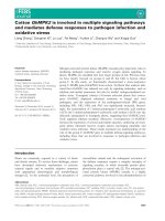
with KRAS and BRAF mutations. Using real-time
PCR methods, the expression level of PIK3CA gene
were analyzed in paired TAs and SSAs. The average
of PIK3CA expression level between tumors and
normal tissues was not significantly different in both
TAs and SSAs (Figure 1). In normal and tumor tissues,
higher expression of PIK3CA gene was found in TAs
than SSAs (p < 0.05). With a gene copy number of 3 or
more defined as amplification, we found the incidence
of PIK3CA amplification in colorectal precancerous
legions was 19.8% (19/96) in the present study (Table
2). Higher frequency of PIK3CA amplification was
found in TAs (25.0%, 16/64) than SSAs (9.4%, 3/32),
however, it did not reach statistical significance (p =
0.07). Similar frequency of PIK3CA gain was found in
TAs and SSAs. Clinicopathological characteristics of
PIK3CA amplification was presented in Table 3. In
TAs, PIK3CA amplification was associated with
left-side (p = 0.025). And mutual exclusivity between
PIK3CA amplification and KRAS mutation was found
in TAs, though it did not get to statistical significance
(p = 0.06). In SSAs, PIK3CA amplification did not have
any clinical significance.
351
Table 1. Clinicopathological characteristics of TAs and SSAs in
present study
Age (mean ± SD)
Sex
Male
Female
Region
Proximal
Distal
MSI
(+)
(-)
Kras mutation
(+)
(-)
Braf mutation
(+)
(-)
TA (n = 64, %)
60.36 ± 10.57
SSA (n = 32, %)
59.03 ± 9.56
43 (67.2)
21 (32.8)
22 (68.8)
10 (31.2)
18 (28.1)
46 (71.9)
7 (21.9)
25 (78.1)
7 (10.9)
57 (89.1)
4 (12.5)
28 (87.5)
15 (23.4)
49 (76.6)
1 (3.1)
31 (96.9)
0 (0)
64 (100)
6 (18.8)
26 (81.2)
p
0.412
0.877
0.51
1.00
0.012
< 0.001
Table 2. Prevalence of PIK3CA content changes in TAs and SSAs
TAs
SSAs
Total
Total
64
32
96
Gain (N, %)
19 (29.7)
11 (34.4)
30 (31.2)
Amplification (N, %)
16 (25.0)
3 (9.4)
19 (19.8)
Table 3. Clinicopathological characteristics of PIK3CA amplification in TAs and SSAs
Total *
Sex
Male
Female
Side
Right
Left
MSI
(+)
(-)
KRAS mutation
(+)
(-)
BRAF mutation
(+)
(-)
TA (n = 64)
25.0% (16/64)
p
SSA (n = 32)
9.4% (3/32)
0.28
20.9% (9/43)
33.3% (7/21)
0.22
13.6% (3/22)
0% (0/10)
0.025
5.6% (1/18)
32.6% (15/46)
0.34
0% (0/7)
12.0% (3/25)
1.00
28.6% (2/7)
24.6% (14/57)
1.00
0% (0/4)
10.7% (3/28)
0.06
6.7% (1/15)
30.6% (15/49)
0.74
0% (0/1)
9.7% (3/31)
25.0% (16/64)
p
0.50
16.7% (1/6)
7.7% (2/26)
p = 0.07 between TA and SSA
*
Discussion
Figure 1. PIK3CA expression level in TAs and SSAs. No significant
difference between normal and tumor samples was found in both TAs and
SPs. Higher PIK3CA amplification was observed in normal and tumor
samples of TAs than SSAs.
This article demonstrates that PIK3CA amplification is important and early event in colorectal precancerous legions, especially in TAs. PIK3CA amplification was found in 19.8% (19/96) of precancerous
legions and this result was in agreement with previous studies [19, 21, 23-27]. PIK3CA amplification has
been reported in 30% of ovarian cancer, 33.1% of lung
squamous cell carcinomas, 32.3% of head and neck
squamous cell carcinomas, 12.2% of endometrial carcinomas, 37.9% of CRC, 15% of anaplastic thyroid
cancer, and 67% of gastric cancer. Jehan et al. [22]
Int. J. Med. Sci. 2015, Vol. 12
showed that PIK3CA amplification was observed in
25.0% of colorectal adenomas and CRCs with PIK3CA
amplification is an independent prognostic marker for
better survival. So, we studied clinicopathological
characteristics of PIK3CA amplification in precancerous legions, comprising of TAs and SSAs. Higher
frequency of PIK3CA amplification was found in TAs
more than SSAs, though it did not reach statistical
significance. In SSAs, PIK3CA amplification was not
associated with any clinicopatholotical characteristics,
however, PIK3CA amplification had various significance in TAs. Higher frequency of PIK3CA amplification was found in left-side TAs and KRAS wild type.
It indicates that PIK3CA amplification may play an
important role in the development of left-side TA independently with KRAS mutation. In many cancers,
PIK3CA amplification was associated with poor
prognosis and cancer development, by activating the
PI3K/Akt signaling pathway aberrantly. Thus, specific genotype-based targeting against the PI3K/Akt
signaling pathway may be an effective therapeutic
strategy for TAs. Its molecular mechanisms should be
studied further to clarify its classification.
Mutual exclusivity between PIK3CA amplification and KRAS mutation was introduced firstly in
previous study by Konopka et al. [24]. It has been
reported inverse relationship between PIK3CA amplification and PIK3CA mutation in various cancers
[19, 21]. Previous study indicated that PIK3CA mutation may play important role in benign and early
stage of colorectal neoplasm [28]. We studied PIK3CA
mutation in 20 patients with TAs, as a preliminary
study, however, PIK3CA mutation was not found
(data not shown). Therefore, we did not include the
analysis of PIK3CA mutation in present study. Previous studies also demonstrated rare PIK3CA mutation in Korean populations with CRCs [29, 30]. Besides the ethnic factor, the absence of PIK3CA mutation might be affected by the morphology of the adenoma. Chang, Chiu [31] demonstrated that the frequency of the PIK3CA mutation was lower in
non-polypoid colorectal neoplasm; however, we had
no record of the morphology of colorectal lesion.
Nevertheless, these data and our result indicated that
PIK3CA amplification is more important factor than
PIK3CA mutation in colorectal carcinogenesis.
Taken together, we studied the PIK3CA amplification in colorectal precancerous legions for the first
time and analyzed their clinicopathological significance. For the first time, a possible role of PIK3CA
amplification in development of CRC is suggested,
and it may drive the development of left-side TAs independently with KRAS mutation. This finding may
have an important implication for the treatment and
the prevention of CRCs.
352
Competing Interests
The authors have declared that no competing
interest exists.
References
1.
2.
3.
4.
5.
6.
7.
8.
9.
10.
11.
12.
13.
14.
15.
16.
17.
18.
19.
20.
21.
22.
23.
Parkin DM, Bray F, Ferlay J, Pisani P. Global cancer statistics, 2002. CA: a
cancer journal for clinicians. 2005; 55: 74-108.
Shim JI, Kim Y, Han MA, Lee HY, Choi KS, Jun JK, et al. Results of colorectal
cancer screening of the national cancer screening program in Korea, 2008.
Cancer research and treatment : official journal of Korean Cancer Association.
2010; 42: 191-8. doi:10.4143/crt.2010.42.4.191.
Fearon ER, Vogelstein B. A genetic model for colorectal tumorigenesis. Cell.
1990; 61: 759-67.
Peinado MA, Malkhosyan S, Velazquez A, Perucho M. Isolation and characterization of allelic losses and gains in colorectal tumors by arbitrarily primed
polymerase chain reaction. Proceedings of the National Academy of Sciences
of the United States of America. 1992; 89: 10065-9.
Kern SE, Fearon ER, Tersmette KW, Enterline JP, Leppert M, Nakamura Y, et
al. Clinical and pathological associations with allelic loss in colorectal carcinoma [corrected]. JAMA : the journal of the American Medical Association.
1989; 261: 3099-103.
Boland CR, Thibodeau SN, Hamilton SR, Sidransky D, Eshleman JR, Burt RW,
et al. A National Cancer Institute Workshop on Microsatellite Instability for
cancer detection and familial predisposition: development of international
criteria for the determination of microsatellite instability in colorectal cancer.
Cancer research. 1998; 58: 5248-57.
Popat S, Hubner R, Houlston RS. Systematic review of microsatellite instability and colorectal cancer prognosis. Journal of clinical oncology : official journal of the American Society of Clinical Oncology. 2005; 23: 609-18.
doi:10.1200/JCO.2005.01.086.
Jass JR. Serrated adenoma and colorectal cancer. The Journal of pathology.
1999; 187: 499-502.
Jass JR, Whitehall VL, Young J, Leggett BA. Emerging concepts in colorectal
neoplasia. Gastroenterology. 2002; 123: 862-76.
Hawkins NJ, Bariol C, Ward RL. The serrated neoplasia pathway. Pathology.
2002; 34: 548-55.
Goldstein NS, Bhanot P, Odish E, Hunter S. Hyperplastic-like colon polyps
that preceded microsatellite-unstable adenocarcinomas. American journal of
clinical pathology. 2003; 119: 778-96. doi:10.1309/DRFQ-0WFU-F1G1-3CTK.
Spring KJ, Zhao ZZ, Karamatic R, Walsh MD, Whitehall VL, Pike T, et al. High
prevalence of sessile serrated adenomas with BRAF mutations: a prospective
study of patients undergoing colonoscopy. Gastroenterology. 2006; 131:
1400-7. doi:10.1053/j.gastro.2006.08.038.
Jass JR, Baker K, Zlobec I, Higuchi T, Barker M, Buchanan D, et al. Advanced
colorectal polyps with the molecular and morphological features of serrated
polyps and adenomas: concept of a 'fusion' pathway to colorectal cancer.
Histopathology. 2006; 49: 121-31. doi:10.1111/j.1365-2559.2006.02466.x.
Samowitz WS, Sweeney C, Herrick J, Albertsen H, Levin TR, Murtaugh MA, et
al. Poor survival associated with the BRAF V600E mutation in microsatellite-stable
colon
cancers.
Cancer
research.
2005;
65:
6063-9.
doi:10.1158/0008-5472.CAN-05-0404.
Walther A, Johnstone E, Swanton C, Midgley R, Tomlinson I, Kerr D. Genetic
prognostic and predictive markers in colorectal cancer. Nature reviews Cancer. 2009; 9: 489-99. doi:10.1038/nrc2645.
Velho S, Oliveira C, Ferreira A, Ferreira AC, Suriano G, Schwartz S, Jr., et al.
The prevalence of PIK3CA mutations in gastric and colon cancer. European
journal of cancer. 2005; 41: 1649-54. doi:10.1016/j.ejca.2005.04.022.
Kato S, Iida S, Higuchi T, Ishikawa T, Takagi Y, Yasuno M, et al. PIK3CA
mutation is predictive of poor survival in patients with colorectal cancer. International journal of cancer Journal international du cancer. 2007; 121: 1771-8.
doi:10.1002/ijc.22890.
Abubaker J, Bavi P, Al-Harbi S, Ibrahim M, Siraj AK, Al-Sanea N, et al. Clinicopathological analysis of colorectal cancers with PIK3CA mutations in Middle
Eastern
population.
Oncogene.
2008;
27:
3539-45.
doi:10.1038/sj.onc.1211013.
Campbell IG, Russell SE, Choong DY, Montgomery KG, Ciavarella ML, Hooi
CS, et al. Mutation of the PIK3CA gene in ovarian and breast cancer. Cancer
research. 2004; 64: 7678-81. doi:10.1158/0008-5472.CAN-04-2933.
Bertelsen BI, Steine SJ, Sandvei R, Molven A, Laerum OD. Molecular analysis
of the PI3K-AKT pathway in uterine cervical neoplasia: frequent PIK3CA
amplification and AKT phosphorylation. International journal of cancer
Journal international du cancer. 2006; 118: 1877-83. doi:10.1002/ijc.21461.
Yamamoto H, Shigematsu H, Nomura M, Lockwood WW, Sato M, Okumura
N, et al. PIK3CA mutations and copy number gains in human lung cancers.
Cancer research. 2008; 68: 6913-21. doi:10.1158/0008-5472.CAN-07-5084.
Jehan Z, Bavi P, Sultana M, Abubaker J, Bu R, Hussain A, et al. Frequent
PIK3CA gene amplification and its clinical significance in colorectal cancer.
The Journal of pathology. 2009; 219: 337-46. doi:10.1002/path.2601.
Baldus SE, Schaefer KL, Engers R, Hartleb D, Stoecklein NH, Gabbert HE.
Prevalence and heterogeneity of KRAS, BRAF, and PIK3CA mutations in
primary colorectal adenocarcinomas and their corresponding metastases.
Int. J. Med. Sci. 2015, Vol. 12
24.
25.
26.
27.
28.
29.
30.
31.
353
Clinical cancer research : an official journal of the American Association for
Cancer Research. 2010; 16: 790-9. doi:10.1158/1078-0432.CCR-09-2446.
Konopka B, Janiec-Jankowska A, Kwiatkowska E, Najmola U, Bidzinski M,
Olszewski W, et al. PIK3CA mutations and amplification in endometrioid
endometrial carcinomas: relation to other genetic defects and clinicopathologic
status of the tumors. Human pathology. 2011; 42: 1710-9.
doi:10.1016/j.humpath.2010.01.030.
Shi J, Yao D, Liu W, Wang N, Lv H, Zhang G, et al. Highly frequent PIK3CA
amplification is associated with poor prognosis in gastric cancer. BMC cancer.
2012; 12: 50. doi:10.1186/1471-2407-12-50.
Hou P, Liu D, Shan Y, Hu S, Studeman K, Condouris S, et al. Genetic alterations and their relationship in the phosphatidylinositol 3-kinase/Akt pathway
in thyroid cancer. Clinical cancer research : an official journal of the American
Association
for
Cancer
Research.
2007;
13:
1161-70.
doi:10.1158/1078-0432.CCR-06-1125.
Suda T, Hama T, Kondo S, Yuza Y, Yoshikawa M, Urashima M, et al. Copy
Number Amplification of the PIK3CA Gene Is Associated with Poor Prognosis
in Non-lymph node metastatic Head and Neck Squamous Cell Carcinoma.
BMC cancer. 2012; 12: 416. doi:10.1186/1471-2407-12-416.
Velho S, Moutinho C, Cirnes L, Albuquerque C, Hamelin R, Schmitt F, et al.
BRAF, KRAS and PIK3CA mutations in colorectal serrated polyps and cancer:
Primary or secondary genetic events in colorectal carcinogenesis? BMC cancer.
2008; 8: 255.
Kwon MJ, Lee SE, Kang SY, Choi YL. Frequency of KRAS, BRAF, and PIK3CA
mutations in advanced colorectal cancers: Comparison of peptide nucleic acid-mediated PCR clamping and direct sequencing in formalin-fixed, paraffin-embedded tissue. Pathology, research and practice. 2011; 207: 762-8.
doi:10.1016/j.prp.2011.10.002.
Kim SY, Shim EK, Yeo HY, Baek JY, Hong YS, Kim DY, et al. KRAS Mutation
Status and Clinical Outcome of Preoperative Chemoradiation With Cetuximab
in Locally Advanced Rectal Cancer: A Pooled Analysis of 2 Phase II Trials.
International journal of radiation oncology, biology, physics. 2013; 85: 201-7.
doi:10.1016/j.ijrobp.2012.03.048.
Chang L-C, Chiu H-M, Shun C-T, Liang J-T, Lin J-T, Chen C-C, et al. Mutational profiles of different macroscopic subtypes of colorectal adenoma reveal
distinct pathogenetic roles for KRAS, BRAF and PIK3CA. BMC gastroenterology. 2014; 14: 221.