Myokines related to leukocyte recruitment are down-regulated in osteosarcoma
Bạn đang xem bản rút gọn của tài liệu. Xem và tải ngay bản đầy đủ của tài liệu tại đây (1.38 MB, 8 trang )
Int. J. Med. Sci. 2018, Vol. 15
Ivyspring
International Publisher
859
International Journal of Medical Sciences
2018; 15(9): 859-866. doi: 10.7150/ijms.24928
Research Paper
Myokines related to leukocyte recruitment are
down-regulated in osteosarcoma
Yu Miao1, Bin Hu2, Qiong Wang3, Qingcheng Yang1, Shumin Zhou2
1.
2.
3.
Department of Orthopedics, Shanghai Jiao Tong University Affiliated Sixth People's Hospital, Shanghai 200233, P.R. China.
Institution of microsurgery for limbs, Shanghai Jiao Tong University Affiliated Sixth People's Hospital, Shanghai 200233, P.R. China.
Department of Oncology, Shanghai Jiao Tong University Affiliated Sixth People's Hospital, Shanghai 200233, P.R. China.
Corresponding authors: Shumin Zhou: , No.600, Yishan Road, Shanghai 200233, P.R. China or Qingcheng Yang: ,
No.600, Yishan Road, Shanghai 200233, P.R. China
© Ivyspring International Publisher. This is an open access article distributed under the terms of the Creative Commons Attribution (CC BY-NC) license
( See for full terms and conditions.
Received: 2018.01.14; Accepted: 2018.04.27; Published: 2018.05.26
Abstract
Myokines are cytokines that are secreted by muscle cells during exercises, muscle development and
pathology. Studies have shown that expression of some individual myokines was altered in tumors.
However, comprehensive analyses of myokines’ expression in osteosarcoma (OS), the most
common malignant tumor in musculoskeletal system, have not been performed. In this study, we
analyzed the expression of 35 myokines in osteosarcoma, peritumoral skeletal muscle, and
cancellous bone by qRT-PCR. Heatmap analysis based on the expression pattern of these myokines
revealed that OS is more likely derived from cancellous bone than peritumoral skeletal muscle.
Thus, we compared the expression of myokines between OS and cancellous bone to reveal a
potential role of myokines in OS development. Our results showed that expression of 19 myokines
in OS was significantly lower than that in cancellous bone. KEGG signaling pathway analysis showed
that these 19 myokines are involved in several important signaling pathways, one of which was
associated with leukocyte recruitment in TNF-α signaling. We verified that expression of these
leukocyte recruitment-related myokines were down-regulated in OS cell line MNNG compared to
those in human BMSC. Downregulation of the myokines related to leukocyte recruitment suggests
that escaping from host immune system may help the occurrence of osteosarcoma.
Key words: myokine screening; osteosarcoma; cancellous bone; peritumoral skeletal muscle; leukocyte
recruitment
Introduction
Osteosarcoma (OS), the most common and
frequent primary malignant solid neoplasm in
musculoskeletal system, is believed to develop from
primitive transformed cells of mesenchymal origins
which are differentiated into osteoblastic lineages and
produces malignant osteoid tissues. Although the
estimated incidence rate is only 5/1,000,000, osteosarcoma (OS) has a peak incidence in teenagers and
young adults [1, 2], which affects patients' life quality
in their remaining years and places heavier burden on
society. The major treatments of OS include tumor
excision surgery, chemotherapy and radiotherapy. In
the past decades, the 5-year survival rates for patients
with localized OS remain approximately 60-70% [3].
Furthermore, the 5-year survival rates for patients
who have remote metastatic niches is only around
20% [4, 5]. The main reasons hindering the progress of
osteosarcoma treatment is the lack of knowledge of
tumorigenesis and biomarkers for early diagnosis [6].
Although numerous studies have been performed to discover the biomarkers for osteosarcoma in
its early stage, little successes have been achieved
[7-9]. Up to now, the main diagnosis methods for
osteosarcoma remain medical imaging and biopsy
[10]. To our knowledge, none of the biomarkers under
studies has been well accepted in clinic worldwide. It
is well known tumor biomarkers are molecules that
are secreted by tumors or specific responses of the
body to the presence of tumors. Therefore,
understanding the responses of osteosarcoma
Int. J. Med. Sci. 2018, Vol. 15
peripheral tissues (e.g. skeletal muscle and cancellous
bone) or their cross-talk may help the discovery of
novel early diagnosis biomarkers of osteosarcoma.
About 20 years ago, skeletal muscle has been
identified as an endocrine and paracrine organ, which
has the ability to produce a variety of humoral
cytokines
and
growth
factors
termed
as
“myokine”[11, 12]. Skeletal muscle can communicate
with other organs including bone, adipose tissue,
brain, liver and pancreas by secreting myokines[13].
These muscle-derived peptides have been proposed
to be involved in anti-inflammatory and metabolic
effects, as well as mediating the preventive effects of
exercise against chronic diseases [14, 15]. A few
studies have focused on the functions and expression
changes of myokines during exercises [16]. For
example, during exercise, IL6 is the first detectable
myokine which have an antiinflammatory effect[17].
Recent studies showed that a few individual myokine
could
mediate
metabolic
changes,
regulate
tumorigenesis and tumor invasion to other remote
organs such as liver and breast [18, 19]. However,
comprehensive analysis of expression for all known
myokines under pathological condition, particularly
in motor system diseases, is very limited [20].
Therefore, in this study, we analyzed the expression
of all known myokines (n=35) in osteosarcoma tissue,
cancellous bone and peritumoral skeletal muscles.
Our results showed that expression of 19 myokines in
OS was significantly lower than that in cancellous
bone. KEGG signaling pathway analysis further
showed that these 19 myokines are involved in
several important signaling pathways, one of which
was associated with leukocyte recruitment in TNF-α
signaling. These results indicated that these
differentially myokines could be potential biomarkers
for the diagnosis of osteosarcoma. Downregulation of
the myokines related to leukocyte recruitment in
TNF-α signaling also suggests that osteosarcoma may
occur by escaping from host innate immune system.
Material and method
Clinical tissues and cells
All the osteosarcoma tissues and peritumoral
muscle tissues were obtained from the patients with
osteosarcoma who received the surgery treatment in
Shanghai Sixth People's Hospital. The normal
cancellous bone tissues were obtained from the
normal distal end of patients with osteosarcoma
treated with amputation, or the patients with severe
trauma who underwent the amputation in Shanghai
Sixth People's Hospital. All the tissues were harvested
for RNA and protein extraction. The human
osteosarcoma cell line MNNG was purchased from
860
the American Type Culture Collection (Manassas, VA,
USA). The BMSC were obtained from Shanghai sixth
People's Hospital (Shanghai, China)
RNA Preparation
Approximately, 100 mg tissues were put in a
grind tube with steel balls and 1 ml Trizol reagent,
and homogenized by a tissue grinder (Scientz-48,
Ningbo, China). About 1 x 10⁷ cells were mixed with 1
ml Trizol reagent. Total RNA from cells, osteosarcoma
tissues, peritumoral muscle tissues, and cancellous
bone tissues was extracted and purified using The
E.Z.N.A.® Total RNA Kit IR6834-01 (OMEGA Bio-tek,
Norcross, GA, U.S.A) according to the manufacturer’s
protocol. The concentration and quality of RNA were
determined by a NanoDrop ND-1000 spectrophotometer (Thermo Fisher Scientific, Waltham, MA, USA).
Reverse‐transcription quantitative PCR
(RT‐qPCR)
About 1 μg of total RNA was reverse transcribed
using the C1000™ Thermal Cycler (Bio-Rad, CA,
USA) according to the manufacturer’s instruction.
Thermal profile consisted of 10 minutes at 25 °C
followed by 60 minutes at 55 °C and 5 minutes at 85
°C. All qPCR reactions were performed in a total
volume of 10 μl mixture, including forward and
reverse primers, cDNA and 1x SYBR® Green PCR
Master Mix (Thermo scientific, Waltham, MA, USA).
Cycling conditions were as follows: 95 °C for 10 min,
followed by 40 cycles of 15 s at 95 °C and 60 s at 72 °C
using a ABI 7900HT machine with software SDS
version 2.3 (Thermo scientific, Waltham, MA, USA).
Primers used in this study are listed in Table S1,
including Eukaryotic Translation Elongation Factor 2
(eEF2) and Glyceraldehyde-3-phosphate Dehydrogenase (GAPDH) as endogenous invariant controls
for data normalization.
Protein analysis
All lysates were obtained by using RIPA Lysis
Buffer (Santa Cruz, Dallas, TX, USA). 30μg proteins
were applied to SDS-PAGE (10% gel) and transferred
to PVDF membranes (Bio-Rad Laboratories, Hercules,
CA), probed with primary antibodies anti-FNDC-5
(1:4000, Abcam), anti-ANGPTL-4 (1:1000, Abcam),
anti-IL-15(1:1000, Abcam), anti-MCP-1 (1:1000,
Abcam) over night at 4℃. After washing, they were
incubated with HRP-labelled anti-mouse (1:3000,
CST) and anti-rabbit (1:3000, CST) secondary
antibodies for 1h at room temperature. Anti-actin Ab
was used as control of protein loading.
KEGG pathway analysis
Kyoto Encyclopedia of Genes and Genomes
(KEGG) pathways were obtained from the website:
Int. J. Med. Sci. 2018, Vol. 15
/>to
determine the differentially expressed genes.
Processes and pathways that showed high enrichment
of upregulated levels of myokines and certain
pathway cascades were displayed.
Statistical analysis
GraphPad Prism v.6.0 (GraphPad software, CA,
USA) and SPSS versions 18.0 (SPSS Inc., Chicago, IL)
were used for statistical analysis. Data are presented
as the mean ± SEM. Myokine gene expression data
were analyzed using a 2-△△CT method and eEF2 was
used as an internal control for each technical replicate
and averaged for each biological replicate. Gene
expression of normal cancellous bone was regarded as
the baseline for the comparison. △CT values from
each group were compared by the non-parametric test
to determine statistically significant differences
between osteosarcoma and cancellation bone. P
values of < 0.05 were considered as statistically
significant.
Result
Myokines are expressed universally in
musculoskeletal tissues
In order to analyze whether myokines are
universally expressed in musculoskeletal system in
OS patients, we analyzed the expression levels of all
35 myokines in OS tissue, peritumoral muscles and
cancellous bones using qRT-PCR. The results showed
that all 35 myokines were universally expressed in
these three tissues (Table S1). Moreover, the myokine
expression patterns in these three tissues can be
861
clearly clustered into two parts in heat map pattern
(Fig. 1). The left part consists of all 8 peritumoral
muscle tissues and 2 OS tissues; and the right part is
formed from other 6 OS tissues and all 8 cancellous
bone tissues. In the right part, bone tissues B4-8 were
clustered, while OS tissues OS1-5 were also clustered.
These data indicated that the myokine expression is
universal in musculoskeletal tissues, and myokine
expression pattern of OS to cancellous bone is more
similar than that to peritumoral tissues.
Most of the myokine expression was
down-regulated in OS compared with those in
cancellous bone
As myokine expression pattern in cancellous
bone and osteosarcoma are close and osteosarcoma is
believed to be derived from transformed bone
marrow mesenchymal cells, we compared the
expression levels of all 35 myokines in these two
tissues in order to identify myokines that are
differentially expressed between osteosarcoma and
cancellous bone tissues. qPCR results showed that 19
genes were significantly down-regulated in osteosarcoma compared with those in cancellous bone
tissues (Fig. 2). These down-regulated genes could be
divided into 5 groups according to their biological
functions (Fig. 2). We also found that 3 myokines
(FNDC5, IL-7 and PAI-1) were upregulated in the
osteosarcoma compared to those in cancellous bone
(Fold changes > 2.0). Although none of them showed
significant difference between these two tissues, the
expression level of myokine FNDC5 was over 20 folds
higher than that in cancellous bone tissues (Fig. 2D).
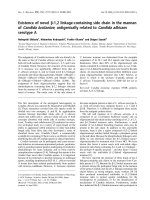
Fig. 1 The relationships of osteosarcoma, cancellous bone and peritumoral skeletal muscle in myokine expression angle. The myokine expression
levels of cancellous bone, peritumoral muscle and osteosarcoma tissues were clearly clustered into two parts. Myokine expression between peritumoral muscle
tissues and cancellous bone tissues differed obviously, whereas the myokine expression in osteosarcoma tissues were remained between them.
Int. J. Med. Sci. 2018, Vol. 15
862
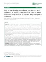
Fig. 2 The myokine expression levels in osteosarcoma tissues compared with cancellous bone tissues. (A) Expression levels of myokine related with
immunological regulation and inflammatory, CCL7, CHI3L1, CX3CL1, DDP4, IL1b, IL4, IL8, IL15, LIF and MCP1 showed significant differences. (B) Expression levels
of myokines related with cell survival and differentiation, DCD, FGF2 and OSM showed significant differences. (C) Expression levels of myokines related with neural
growth, BDNF showed significant differences. (D) Expression levels of myokines related with metabolism, ANGPTL4, Fam132b and FGF21 showed significant
differences. (E) Myokines with other functions, TNF-α showed significant differences. (n = 8, **P < 0.01, *P < 0.05) (F) The protein level of ANGPTL4, IL15 and MCP1
in three randomly chosen OS tissues and cancellous bone tissues.
In order to verify the expressions of these
myokines in their protein levels, representative OS
and cancellous bone tissue lysis were analyzed by
antibodies against selected myokines. The results
showed that ANGPTL4, IL15 and MCP1 proteins
were indeed down-regulated in OS tissues, whereas
FNDC5 was up-regulated (Fig. 2F).
(ko04657) and ‘TNF signaling pathway’ (ko04668).
Cytokine-cytokine receptor interaction has a very
broad definition, IL-17 signaling is mainly reported in
the immune system and TNF signaling is associated
with leukocyte recruitment and Inflammatory.
KEGG analysis revealed pathways that are
associated with OS
We are most interested in TNF signaling
pathway as it may suggest potential inhibition of
immune system in osteosarcoma tissues. To verify the
TNF signaling pathway, we further determined the
expression of genes that are associated with
inflammatory (IL1b, IL6, IL15, LIF), leukocyte
recruitment (MCP1, CCL5, CCL20, CXCL1, CXCL2,
CXCL3, CXCL10, CX3CX1) and the receptor of TNF-α
(TNFR1) in osteosarcoma cell line MNNG using
qPCR. Human BMSC cells, which is from normal
In order to identify the molecular mechanism
underlying the changes in myokine expression
between OS tissue and normal cancellous bone, we
performed a KEGG pathway analyses on 19 genes
whose expression significantly differed in these two
tissues. The top 10 signaling pathway are displayed in
Fig. 3a. and the top 3 are ‘Cytokine-cytokine receptor
interaction’(ko04060), ‘IL-17 signaling pathway’
Verification of the TNF signaling pathway in
OS cell line
Int. J. Med. Sci. 2018, Vol. 15
cancellous bone, are used as a normal control. Not
surprisingly, the result showed that majority of these
genes (with three exceptions: CX3CL1, CXCL10 and
LIF) were significantly down-regulated in MNNG cell
compared to that in human BMSC cells (Fig. 4A).
Taken together, these results verified that TNF
signaling pathway is down-regulated in OS.
Down-regulation of myokines related to leukocytes
recruitment in TNF signaling may protect OS from
being attacked by leukocytes (Fig. 4B).
Discussion
In the last decades, myokines are identified as
cytokines and growth factors that exert their functions
through endocrine or paracrine pattern [11, 21]. The
function of myokines has been widely studied in
physiological processes, such as exercise [16], fasting
863
and temperature keeping [22], pathological processes,
such as diabetes [23, 24], and obesity [25]. More
importantly, many studies have revealed the role of
some individual myokines in various type of cancers,
such as breast cancer, prostate cancer and so on
[26-28]. However, comprehensive analysis of all
known myokines has not been performed under
pathological condition, particularly in OS. To our
knowledge, this is the first study that identified all
known myokines that are differentially expressed in
osteosarcoma, peritumoral skeletal muscle and
cancellous bone, and explored the potential molecular
mechanisms underlying these differences in
osteosarcoma, and cancellous bone, which may
become potential biomarkers for the diagnosis of
osteosarcoma.
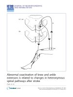
Fig. 3 TNF-α signaling pathway was found to be inhibited in osteosarcoma. (A) Top 10 signaling pathways discovered by KEGG were displayed basing on
the significantly differentially expressed myokines. (B) Significant changes in myokine expression levels were observed in osteosarcoma compared to cancellous bone
in the downstream of TNF signaling pathway(ko04668). Hit myokines are labeled with grey boxes.
Int. J. Med. Sci. 2018, Vol. 15
Fig. 4 Verification of the down-regulation of TNF-α pathway. (A)
Cytokines involved in inflammatory and leukocyte recruitment in the TNF
signaling pathway map (ko04668) were detected by qPCR in osteosarcoma cell
line MNNG and human BMSC cell. CCL20, CCL5, CXCL1, CXCL2, CXCL3,
IL15, IL1b, IL6, MCP1 and TNFRSF1 were significantly decreased (**P < 0.01, *P
< 0.05). (B) The diagram of how the inhibition of TNF protects osteosarcoma
cells from host immune system.
In this study, the expression of 35 myokines (to
the best of our knowledge, these are almost all the
myokine genes) were analyzed in the osteosarcoma
tissues, normal cancellous bone tissues and
peritumoral muscle tissues. It is well known that bone
and skeletal muscle are both derived from somatic
mesoderm during embryonic development. Muscle
pouch has also been fully used in ectopic bone
formation induced by BMPs [29]; hence, we thought
864
that there might be some similarity of myokine
expression between these three tissues [30, 31]. As
expected, the results from qPCR screening showed a
universal expression of myokines in OS, peritumoral
muscle and cancellous bones. Besides, the clustered
heat map (Fig. 1) showed that the expression of
myokines in osteosarcoma is more similar to that in
the cancellous bone compared with that in
peritumoral muscle tissues. This indicates that
myokines can be used as a parameter to tell the
relations between musculoskeletal tissues. Based on
the different expression profile between cancellous
bone and osteosarcoma, the result suggests that the
inhibition of the TNF-α signaling pathway probably
protects osteosarcoma cells from the attacking of
leukocytes according to the KEGG signaling pathway
analysis.
As known to all, tumor necrosis factor alpha
(TNF-α), which is mainly secreted by activated
macrophages and activated T lymphocytes, has been
demonstrated to play an important role in
inflammatory, apoptosis, angiogenesis, and cell
proliferation via the regulation of various signaling
pathways, such as MAPK signaling pathway [32, 33].
On the other hand, it has the ability to cause apoptosis
of tumor-associated endothelial cells by clustering
death domain-containing proteins, leading to caspase
activation [34], ultimately leading to the complete
destruction of the tumor vasculature [35]. With regard
to the functions of TNF-α in osteosarcoma, some
studies showed that TNF-α and inflammatory
cytokines are required for the tumorigenesis and
inducing the migration and invasion of osteosarcoma
[36-38], whereas some diverse conclusions were
observed in other studies. A recent study reported
that TNF-α were upregulated when the tumorigenesis
gene HOTAIR was knocked down in osteosarcoma to
inhibit the proliferation and differentiation [39].
Moreover, Pahl et. al. also showed that human
macrophages which take a major responsibility for
TNF-α secretion can be induced to exert direct
anti-tumor activity against osteosarcoma cells [40].
Consistent with this result, our study also indicates
that TNF signaling is probably attenuated to reduce
the expressions of leukocyte-related cytokines in
osteosarcoma cells, which protects osteosarcoma cells
from the attack of leukocytes from host. This finding
enriches our knowledge of how osteosarcoma protect
themselves from host immune system, which could
offer a novel approach to develop osteosarcoma
therapies in the future.
In conclusion, we demonstrated that the
expression of myokines is universal in OS,
peritumoral muscle and cancellous bone and the
relation between cancellous bone and OS is closer
Int. J. Med. Sci. 2018, Vol. 15
than that between peritumoral muscle and OS in
terms of myokines’ expression pattern. Moreover, we
also indicate that the TNF-α signaling pathway is
inhibited in OS tissue and cell, resulting in
suppression of downstream cytokines related to
inflammation and leukocyte recruitment in OS. Our
results also suggest that myokines that are associated
with TNF signaling pathway could be potential
biomarkers for the diagnosis of OS.
Supplementary Material
Table S1. />
Acknowledgments
This work was supported by the Shanghai
Jiaotong University Medical and Industry cross fund
(YG2017MS21) and National Natural Science
Foundation of China (No.81501939).
Ethics Committee Approval
This study was approved by the Ethics
Committee of Shanghai Jiao Tong University
Affiliated Sixth People's Hospital and performed
according to the Principles of the Declaration of
Helsinki.
Competing Interests
The authors have declared that no competing
interest exists.
References
1.
Botter SM, Neri D, Fuchs B. Recent advances in osteosarcoma. Current
opinion in pharmacology. 2014;16:15-23.
2. Ottaviani G, Jaffe N. The epidemiology of osteosarcoma. Cancer
treatment and research. 2009;152:3-13.
3. Longhi A, Fabbri N, Donati D, Capanna R, Briccoli A, Biagini R, et al.
Neoadjuvant chemotherapy for patients with synchronous multifocal
osteosarcoma: results in eleven cases. Journal of chemotherapy.
2001;13(3):324-30.
4. Bacci G, Briccoli A, Rocca M, Ferrari S, Donati D, Longhi A, et al.
Neoadjuvant chemotherapy for osteosarcoma of the extremities with
metastases at presentation: recent experience at the Rizzoli Institute in 57
patients treated with cisplatin, doxorubicin, and a high dose of
methotrexate and ifosfamide. Annals of oncology : official journal of the
European Society for Medical Oncology. 2003;14(7):1126-34.
5. Chen L, Wang Q, Wang GD, Wang HS, Huang Y, Liu XM, et al. miR-16
inhibits
cell
proliferation
by
targeting
IGF1R
and
the
Raf1-MEK1/2-ERK1/2 pathway in osteosarcoma. FEBS letters.
2013;587(9):1366-72.
6. Marina N, Gebhardt M, Teot L, Gorlick R. Biology and therapeutic
advances for pediatric osteosarcoma. The oncologist. 2004;9(4):422-41.
7. Fei D, Li Y, Zhao D, Zhao K, Dai L, Gao Z. Serum miR-9 as a prognostic
biomarker in patients with osteosarcoma. The Journal of international
medical research. 2014;42(4):932-7.
8. Zhou G, Lu M, Chen J, Li C, Zhang J, Chen J, et al. Identification of
miR-199a-5p in serum as noninvasive biomarkers for detecting and
monitoring osteosarcoma. Tumour biology : the journal of the
International Society for Oncodevelopmental Biology and Medicine.
2015;36(11):8845-52.
9. Kubo T, Shimose S, Matsuo T, Fujimori J, Arihiro K, Ochi M.
Interferon-alpha/beta receptor as a prognostic marker in osteosarcoma.
The Journal of bone and joint surgery American volume.
2011;93(6):519-26.
10. Moore DD, Luu HH. Osteosarcoma. Cancer treatment and research.
2014;162:65-92.
865
11. Karstoft K, Pedersen BK. Skeletal muscle as a gene regulatory endocrine
organ. Current opinion in clinical nutrition and metabolic care.
2016;19(4):270-5.
12. Pedersen BK, Akerstrom TC, Nielsen AR, Fischer CP. Role of myokines
in exercise and metabolism. Journal of applied physiology.
2007;103(3):1093-8.
13. Petersen AM, Pedersen BK. The anti-inflammatory effect of exercise.
Journal of applied physiology. 2005;98(4):1154-62.
14. Benatti FB, Pedersen BK. Exercise as an anti-inflammatory therapy for
rheumatic diseases-myokine regulation. Nature reviews Rheumatology.
2015;11(2):86-97.
15. Pedersen BK, Saltin B. Exercise as medicine - evidence for prescribing
exercise as therapy in 26 different chronic diseases. Scandinavian journal
of medicine & science in sports. 2015;25 Suppl 3:1-72.
16. Huh JY, Siopi A, Mougios V, Park KH, Mantzoros CS. Irisin in response
to exercise in humans with and without metabolic syndrome. The
Journal of clinical endocrinology and metabolism. 2015;100(3):E453-7.
17. Steensberg A, Fischer CP, Keller C, Moller K, Pedersen BK. IL-6 enhances
plasma IL-1ra, IL-10, and cortisol in humans. American journal of
physiology Endocrinology and metabolism. 2003;285(2):E433-7.
18. Pedersen BK, Febbraio MA. Muscle as an endocrine organ: focus on
muscle-derived
interleukin-6.
Physiological
reviews.
2008;88(4):1379-406.
19. Kjaer M, Pollack SF, Mohr T, Weiss H, Gleim GW, Bach FW, et al.
Regulation of glucose turnover and hormonal responses during electrical
cycling in tetraplegic humans. The American journal of physiology.
1996;271(1 Pt 2):R191-9.
20. Paulson TA, Bishop NC, Smith BM, Goosey-Tolfrey VL.
Inflammation-mediating cytokine response to acute handcycling
exercise with/without functional electrical stimulation-evoked
lower-limb cycling. Journal of rehabilitation research and development.
2014;51(4):645-54.
21. Lightfoot AP, Cooper RG. The role of myokines in muscle health and
disease. Current opinion in rheumatology. 2016;28(6):661-6.
22. Li X, Fang W, Hu Y, Wang Y, Li J. Characterization of fibronectin type III
domain-containing protein 5 (FNDC5) gene in chickens: Cloning, tissue
expression, and regulation of its expression in the muscle by fasting and
cold exposure. Gene. 2015;570(2):221-9.
23. Akerstrom TC, Krogh-Madsen R, Petersen AM, Pedersen BK. Glucose
ingestion during endurance training in men attenuates expression of
myokine receptor. Experimental physiology. 2009;94(11):1124-31.
24. Gray SR, Kamolrat T. The effect of exercise induced cytokines on insulin
stimulated glucose transport in C2C12 cells. Cytokine. 2011;55(2):221-8.
25. Peterson JM, Mart R, Bond CE. Effect of obesity and exercise on the
expression of the novel myokines, Myonectin and Fibronectin type III
domain containing 5. PeerJ. 2014;2:e605.
26. Hojman P, Dethlefsen C, Brandt C, Hansen J, Pedersen L, Pedersen BK.
Exercise-induced muscle-derived cytokines inhibit mammary cancer cell
growth. American journal of physiology Endocrinology and metabolism.
2011;301(3):E504-10.
27. Gannon NP, Vaughan RA, Garcia-Smith R, Bisoffi M, Trujillo KA. Effects
of the exercise-inducible myokine irisin on malignant and non-malignant
breast epithelial cell behavior in vitro. International journal of cancer.
2015;136(4):E197-202.
28. Hayes BD, Brady L, Pollak M, Finn SP. Exercise and Prostate Cancer:
Evidence and Proposed Mechanisms for Disease Modification. Cancer
epidemiology, biomarkers & prevention : a publication of the American
Association for Cancer Research, cosponsored by the American Society
of Preventive Oncology. 2016;25(9):1281-8.
29. Scott MA, Levi B, Askarinam A, Nguyen A, Rackohn T, Ting K, et al.
Brief review of models of ectopic bone formation. Stem cells and
development. 2012;21(5):655-67.
30. Braun T, Gautel M. Transcriptional mechanisms regulating skeletal
muscle differentiation, growth and homeostasis. Nature reviews
Molecular cell biology. 2011;12(6):349-61.
31. DiGirolamo DJ, Kiel DP, Esser KA. Bone and skeletal muscle: neighbors
with close ties. Journal of bone and mineral research : the official journal
of the American Society for Bone and Mineral Research.
2013;28(7):1509-18.
32. Wajant H. The role of TNF in cancer. Results and problems in cell
differentiation. 2009;49:1-15.
33. Wang X, Lin Y. Tumor necrosis factor and cancer, buddies or foes? Acta
pharmacologica Sinica. 2008;29(11):1275-88.
34. Lin Y, Devin A, Rodriguez Y, Liu ZG. Cleavage of the death domain
kinase RIP by caspase-8 prompts TNF-induced apoptosis. Genes &
development. 1999;13(19):2514-26.
35. Lejeune FJ, Lienard D, Matter M, Ruegg C. Efficiency of recombinant
human TNF in human cancer therapy. Cancer immunity. 2006;6:6.
Int. J. Med. Sci. 2018, Vol. 15
866
36. Mori T, Sato Y, Miyamoto K, Kobayashi T, Shimizu T, Kanagawa H, et al.
TNFalpha promotes osteosarcoma progression by maintaining tumor
cells in an undifferentiated state. Oncogene. 2014;33(33):4236-41.
37. Rutkowski P, Kaminska J, Kowalska M, Ruka W, Steffen J. Cytokine and
cytokine receptor serum levels in adult bone sarcoma patients:
correlations with local tumor extent and prognosis. Journal of surgical
oncology. 2003;84(3):151-9.
38. Liu C, Zhao P, Yang Y, Xu X, Wang L, Li B. Ampelopsin suppresses
TNF-alpha-induced migration and invasion of U2OS osteosarcoma cells.
Molecular medicine reports. 2016;13(6):4729-36.
39. Zheng H, Min J. Role of Long Noncoding RNA HOTAIR in the Growth
and Apoptosis of Osteosarcoma Cell MG-63. BioMed research
international. 2016;2016:5757641.
40. Pahl JH, Kwappenberg KM, Varypataki EM, Santos SJ, Kuijjer ML,
Mohamed S, et al. Macrophages inhibit human osteosarcoma cell growth
after activation with the bacterial cell wall derivative liposomal muramyl
tripeptide in combination with interferon-gamma. Journal of
experimental & clinical cancer research : CR. 2014;33:27.