Circulating microRNA panel as a novel biomarker to diagnose bisphosphonate-related osteonecrosis of the jaw
Bạn đang xem bản rút gọn của tài liệu. Xem và tải ngay bản đầy đủ của tài liệu tại đây (985.68 KB, 8 trang )
Int. J. Med. Sci. 2018, Vol. 15
Ivyspring
International Publisher
1694
International Journal of Medical Sciences
2018; 15(14): 1694-1701. doi: 10.7150/ijms.27593
Research Paper
Circulating microRNA Panel as a Novel Biomarker to
Diagnose Bisphosphonate-Related Osteonecrosis of the
Jaw
Rui Yang1*, Yurong Tao2*, Chao Wang1, Yi Shuai3, Lei Jin3
1.
2.
3.
Department of Stomatology, PLA Army General Hospital, Beijing, 100000, People’s Republic of China;
Department of Gastroenterology, PLA Army General Hospital, Beijing, 100000, People’s Republic of China;
Department of Stomatology, Nanjing General Hospital of Nanjing Military Command, Nanjing, Jiangsu 210002, People’s Republic of China.
*These authors contributed equally to the study.
Corresponding authors: Lei Jin, MD. PhD. Tel: +86-25-80861166, Fax: +86-25-80863661, E-mail: ; Yi Shuai, MD. PhD. Tel: +86-25-80861166, Fax:
+86-25-80863661, E-mail:
© Ivyspring International Publisher. This is an open access article distributed under the terms of the Creative Commons Attribution (CC BY-NC) license
( See for full terms and conditions.
Received: 2018.05.31; Accepted: 2018.11.02; Published: 2018.11.22
Abstract
There is no defined biomarker for BRONJ diagnosis with satisfactory performance in clinic. In this study,
we established the BRONJ model and selected 7 microRNAs as candidate for BRONJ diagnosis from
microRNA microarray reported by other research. Dysregulated microRNAs during BRONJ were
detected and validated in two independent animal experiments using serum samples. In the first part,
serum miR-21, miR-23a and miR-145 were significantly altered in between BRONJ and control group.
And
an
Indice
was
constructed
as
-0.032+(0.154×miR-21)+(0.145×miR-23a)+
(-0.700×miR-145) using logistic regression model to improve diagnostic performance. The performance
of Indice to differentiate BRONJ subjects from control group was analyzed as AUC of 0.82 (95% CI,
0.72-0.92) or 0.85 (95% CI, 0.73-0.97) in the first or second part. Moreover, the predictive performance
of Indice to discriminate BRONJ-1w and BRONJ-4w from control group was displayed as AUC of 0.65
(95% CI, 0.47-0.84) or 0.75 (95% CI, 0.60-0.91), which was better than individual circulating microRNAs.
In addition, the expressions of candidate microRNAs were validated in human samples. Consequently, we
investigated a combined Indice constructed with circulating microRNAs for BRONJ diagnosis and
prediction.
Key words: bisphosphonate-related osteonecrosis of the jaw, circulating microRNA, biomarker, diagnosis
Introduction
Bisphosphonates are commonly known as
powerful inhibitors of osteoclastogenesis, which have
been used to prevent the osteoporotic bone loss and
reduce the risk of osteoporotic fracture in patients
suffered from postmenopausal osteoporosis[1].
Although bisphosphonates markedly ameliorate
osteoporosis, their side-effects largely limit the clinical
application of these drugs for osteoporosis treatment.
Bisphosphonate-related osteonecrosis of the jaw
(BRONJ) has been recognized as a rare but severe
adverse event associated with bisphosphonates
administration[2]. It has been reported that oral and
maxillofacial surgery may obviously increase the risk
of such a drug-related complication, which mainly
attributes to impaired oral wound healing[3]. In
addition, the risk of BRONJ is positive related with
the dose and accumulation of bisphosphonates
exposure[4].
BRONJ has been reported for about fifteen years.
However, the exact mechanism of this drug-related
disease seems to be multi-factorial and remains
elusive, resulting in management failure of BRONJ.
Apart from age, sex, smoking, oral hygiene, infection
and systemic diseases, genetic background has been
frequently reported to be a predisposing element for
initiation of osteonecrosis of the jaw (ONJ)[5, 6].
Emerging evidences showed genetic association of
diverse genes dysregulation with BRONJ[7-9],
Int. J. Med. Sci. 2018, Vol. 15
suggesting that altered gene expressions might be
potential biomarkers for BRONJ diagnosis. In
addition, since BRONJ is closely related with bone
metabolic disorders, bone turnover markers have
been emerging to support diagnosis of BRONJ[10-13].
Nevertheless, inconsistent diagnostic performances
were observed in various researches and no approved
clinical guide has been established to manage BRONJ.
On account of the increasing usage of anti-resorptive
pharmaceuticals like bisphosphonates for various
bone disorders, it is essential to research and develop
specific and stable biomarkers to identify subjects at
high risk of developing BRONJ.
Recently, a novel approach has been proposed to
diagnose diseases using circulating microRNAs,
which is a type of microRNAs with specificity and
stability existing in body fluids[14]. The diagnostic
performances of circulating microRNAs have been
validated in numerous diseases, including cancers
[15], heart diseases[16], osteoporosis[17], etc. However, no research has been reported to diagnose
BRONJ using circulating microRNAs. A recent
research described an altered microRNA expression
profile in multiple myeloma patients with BRONJ,
suggesting that post-transcriptional regulation might
be crucial for BRONJ development[18]. They obtained
total RNAs of circulating lymphocytes for microRNA
analysis from healthy subjects and multiple myeloma
patients with BRONJ. A class of fourteen microRNAs
markedly elevated in patients with BRONJ, including
miR-16-1, miR-21, miR-23a, miR-28, miR-101-1,
miR-124-1, miR-129-1, miR-139, miR-145, miR-149,
miR-202, miR-221, miR-424 and miR-520[18]. Most of
aforementioned microRNAs have been revealed to
regulate bone metabolism and influence bone remodeling, indicating that microRNA signatures might
exert their specific regulation on osteoblastogenesis
and osteoclastogenesis in BRONJ. Circulating
microRNAs are closely related to tissue or cell specific
microRNAs, thus the altered microRNAs profile
might be a promising resource for circulating
microRNA biomarkers study.
In this study, we investigated three discriminative circulating microRNAs and a combined
microRNAs panel based on the data from microRNA
microarray of Caterina Musolino’s study[18] to
propose a novel strategy for diagnosing BRONJ and
alert its initiation.
Materials and Methods
Ethics
All animals were purchased from Beijing Vital
River Laboratory Animal Technology Co., Ltd. All the
1695
animal study protocols were approved by the Animal
Care Committee of the PLA Army General Hospital,
Beijing, China, which was on the basis of NIH Guide
for the Care and Use of Laboratory Animals. The
collection and usage of human sera were approved by
the Institutional Review Board for Human Subjects
Research of PLA Army General Hospital (Ethic NO.
2018-50). All the participants were provided written
informed consents for their donation of sera in this
research.
Animal groups and model establishment
A total of 140 female Sprague-Dawley rats (10-12
months old, 240-280 g) were involved in this study.
All the rats were separated into two parts, 60 rats
were used for the first part, while the rest was used
for the second part. In the first part, 60 rats were
equally divided into control group and BRONJ group,
while 80 rats were equally divided into four groups in
the second part, namely control group, BRONJ-1w
group, BRONJ-4w group and BRONJ-8w group. The
model of BRONJ was established according to the
protocols reported by R. Nicole Howie and his
colleagues [19]. Briefly, rats in BRONJ group were
weekly administrated with zoledronate (LifeSciences,
USA) at a dose of 80 μg/kg body weight (iv.) for 13
weeks, which was followed by first and second
molars extraction on the left side. Meanwhile, rats in
control group were intravenously injected with
phosphate-buffered saline. All the operations were
conducted under anesthesia. All the rats were housed
under specific pathogen-free conditions with a
temperature of 24°C, cycles of 12 h light/12 h dark,
and humidity of 50%–55%. The jawbones were
obtained for microCT analysis. After microCT
scanning, jawbones were decalcified, and then for
H&E staining and TRAP staining (Sigma-Aldrich)
according to the protocol introduction.
Whole blood collection and serum preparation
For rats sera, all the rats were fasted half day
before blood collection. 5 mL venous blood was
collected from rats’ abdominal vein under anesthesia
in the morning. For human sera, fasting blood
samples (5 mL) were collected via ulnar vein puncture
and imported into the sterile vacuous dry tube
without any anticoagulation agents, in the morning
(8:00 am to 12:00 am). Eleven control participants and
six BRONJ patients were recruited (Table S1). The
blood samples were stored for 30 minutes at room
temperature, then were centrifuged at 4℃, 1000×g,
for 15 min to permit the completely dissociation of cell
and cell debris free serum.
Int. J. Med. Sci. 2018, Vol. 15
Circulating microRNAs isolation, cDNA
preparation and q-RT-PCR analysis
Total RNA was isolated from 200 μL serum
using mirVana Paris Kit (Ambion, USA) following the
provided protocol. A spike-in reference of Lyophilized C.elegans miR-39 miRNA mimic (Qiagen,
Germany) was used to normalize the data according
to the manufacturer’s instruction. The microRNAs
were reversed to cDNA using miScript II RT Kit
(Qiagen, Germany).
Q-RT-PCR analysis of microRNAs was conducted using miScript SYBR Green PCR Kit (Qiagen,
Germany) with a 7500 Real-Time PCR System
(LifeSciences, USA) according to the manufacturer’s
protocol. The reaction procedure was set as follows:
Step 1: 95℃ 30 s; Step 2: PCR reaction, GO TO: 39 (40
cycles), 95℃ 5 s, 60℃ 30 s; Step 3: Melt Curve.
Relative expressions of candidate microRNAs were
normalized by the level of C.elegans miR-39 miRNA
mimic using 2–Δct method. Forward primers sequences
for candidate microRNAs were displayed in Table S2,
and the universal reverse primer was supplied with
the kit.
Statistical analysis
All the data were calculated in SPSS 13.0. All the
figures were graphed in GraphPad Prism 7. Student’s
t test or non-parametric test was used for comparison
of control and BRONJ groups. The Kruskal-Wallis test
or non-parametric test was used for pairwise
comparisons of control, BRONJ-1w, BRONJ-4w and
BRONJ-8w groups. The combined Indice was
developed using a logistic regression model based on
the data from the first part. Receiver operating
characteristic (ROC) curve and area under ROC curve
(AUC) were used for the diagnostic performance
exhibition of each microRNA and Indice. The cutoff
points were defined according to the designed
sensitivity of 80.00%. The threshold value for
statistical significance was set at p<0.05.
Results
Establishment of the BRONJ model
The jaws of Control and BRONJ group were all
scanned by microCT. The data revealed that all the
rats in the BRONJ group developed necrosis of the
jaw. Severe lesions were observed in the jaws of
BRONJ group, whereas the Control group showed a
complete wound healing (Fig. S1A, B). Furthermore,
the HE staining confirmed the data of microCT
scanning. Bone lesions and inflammatory infiltration
were observed in the sections of BRONJ group,
however the Control group showed healthy structure
(Fig. S1C, D). In addition, much fewer TRAP positive
1696
areas were observed in BRONJ group compared to
Control group (Fig. S1E, F). The aforementioned
results suggested that the BRONJ model were
successfully established.
Selection of candidate circulating miRNAs
(first part)
Seven microRNAs (miR-21, miR-23a, miR-28,
miR-124-1, miR-129-1, miR-145 and miR-149) from
Caterina Musolino’ study were selected to analyze
candidate circulating microRNAs in BRONJ, according to the criteria of high preservation and correlation
with bone metabolism. Among the candidate
microRNAs, miR-21 and miR-23a were increased in
serum of BRONJ compared to control group, while
miR-145 was decreased. Whereas, miR-28, miR-129-1
and miR-149 showed no difference in serum between
BRONJ and control group, and miR-124-1 could not
detected in serum. The three altered microRNAs
(miR-21, miR-23a and miR-145) were then analyzed
using ROC curve system. However, miR-21, miR-23a
and miR-145 presented a moderate diagnostic
performance with the respective AUC value of 0.70
(95% CI, 0.57-0.84), 0.72 (95% CI, 0.59-0.85) or 0.65
(95% CI, 0.51-0.79). The diagnostic information was
displayed in Figure 1, 2 and Table 1. In addition,
miR-21, miR-23a and miR-145 were also detected in
the human derived sera to validate the performance.
Similar to aforementioned results, miR-21 and
miR-23a were elevated in BRONJ compared to control
group, while miR-145 was declined (Figure S2).
Diagnostic performance of combined Indice in
BRONJ diagnosis (first and second part)
The combined Indice was developed as
-0.032+(0.154 × miR-21)+(0.145 × miR-23a)+(-0.700 ×
miR-145) according to a logistical regression model
(first part). The AUC of Indice was 0.82 (95% CI,
0.72-0.92) to differentiate BRONJ from control group,
which was highly improved compared with individual microRNA, as well as the diagnostic specificity
(63.33%) and accuracy (71.67%) based on a designed
sensitivity of 80.00%. The diagnostic information was
presented in Figure 1, 2 and Table 1.
Another independent experiment (second part)
was conducted to validate the diagnostic performance
of candidate microRNAs and combined Indice in
BRONJ diagnosis. In accordance with the first part,
the AUC (0.85), specificity (70.00%) and accuracy
(75.00%) of combined Indice also outclassed those of
miR-21, miR-23a and miR-145, suggesting a better
diagnostic performance of combined Indice compared
with individual microRNAs. The diagnostic information was presented in Figure 3 and Table 2.
Int. J. Med. Sci. 2018, Vol. 15
1697
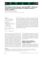
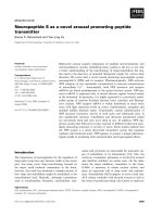
Figure 1. The normalized expressions of rno-miR-21, rno-miR-23a, rno-miR-28, rno-miR-124-1, rno-miR-129-1, rno-miR-145, rno-miR-149 and combined Indice in
the sera of the control and BRONJ groups (first part) (*p<0.05, **p<0.01, *** p<0.001, NS: no significance).
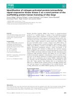
Figure 2. ROC curves of rno-miR-21, rno-miR-23a, rno-miR-28, rno-miR-124-1, rno-miR-129-1, rno-miR-145, rno-miR-149 and combined Indice in the sera of the
control and BRONJ groups (first part).
Table 1. Diagnostic performance of individual microRNA and Indice on BRONJ (first part)
miRNA
miR-21
miR-23a
miR-145
Indice
Cutoff
value
1.42
2.23
0.85
0.64
AUC
(95% CI)
0.70 (0.57-0.84)
0.72 (0.59-0.85)
0.65 (0.51-0.79)
0.82 (0.72-0.92)
p value
0.007
0.004
0.041
<0.001
Designed
Sensitivity# (%)
80.00
80.00
80.00
80.00
Specificity (%) Accuracy (%) True
positive
46.67
63.34
24
36.67
58.34
24
33.33
56.67
24
63.33
71.67
24
True
negative
14
11
10
19
False
positive
16
19
20
11
False
negative
6
6
6
6
Note: Designed sensitivity#: the performance was considered by defining a cutoff value corresponding to fixing the sensitivity to 80.00%.
Int. J. Med. Sci. 2018, Vol. 15
1698
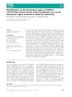
Figure 3. The normalized expressions and ROC curves of rno-miR-21, rno-miR-23a, rno-miR-145 and combined Indice in the sera of the control, BRONJ-1w,
BRONJ-4w and BRONJ-8w groups (second part) (*p<0.05, **p<0.01, *** p<0.001).
Table 2. Diagnostic and predictive performance of individual microRNA and Indice on BRONJ progression (second part)
miRNA
Cutoff
value
Control Vs BRONJ-1w
miR-21
0.21
miR-23a
0.32
miR-145
0.80
Indice
-0.37
Control Vs BRONJ-4w
miR-21
0.12
miR-23a
0.30
miR-145
0.53
Indice
-0.25
Control Vs BRONJ-8w
miR-21
0.24
miR-23a
0.37
miR-145
0.49
Indice
-0.09
AUC
(95% CI)
p value
Designed
Sensitivity# (%)
Specificity
(%)
Accuracy (%)
True
positive
True
negative
False
positive
False
negative
0.61 (0.44-0.79)
0.64 (0.48-0.82)
0.65 (0.47-0.83)
0.65 (0.47-0.84)
0.224
0.099
0.108
0.048
80.00
80.00
80.00
80.00
30.00
40.00
50.00
55.00
55.00
60.00
65.00
67.50
16
16
16
16
6
8
10
11
14
12
10
9
4
4
4
4
0.73 (0.55-0.91)
0.71 (0.55-0.87)
0.67 (0.50-0.85)
0.75 (0.60-0.91)
0.013
0.024
0.049
0.006
80.00
80.00
80.00
80.00
25.00
40.00
55.00
55.00
52.50
60.00
67.50
67.50
16
16
16
16
5
8
11
11
15
12
9
9
4
4
4
4
0.74 (0.57-0.90)
0.73 (0.57-0.89)
0.71 (0.54-0.88)
0.85 (0.73-0.97)
0.011
0.010
0.021
<0.001
80.00
80.00
80.00
80.00
40.00
40.00
65.00
70.00
60.00
60.00
72.50
75.00
16
16
16
16
8
8
13
14
12
12
7
6
4
4
4
4
Note: Designed sensitivity#: the performance was considered by defining a cutoff value corresponding to fixing the sensitivity to 80.00%.
Int. J. Med. Sci. 2018, Vol. 15
Predictive performance of combined Indice in
BRONJ progress (second part)
Apart from diagnosis, prediction of BRONJ
initiation might also significant for BRONJ management. Therefore, this study tried to distinguish
different phases of BRONJ development. Only miR-21
could distinguish BRONJ-4w from control group,
whereas none of these three microRNAs could
distinguish BRONJ-1w from control group. However,
the Indice was gradually increased among control,
BRONJ-1w, BRONJ-4w and BRONJ-8 group, meanwhile the combined Indice effectively differentiated
BRONJ-1w and BRONJ-4w from control group with
respective AUC of 0.65 (95% CI, 0.47-0.94) or 0.75 (95%
CI, 0.60-0.91), indicating that combined Indice might
be a potential predictor of BRONJ progress. The
predictive information was presented in Figure 3 and
Table 2.
Discussion
Bisphosphonate-related osteonecrosis of the jaw
has been reported for more than ten years. However,
there is no certain identification of biomarkers for
BRONJ diagnosis. In current study, we evaluated
seven circulating microRNAs and found three of them
(miR-21, miR-23a and miR-145) markedly differing
between control and BRONJ groups. Nevertheless,
none of the AUC values was greater than 0.80,
indicating a moderate diagnostic effect of the three
selected microRNAs. Therefore, we further
investigated a combined Indice (-0.032+(0.154 ×
miR-21)+(0.145×miR-23a)+(-0.700×miR-145)) based
on a logistic regression model, which highly
improved diagnostic efficiency of BRONJ with an
AUC of 0.82 (95% CI, 0.72-0.92) , specificity of 63.33%,
and accuracy of 71.67% compared to individual
microRNAs. The expressions of miR-21, miR-23a and
miR-145 were detected using human samples, which
were consistent with aforementioned observations,
suggesting a conserved role of these three microRNAs
in BRONJ initiation and development. There were
only 11 healthy controls and 6 BRONJ patients being
included, the broader variation of miRNA expressions
in human samples was partially credit to the
individual difference and small sample size. In
addition, a validation study was absent for human
sample analysis because of the limited sample size.
Therefore a larger sample size is needed for further
study. Moreover, the diagnostic performance of
Indice has been validated using an independent
animal experiment, suggesting that the combined
Indice is a potent biomarker for BRONJ diagnosis. In
addition, although individual microRNA failed to
predict BRONJ initiation after 1 week or 4 weeks
induction, the Indice effectively distinguished
1699
BRONJ-1w and BRONJ-4w from control subjects. To
our knowledge, it is the first time to investigated the
circulating microRNA formed Indice to be a
promising biomarker for diagnosing or predicting
BRONJ initiation and development.
One of the likely etiologies of BRONJ is
destructive bone remodeling triggered by an
imbalance of bone resorption and bone formation.
Therefore, bone turnover markers have been reported
to diagnose BRONJ, including C-terminal telopeptide
of type I collagen (CTx), N-telopeptide of type I
collagen (NTX), tartrateresistant acid phosphatase
isoform 5b (TRACP 5b), receptor activator for nuclear
factor-κ B ligand (RANKL)/osteoprotegerin (OPG),
total alkaline phosphatase (tALP) and bone-specific
alkaline phosphatase (BAP)[10-13]. However, their
diagnostic performance has not been well defined.
According to Jin-Woo Kim’ study, only serum TRACP
5b showed an AUC of 0.807, whereas other serum
markers showed poor performance[12]. Similarly,
Antonia Kolokythas and colleagues observed an
elevation of salivary NTX in medication-related
osteonecrosis of the jaws[13]. Additionally, Vivek
Thumbigere-Math and colleagues revealed that none
of the reported bone turnover markers differentiate
BRONJ from healthy control[11]. Moreover, Vivek
Thumbigere-Math’s research also investigated a
minor augment of angiogenic marker-vascular endothelial growth factor (VEGF) in BRONJ compared to
healthy control[11]. However, no comprehensive
diagnostic evaluation and no validated experiment
were provided in the previous studies. In our system,
we systemically investigated diagnostic performance
of Index with a high AUC to distinguish BRONJ from
control group using two independent experiments.
Prediction and early intervention might be
beneficial for BRONJ management. Jin-Woo Kim and
colleagues reported a sharp decrease of CTx, RANKL
and TRACP 5b in BRONJ subjects (6 weeks after
BRONJ induction) compared to normal control,
suggesting a potential role for alerting BRONJ[12].
However, six weeks after BRONJ induction seems a
little late for BRONJ therapy. In our study, we investigated circulating microRNAs constructed Indice to
forecast BRONJ initiation only one week after BRONJ
induction, which is prospected to be a predictive
marker for BRONJ.
Although miR-21 has been reported to be a
crucial regulator in bone metabolism, its detailed
mechanisms remain complex. According to the
researches, miR-21 improved osteogenic differentiation by targeting Smad7, Spry1 and PLAP1[20-22].
Apart from influence on osteblastogenesis, miR-21
also plays a critical role in up-regulation of
osteoclastogensis via augmenting RANKL and
Int. J. Med. Sci. 2018, Vol. 15
suppressing OPG expression[23]. In addition, miR-21
also displayed positive correlation with particleinduced osteolysis pathogenesis[24], while knocking
down of miR-21 resulted in osteoclastogenesis
restriction[25]. In our system, circulating miR-21 was
up-regulated during BRONJ progress, which was
consistent with the miR-21 variation in pro-osteoclastogenesis. MiR-23a has been reported to suppress
osteoblastic differentiation by regulating TGF-β
signaling, Tmem64 or LRP5[26-28]. Furthermore,
miR-23a targeting LRP5 was also closely related with
steroid-associated femoral head necrosis, meanwhile
miR-23a inhibitor ameliorated osteonecrosis in an
animal model[29]. MiR-145 not only has been
validated to be increased during osteoblast differentiation[30], but also to inhibit monocytes related
osteoclastogenesis induced by RANKL[31]. However,
silencing of miR-145 resulted in rescued steroidinduced avascular necrosis of the femoral head[32]. In
our study, an elevation of serum miR-21, miR-23a,
and a reduction of serum miR-145 were observed in
BRONJ subjects, which were corresponding to the
cellular functions reported by previous researches.
However, the correlation of intracellular microRNAs
and circulating microRNAs in BRONJ development
remains unknown, as well as the detailed mechanisms. Further studies are required to understand the
role of these microRNAs in BRONJ development.
However, some limitations of current study
should be considered and further modified. Firstly, a
high throughput sequencing of serum microRNAs
might be preferable to obtain data base and
bioinformatics rather than selection from literature
review. Secondly, a target gene prediction and gain or
loss experiments should be conducted to reveal the
detailed mechanisms of dysregulated circulating
microRNAs in BRONJ development.
Supplementary Material
Supplementary figures and tables.
/>
Abbreviations
BRONJ: bisphosphonate-related osteonecrosis of
the jaw; ONJ: osteonecrosis of the jaw; ROC: receiver
operating characteristic; AUC: curve and area under
ROC curve; CTx: C-terminal telopeptide of type I
collagen; NTX: N-telopeptide of type I collagen;
TRACP 5b: tartrateresistant acid phosphatase isoform
5b; RANKL: receptor activator for nuclear factor-κ B
ligand; OPG: osteoprotegerin; tALP: total alkaline
phosphatase; BAP: bone-specific alkaline phosphatase; VEGF: vascular endothelial growth factor.
1700
Acknowledgements
This work was financially supported by grants
from the Jiangsu Province “333” High Level Talents
Cultivation Project (Grant No: BK2016544), Jiangsu
Province Medical Key Talents Project (Grant No:
ZDRCA2016095) and Military Medical Science and
Technology Youth Cultivation Project (Grant No:
17QNP054).
We thank Shengqi Zang, Yulin An, Xiaolei Shi,
Lei Zhu and Rui Mu for intellectual support of BRONJ
related information in Department of Stomatology,
Nanjing General Hospital of Nanjing Military
Command, Nanjing, Jiangsu, People’s Republic of
China.
Authorship
Lei Jin, Yi Shuai, Rui Yang and Yurong Tao
conceived and designed the study. Lei Jin and Yi
Shuai supervised the project. Rui Yang and Yurong
Tao conducted the experiments, analyzed the data
and wrote the manuscript. Chao Wang took part in
sample collection and provided some technical
supports. Lei Jin and Yi Shuai reviewed and revised
the manuscript.
Competing Interests
The authors have declared that no competing
interest exists.
References
1.
Cosman F, de Beur SJ, LeBoff MS, Lewiecki EM, Tanner B, Randall S, et al.
Clinician's Guide to Prevention and Treatment of Osteoporosis. Osteoporos
Int. 2014; 25: 2359-81.
2. Spanou A, Lyritis GP, Chronopoulos E, Tournis S. Management of
bisphosphonate-related osteonecrosis of the jaw: a literature review. Oral Dis.
2015; 21: 927-36.
3. Bodem JP, Kargus S, Eckstein S, Saure D, Engel M, Hoffmann J, et al. Incidence
of bisphosphonate-related osteonecrosis of the jaw in high-risk patients
undergoing surgical tooth extraction. J Craniomaxillofac Surg. 2015; 43: 510-4.
4. Kuroshima S, Sasaki M, Nakajima K, Tamaki S, Hayano H, Sawase T.
Prevalence of bisphosphonate-related osteonecrosis of the jaw-like lesions is
increased in a chemotherapeutic dose-dependent manner in mice. Bone. 2018;
112: 177-86.
5. Kim JH, Ko YJ, Kim JY, Oh Y, Hwang J, Han S, et al. Genetic investigation of
bisphosphonate-related osteonecrosis of jaw (BRONJ) via whole exome
sequencing and bioinformatics. PLoS One. 2015; 10: e0118084.
6. Kim KY, Zhang X, Cha IH. Identifying a combined biomarker for
bisphosphonate-related osteonecrosis of the jaw. Clin Implant Dent Relat Res.
2018; 20: 191-8.
7. Arduino PG, Menegatti E, Scoletta M, Battaglio C, Mozzati M, Chiecchio A, et
al. Vascular endothelial growth factor genetic polymorphisms and haplotypes
in female patients with bisphosphonate-related osteonecrosis of the jaws. J
Oral Pathol Med. 2011; 40: 510-5.
8. Basi DL, Hughes PJ, Thumbigere-Math V, Sabino M, Mariash A, Lunos SA, et
al. Matrix metalloproteinase-9 expression in alveolar extraction sockets of
Zoledronic acid-treated rats. J Oral Maxillofac Surg. 2011; 69: 2698-707.
9. Manzano-Moreno FJ, Ramos-Torrecillas J, Melguizo-Rodriguez L,
Illescas-Montes R, Ruiz C, Garcia-Martinez O. Bisphosphonate Modulation of
the Gene Expression of Different Markers Involved in Osteoblast Physiology:
Possible Implications in Bisphosphonate-Related Osteonecrosis of the Jaw.
International journal of medical sciences. 2018; 15: 359-67.
10. Bagan L, Jimenez Y, Leopoldo M, Rubert A, Bagan J. Serum levels of RANKL
and OPG, and the RANKL/OPG ratio in bisphosphonate-related
osteonecrosis of the jaw: Are they useful biomarkers for the advanced stages of
osteonecrosis? Med Oral Patol Oral Cir Bucal. 2017; 22: e542-e7.
11. Thumbigere-Math V, Michalowicz BS, Hughes PJ, Basi DL, Tsai ML, Swenson
KK, et al. Serum Markers of Bone Turnover and Angiogenesis in Patients With
Bisphosphonate-Related Osteonecrosis of the Jaw After Discontinuation of
Int. J. Med. Sci. 2018, Vol. 15
12.
13.
14.
15.
16.
17.
18.
19.
20.
21.
22.
23.
24.
25.
26.
27.
28.
29.
30.
31.
32.
1701
Long-Term Intravenous Bisphosphonate Therapy. J Oral Maxillofac Surg.
2016; 74: 738-46.
Kim JW, Cha IH, Kim SJ, Kim MR. Biomarkers for Bisphosphonate-Related
Osteonecrosis of the Jaw. Clin Implant Dent Relat Res. 2016; 18: 281-91.
Kolokythas A, Karras M, Collins E, Flick W, Miloro M, Adami G. Salivary
Biomarkers Associated With Bone Deterioration in Patients With
Medication-Related Osteonecrosis of the Jaws. J Oral Maxillofac Surg. 2015; 73:
1741-7.
Turchinovich A, Weiz L, Burwinkel B. Extracellular miRNAs: the mystery of
their origin and function. Trends Biochem Sci. 2012.
Zhou J, Yu L, Gao X, Hu J, Wang J, Dai Z, et al. Plasma microRNA panel to
diagnose hepatitis B virus-related hepatocellular carcinoma. J Clin Oncol.
2011; 29: 4781-8.
Egea V, Schober A, Weber C. Circulating miRNAs: messengers on the move in
cardiovascular disease. Thromb Haemost. 2012; 108: 590-1.
Hackl M, Heilmeier U, Weilner S, Grillari J. Circulating microRNAs as novel
biomarkers for bone diseases - Complex signatures for multifactorial diseases?
Mol Cell Endocrinol. 2016; 432: 83-95.
Musolino C, Oteri G, Allegra A, Mania M, D'Ascola A, Avenoso A, et al.
Altered microRNA expression profile in the peripheral lymphoid
compartment of multiple myeloma patients with bisphosphonate-induced
osteonecrosis of the jaw. Ann Hematol. 2018.
Howie RN, Borke JL, Kurago Z, Daoudi A, Cray J, Zakhary IE, et al. A Model
for Osteonecrosis of the Jaw with Zoledronate Treatment following Repeated
Major Trauma. PLoS One. 2015; 10: e0132520.
Li H, Yang F, Wang Z, Fu Q, Liang A. MicroRNA-21 promotes osteogenic
differentiation by targeting small mothers against decapentaplegic 7.
Molecular medicine reports. 2015; 12: 1561-7.
Yang N, Wang G, Hu C, Shi Y, Liao L, Shi S, et al. Tumor necrosis factor alpha
suppresses the mesenchymal stem cell osteogenesis promoter miR-21 in
estrogen deficiency-induced osteoporosis. J Bone Miner Res. 2013; 28: 559-73.
Li C, Li C, Yue J, Huang X, Chen M, Gao J, et al. miR-21 and miR-101 regulate
PLAP-1 expression in periodontal ligament cells. Molecular medicine reports.
2012; 5: 1340-6.
Pitari MR, Rossi M, Amodio N, Botta C, Morelli E, Federico C, et al. Inhibition
of miR-21 restores RANKL/OPG ratio in multiple myeloma-derived bone
marrow stromal cells and impairs the resorbing activity of mature osteoclasts.
Oncotarget. 2015; 6: 27343-58.
Zhou Y, Liu Y, Cheng L. miR-21 expression is related to particle-induced
osteolysis pathogenesis. J Orthop Res. 2012; 30: 1837-42.
Hu CH, Sui BD, Du FY, Shuai Y, Zheng CX, Zhao P, et al. miR-21 deficiency
inhibits osteoclast function and prevents bone loss in mice. Sci Rep. 2017; 7:
43191.
Zeng HC, Bae Y, Dawson BC, Chen Y, Bertin T, Munivez E, et al. MicroRNA
miR-23a cluster promotes osteocyte differentiation by regulating TGF-beta
signalling in osteoblasts. Nat Commun. 2017; 8: 15000.
Guo Q, Chen Y, Guo L, Jiang T, Lin Z. miR-23a/b regulates the balance
between osteoblast and adipocyte differentiation in bone marrow
mesenchymal stem cells. Bone research. 2016; 4: 16022.
Li T, Li H, Wang Y, Li T, Fan J, Xiao K, et al. microRNA-23a inhibits osteogenic
differentiation of human bone marrow-derived mesenchymal stem cells by
targeting LRP5. Int J Biochem Cell Biol. 2016; 72: 55-62.
Dong Y, Li T, Li Y, Ren S, Fan J, Weng X. MicroRNA-23a-3p inhibitor
decreases osteonecrosis incidence in a rat model. Molecular medicine reports.
2017; 16: 9331-6.
Sun K, Wang J, Liu F, Ji Z, Guo Z, Zhang C, et al. Ossotide promotes cell
differentiation of human osteoblasts from osteogenesis imperfecta patients by
up-regulating miR-145. Biomed Pharmacother. 2016; 83: 1105-10.
Yu FY, Xie CQ, Sun JT, Peng W, Huang XW. Overexpressed miR-145 inhibits
osteoclastogenesis in RANKL-induced bone marrow-derived macrophages
and ovariectomized mice by regulation of Smad3. Life Sci. 2018; 202: 11-20.
Tian ZJ, Liu BY, Zhang YT, Chen XZ, Qiao GY, Wang S, et al. MiR-145
silencing promotes steroid-induced avascular necrosis of the femoral head
repair via upregulating VEGF. Eur Rev Med Pharmacol Sci. 2017; 21: 3763-9.