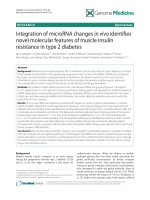
Regulation of DMT1 on bone microstructure in type 2 diabetes
Bạn đang xem bản rút gọn của tài liệu. Xem và tải ngay bản đầy đủ của tài liệu tại đây (1.49 MB, 9 trang )
Int. J. Med. Sci. 2015, Vol. 12
Ivyspring
International Publisher
441
International Journal of Medical Sciences
2015; 12(5): 441-449. doi: 10.7150/ijms.11986
Research Paper
Regulation of DMT1 on Bone Microstructure in Type 2
Diabetes
Wei-Lin Zhang†, Hong-Zheng Meng†, Mao-Wei Yang
Department of Orthopedics, the First Hospital of China Medical University, Shenyang, Liaoning, China
†
These authors contribute equally to this work.
Corresponding author: Mao-Wei Yang, Department of Orthopedics, The First Hospital of China Medical University, 155 North Nanjing
Street, Shenyang, Liaoning, 110001, China. E-mail: ; FAX: +86 24 83283360; Phone: +86 24 83283360
© 2015 Ivyspring International Publisher. Reproduction is permitted for personal, noncommercial use, provided that the article is in whole, unmodified, and properly cited.
See for terms and conditions.
Received: 2015.02.25; Accepted: 2015.05.18; Published: 2015.05.26
Abstract
Diabetic osteoporosis is gradually attracted people's attention. However, the process of bone
microstructure changes in diabetic patients, and the exact mechanism of osteoblast iron overload
are unclear. Therefore, the present study aimed to explore the function of DMT1 in the pathological process of diabetic osteoporosis. We build the type two diabetes osteoporosis models with
SD rats and Belgrade rats, respectively. Difference expression of DMT1 was detected by using the
method of immunohistochemistry and western blotting. Detection of bone microstructure and
biomechanics and iron content for each group of samples. We found that DMT1 expression in type
2 diabetic rats was higher than that in normal rats. The bone biomechanical indices and bone
microstructure in the rat model deficient in DMT1 was significantly better than that in the normal
diabetic model. The loss of DMT1 can reduce the content of iron in bone. These findings indicate
that DMT1 expression was enhanced in the bone tissue of type 2 diabetic rats, and plays an important role in the pathological process of diabetic osteoporosis. Moreover, DMT1 may be a
potential therapeutic target for diabetic osteoporosis.
Key words: DMT1, type 2 diabetes, osteoporosis, biomechanics, micro-CT
Introduction
Due to economic developments, the incidence of
chronic diseases such as diabetes is rising year by
year. Various complications caused by diabetes significantly affect human health. A survey suggested
that the risk of fractures in patients with diabetes is
much higher than that in patients without diabetes[1].
A variety of complications in diabetics related to
fractures affect the patient's quality of life and health,
and result in a heavy economic burden and effects on
society. Therefore, a study on the mechanism of diabetic bone microstructure changes is necessary to
prevent fractures.
The relationship between iron overload and osteoporosis has previously been confirmed. Weiss G
found excessive deposition of iron in patients with
hemochromatosis, and 63% of patients developed
osteoporosis[2]. Chen B and other researchers found
that iron overload had an inhibitory effect on osteogenesis[3]. However, the process of bone microstructure changes in diabetic patients, and the exact
mechanism of osteoblast iron overload are unclear.
Divalent metal ion transporter 1 (DMT1) transports metal ions across membranes in mammals. The
transporter is widely distributed in the human body.
Studies have shown that DMT1 transports iron into
epithelial cells in the intestinal membrane[4]. In Belgrade rats with DMT1 gene mutations, iron was not
transferred into the cytoplasm, and the iron eventually returned to the cell surface, demonstrating that
DMT1 is necessary for iron to be released into the
cytoplasm[5].
The mechanism involved in bone microstructure
changes in diabetes may be related to increased expression of DMT1in bone tissue, which promotes the
Int. J. Med. Sci. 2015, Vol. 12
release of iron ions from osteoblasts, causing iron
overload in cells, leading to bone microstructure
changes resulting in increased bone fragility and an
increased risk of fracture. In the present study, Belgrade rats were used to verify the above hypothesis,
which if confirmed will provide a new method way
for studying the theory of diabetic osteoporosis, and
provide potential therapeutic targets for the clinical
treatment of osteoporosis.
Materials and Methods
The experimental design fully complies with the
randomized controlled trial principle
Ethics Statement
The institutional Ethics Review Board of the First
Hospital of China Medical University approved the
study. The using of animal in our experiments is consistent with ethical requirements. All activities associated with this research project will be performed in
accordance with the First Hospital of China Medical
University Institutional Guidelines and Clinical Regulations.
Experimental animals
Male SPF SD rats, 3-months old, weighing 200±
20g were purchased from China Medical University,
Department of Experimental Animals(Animal Certificate Number: SCXK (Liaoning) 2008-0005). Male
Belgrade rats, the Belgrade rat is an animal model of
DMT1 deficiency, 3-months old, weighing 200± 20g
were purchased from the Rat Resource & Research
Center at the University of Missouri, USA. 20 SD rats
(10 rats were used to establish a type 2 diabetes model
and 10 used for comparison) were used to determine
the differential expression of DMT1. 30 SD rats and 15
Belgrade rats (15 SD rats were used to establish a
normal type 2 diabetes model group, 15 SD rats were
included in the sham group, 15 Belgrade rats were
used to establish a type 2 diabetes Belgrade model
group) were included to determine the targets of
biomechanics and bone microstructure.
Models and specimen collection
The rats received a high-fat diet for two months
and were allowed water for 12 hours/day. The rats
were given an intraperitoneal injection of streptozotocin (STZ) at a dose of 30mg/kg. After 72 hours,
fasting plasma glucose > 7.8mmol/Land reduced
insulin sensitivity were observed and the models
were successfully established[6]. The rats not used for
modeling were fed a normal diet. All rats were
housed under standard laboratory conditions and
maintained under controlled temperature (22 ± 3℃)
and humidity conditions with a daily cycle of 12 h
442
light and 12 h dark. The weight of the rats was maintained between 220g and 270g, and blood glucose was
maintained between 5mmol/Land 18mmol/L. Rats
with values outside these ranges were eliminated. The
rats were fed up to 8 months of age, killed by cervical
dislocation, the tibia was immediately removed aseptically and placed into fresh4% phosphate buffered
formalin solution, and stored at 4℃ in a refrigerator.
Immunohistochemistry
Tissue sections (5µm) were incubated with rabbit
anti-rat DMT1 (1:800; Santa Cruz Biotechnology)
primary antibody. Horseradish peroxidase-labeled
goat anti-rabbit (1:400; Santa Cruz Biotechnology)
secondary antibody was used. Semi-quantitative
analysis was performed at 200× magnification per
visual field (0.145 mm2) for DMT1 extravasation, using imaging software (ImagePro Plus 6.0; Media Cybernetics, Bethesda, MD, USA). The mean IOD values
were analyzed and averaged. The semi quantitative
analysis of immunohistochemical results on the basis
of the positive cell percentage ratio and tinting
strength.-and± judged as the negative, + and + +
judged as positive, the positive cells : 0% recorded as 0
points, ≤25% recorded as 1 points, 26-50% recorded as
2 points, 51-75% recorded as 3 points, >75% recorded
as 4 points. Coloring intensity: no color recorded as 0
points, Light yellow recorded as 1 point, Claybank
recorded as 2 points, Brown recorded as 3 points. Two
results are combined: 0 points for (-), 2-3 points for (
±), 4-5 points for (+),6-7 points for (+ +).
Bone biomechanical test
The rat tibias were analysis by Biomechanical
properties with 858 Mini Bionix materials testing
systems. The tibias were placed on rheometer
three-point bending test , loading rate was 0.01mm.s-1,
a span of 15mm. Amount of inner and outer middle
backbone by the load , degree of conversion and draw
radial stress-strain curve. We got maximum strength
and elastic modulus through this curve.
Micro-CT scan
Fixed the handle good right distal femur (truncated) along the long axis perpendicular to the specimen in the sample holder, viva CT 40 to select scan
parameters : image matrix of 1024 × 1024, Integration
time (integration time) for 200 ms, energy/intensity
for 70 kVp, 114 μA, 8W. After the scan is complete,
select from the distal growth plate 1.0mm, 3.0mm
thickness of the bone tissue is interested in cancellous
bone area (region of interest, ROI) line of reconstruction, the lowest threshold of 190 extracts image information. After recombinant images using Micro-CT
comes with software for quantitative analysis. Physi
Int. J. Med. Sci. 2015, Vol. 12
cal parameters as follows: bone mineral density
(BMD), bone volume/total volume (BV/TV), trabecular number (Tb.N), trabecular thickness (Tb.Th).
Haematoxylin and eosin (HE) staining
Tissues were fixed in 4% paraformaldehyde/
phosphatebuffered saline (PBS), post-fixed with the
same fixative and cut into 16 μm sections on a freezing
microtome. Standard HE staining was performed and
degeneration grade was scored by two independent
observers as previously reported[7].
Western blotting
Proteins of NP tissue and cultured cells were
lysed by Lysis Buffer containing PMSF on ice. The
extracted proteins were electrophoresed through 12%
SDS polyacrylamide gels and transferred to a PVDF
membrane (Invitrogen). After being blocked in PBS
containing 5% fat-free milk powder for 1 h, antibodies
against DMT1 (Abcam, Cambridge, MA,USA) were
used to detect the proteins. Goat anti-rabbit immunoglobulin conjugated to horseradish peroxidase (Sigma, St. Louis, MO, USA) was used as the secondary
antibody. Signals were detected using Pierce ECL
western blotting substrate (Pierce Biotechnology,
Rockford, CA, USA).
Tibia detect iron content
The tibia dried to constant weight, referred to the
dry weight, is placed 650℃calcined to the powder in a
muffle furnace, grind with a mortar sufficiently Pieces, accurately weighed sample taken post-ash 0.05g,
was added into 1mL 1: 1 HNO3, 0.2 mL 1: 1 HCl solution a mixed solution of Solution with ultrapure water
(18.2 megohms) volume to 10 mL, and then fully dissolve ultrasound to translucent, in inductively coupled plasma Daughter emission spectrometer
(ICP-AES, the US Perkin Elmer Corporation, Model :
Optima 2100DV) adopted on Determination of iron
content by atomic absorption spectrometry.
Plasma measurements
Venous blood (tail vein) was collected before
experimentation to measure fasting concentrations of
blood glucose (FBG) (Roach blood glucose instrument). Intraocular angular vein blood (2.5-4mL) was
collected for measurement of fasting plasma insulin
(FINS) by radioimmunoassay (3v-diagnostic Bioengineer, Shandong, China) and plasma estrogen by
ELISA (Rat Estrogen/E ELISA Kit, 3v-Diagnostic Bioengineer, Shandong, China). The insulin sensitivity
index (ISI) was calculated using the formula
(1/FBG×FINS)[8].
Statistical analysis
Two-group comparisons were performed using
443
Student’s t-test. Multiple group parameters comparisons were performed using one-way analysis of variance followed by Turkey’s post-test. A P value less
than 0.05 was considered statistically significant. The
statistical analysis was performed using the SPSS statistical package (SPSS, Chicago, IL, USA).
Results
Correlation between DMT1 and diabetic
osteoporosis
The tibias removed from SD rats which were
used as a model of type II diabetic osteoporosis were
analyzed by immunohistochemistry and western
blotting. The expression level of DMT1 in the type 2
diabetic osteoporosis model was found to be higher
than that in normal rats (Figure 1).
Evaluation of the type 2 diabetic osteoporosis
model
Fasting plasma glucose and fasting plasma insulin were determined in each group of rats, and the
insulin sensitivity index (ISI) was calculated. We
found that the normal model group and the Belgrade
model group conformed to the standard, and were
regarded as successful models (Figure 2, Table 1).
Table 1. Each group of data
sham
0.277±0.010
79.230±10.200
139.900±12.000
5.460±1.000
normal
0.207±0.017
150.230±11.500
106.100±9.800
5.310±0.800
belgrade
0.245±0.004
65.510±13.600
122.500±10.300
5.030±0.600
25.100±1.400
44.400±5.300
1.770±0.200
19.100±2.600
23.300±5.200
1.210±0.100
22.210±1.600
33.300±5.200
1.520±0.150
FINS (U/L)
23.5400±12.200
79.300±17.000
80.000±15.000
FBG (mmol/L)
4.300±15.000
8.300±2.000
8.500±1.600
BMD (g/cm2)
Iron content (μg/g)
MaxStrength (MPa)
ElasticModulus
(KPa)
Tb.N (cm-1)
BV/TV (%)
Tb.Th (cm-1)
Effects of DMT1 on bone mineral density and
iron content
The bone mineral density and iron content in
each rat tibia were determined. We found that the
bone mineral density in the Belgrade model group
and normal model group was lower than that in the
sham group. However, bone mineral density in the
Belgrade model group was higher than that in the
normal model group. The iron content in the normal
model group was higher than that in the sham group,
and the iron content in the Belgrade model group was
lower than that in the sham group (Figure 3, Table 1).
Effects of DMT1 on bone microstructure
The rat tibias were scanned and analyzed using
micro-CT. Bone microstructure in the Belgrade model
Int. J. Med. Sci. 2015, Vol. 12
group was better than that in the normal model
group, however, bone microstructure in both model
groups was worse than that in the sham group. The
results of HE staining confirmed these findings (Figure 4, 5, Table 1).
Effects of DMT1 on bone biomechanics
Using the MaxStrength and ElasticModulus
tests on the tibia from each group of rats, we found
that bone biomechanics in the Belgrade model group
was better than those in the normal model group, but
the two groups were worse in terms of the MaxStrength indices than the sham group. However, no
statistical differences of in the ElasticModulus test
were observed between the groups (Figure 6, Table 1).
444
Discussion
In the type 2 diabetes Belgrade rat model, we
determined the function of DMT1 in diabetic osteoporosis, and found the following: (1) DMT1 expression in type 2 diabetic rats was higher than that in
normal rats. (2) The bone biomechanical indices in the
rat model deficient in DMT1 were significantly better
than those in the normal diabetic model.(3) Bone microstructure in the rat model deficient in DMT1 was
significantly better than that in the normal diabetic
model. (4) The loss of DMT1 can reduce the content of
iron in bone.
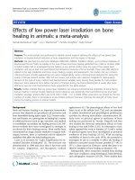
Figure 1. Correlation between DMT1 and diabetic osteoporosis. (A) Western blot analysis shows expression of DMT1 in diabetic osteoporosis group is
stronger than normal rats group. (B) The situation of DMT1 expression in bone tissue using immunohistochemical method. Diabetic osteoporosis group
significantly was stronger than normal rats group. Scale bars, 20μm. n=10 per group. Data are means ± SD. *P < 0.05.
Int. J. Med. Sci. 2015, Vol. 12
445
Figure 2. Evaluation of the type 2 diabetic osteoporosis model. No significant difference between Belgrade model group and normal model group, but
compared with sham group had significant difference, n=10 per group. Data are means ± SD. *P < 0.05.
Figure 3. Effects of DMT1 on bone mineral density and iron content. There were statistically significant differences between the each group. The bone
mineral density of Belgrade model group was higher than normal model group, but the two groups were lower than those in sham group. Iron content of
Belgrade model group was the lowest. Iron content of normal model group was the highest, n=10 per group. Data are means ± SD. *P < 0.05.
Int. J. Med. Sci. 2015, Vol. 12
446
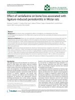
Figure 4. Effects of DMT1 on bone microstructure. Through the HE staining we observed the number and thickness of trabecular bone. Belgrade model
group was better than normal model group, but the two groups were worse than those in sham group. This result can also be verified by micro-CT image.
Scale bars, 100μm.
Figure 5. Effects of DMT1 on bone microstructure. According to the results of BV/TV, Tb.N, Tb.Th, Belgrade model group was better than normal model
group, but the two groups were worse than those in sham group, n=10 per group. Data are means ± SD. *P < 0.05.
Int. J. Med. Sci. 2015, Vol. 12
447
Figure 6. Effects of DMT1 on bone biomechanics. According to the results of MaxStrength and ElasticModulus tests, Belgrade model group was better
than normal model group, but the two groups were worse than those in sham group, n=10 per group. Data are means ± SD. *P < 0.05.
Diabetic osteoporosis belongs to secondary osteoporosis, which is a serious diabetes complications
in human skeletal system, the concept was first proposed in 1948[9]. It has recently become apparent that
the risk of osteoporosis-related bone fracture is increased in both type 1 and type 2 diabetic patients[10,
11]. Type 1 diabetes is generally associated with a
reduction in BMD, but the change of BMD in type 2
diabetic osteoporosis are not obvious [12], so the
pathogenesis of osteoporosis in type 2 diabetes compared with type 1 diabetes osteoporosis is more complex, and the incidence of type 2 diabetes is higher
than the incidence of type 1 diabetes, so this study
focuses on type 2 diabetes.
When the binding capacity of transferritin is
overwhelmed by a high iron concentration in the circulation and tissues, free iron is deposited in tissues
and thus creates a pathological condition called iron
overload[13]. Existing research shows that iron overload is associated with many diseases, such as hemochromatosis, sickle cell disease, and liver
diseases[14-16]. However, research has also suggested
that iron overload is associated with bone metabolism
abnormalities, such as osteopenia, osteoporosis, and
osteomalacia[17-20]. However, the mechanism of iron
overload in osteoporosis has not been extensively
studied. The present study, for the first time, showed
a difference in DMT1 expression between diabetic rats
and normal rats. This discovery is of great significance
in determining the mechanism of iron overload.
DMT1 is the major apical transporter responsible
for intestinal Fe2+ absorption, and is also ubiquitously
expressed in the endosomalcompartments where it is
responsible for Fe2+ export from the endosome during
the transferrin cycle[21, 22]. Thus, DMT1 expression is
closely related to iron overload. DMT1 is not only
involved in iron and manganese metabolism, but is
also involved in the uptake of other metals. Research
has shown that DMT1 also participates in Cu2+ and
Cd2+ transport[23, 24]. Due to the characteristics of
DMT1, there are currently many research studies being carried out. However, to date, there is no relevant
research on the relationship between DMT1 and osteoporosis. For the first time, we found that DMT1
affects the biological characteristics of bone and bone
microstructure. And we confirmed the effect of DMT1
on iron content in bone tissue. This discovery has
highlighted the important role of DMT1 in the process
of osteoporosis.
Belgrade rats were described for the first time in
1966 and were the offspring of an X-irradiated albino
rat in Belgrade, Yugoslavia[25]. The Belgrade rat is an
animal model of DMT1deficiency.This deficiency is
due to a glycine-to-arginine substitution (G185R) in
the fourth putative transmembranedomain of DMT1
resulting in loss of activity of the transporter[26].
There are multiple examples where the Belgrade rat,
as a model of iron deficiency, has been useful in
characterizing not only the role of DMT1 in the
transport of this metal, but also its contribution to
pathologies of intermediary metabolism, its protective
role in detoxification of the lungs, its participation in
neurotoxicity of airborne metal uptake by the olfactory pathway, in the development of the kidneys, in
promoting altered renal function, in brain iron metabolism and in hepatic iron handling[27-29]. How
Int. J. Med. Sci. 2015, Vol. 12
ever, the reason why these rats were chosen for this
study was due to their stable lack of DMT1. In addition, interference and individual differences are small,
and these rats have many other advantages. This
study is the first to establish a diabetic osteoporosis
model using Belgrade rats. This is of significant importance in the study of DMT1 and osteoporosis.
Laboratory animals have played a key role in the
unprecedented recent improvements in the management of osteoporosis. Animal models of osteoporosis
involve a variety of animals and a variety of methods.
Each model has its own advantages, disadvantages
and scope of use[30, 31]. This study used a rat model
of diabetes mellitus combined with osteoporosis established using intralipid and a small dose of streptozotocin. Because our research goal was to explore
the mechanism of type 2 diabetes complicated by osteoporosis, the model needed to imitate the pathological process of type 2 diabetes. This model was
helpful in our study. Three-month old rats are sexually mature, 6-monthold rats have mature bones,
and 17-month old rats are relatively old[32]. We used
8-monthold rats, as the rat bones were fully mature
and completely affected by diabetes. The bone characteristics of these rats sufficiently reflected the effects
of the differential expression of DMT1. Type 1 diabetes is the absolute lack of insulin, and the main characteristic of type 2 diabetes is insulin resistance. We
tested the ISI in order to assess whether this model is
type 2 diabetes model.
There are several limitations in the present
study. For example, although our data demonstrated
the relationship between DMT1 and osteoporosis, we
did not perform in vitro experiments. This study focused on the effects of DMT1 on diabetic osteoporosis,
which has laid the foundation to explore the specific
mechanism involved and indicate the direction for
future research. Further in-depth studies on this topic
are required.
In conclusion, DMT1 expression was enhanced
in the bone tissue of type 2 diabetic rats, and plays an
important role in the pathological process of diabetic
osteoporosis. Moreover, DMT1 may be a potential
therapeutic target for diabetic osteoporosis.
Acknowledgement
This study was supported by the Chinese National Natural Science Foundation Project, Fund of
liaoning province department of education and
Shenyang municipal science and technology fund
(81471094, 81170808, L2013301 and F12-277-1-47).
Author Contributions
WLZ, HZM, MWY conceived of the study, participated in the design of the study and performed the
448
statistical analyses. All authors carried out the experiments. WLZ drafted the manuscript with the help
of HZM and YMW. All authors have read and approved the final manuscript
Competing Interests
The authors declare no competing financial interests.
References
1.
2.
3.
4.
5.
6.
7.
8.
9.
10.
11.
12.
13.
14.
15.
16.
17.
18.
19.
Jepsen KJ, Schlecht SH. Biomechanical mechanisms: resolving the apparent
conundrum of why individuals with type II diabetes show increased fracture
incidence despite having normal BMD. Journal of bone and mineral research :
the official journal of the American Society for Bone and Mineral Research.
2014; 29: 784-6. doi:10.1002/jbmr.2189.
Weiss G. Genetic mechanisms and modifying factors in hereditary
hemochromatosis. Nature reviews Gastroenterology & hepatology. 2010; 7:
50-8. doi:10.1038/nrgastro.2009.201.
Chen B, Yan YL, Liu C, Bo L, Li GF, Wang H, et al. Therapeutic effect of
deferoxamine on iron overload-induced inhibition of osteogenesis in a
zebrafish model. Calcified tissue international. 2014; 94: 353-60.
doi:10.1007/s00223-013-9817-4.
Andrews NC, Schmidt PJ. Iron homeostasis. Annual review of physiology.
2007; 69: 69-85. doi:10.1146/annurev.physiol.69.031905.164337.
Sheftel AD, Mason AB, Ponka P. The long history of iron in the Universe and
in health and disease. Biochimica et biophysica acta. 2012; 1820: 161-87.
doi:10.1016/j.bbagen.2011.08.002.
Li B, Wang Y, Liu Y, Ma J, Li Y. Altered gene expression involved in insulin
signaling pathway in type II diabetic osteoporosis rats model. Endocrine. 2013;
43: 136-46. doi:10.1007/s12020-012-9757-1.
Sive JI, Baird P, Jeziorsk M, Watkins A, Hoyland JA, Freemont AJ. Expression
of chondrocyte markers by cells of normal and degenerate intervertebral discs.
Molecular pathology : MP. 2002; 55: 91-7.
Vangipurapu J, Stancakova A, Kuulasmaa T, Soininen P, Kangas AJ,
Ala-Korpela M, et al. Association between liver insulin resistance and
cardiovascular risk factors. Journal of internal medicine. 2012; 272: 402-8.
doi:10.1111/j.1365-2796.2012.02540.x.
Takamoto I, Kadowaki T. [Diabetes and osteoporosis]. Clinical calcium. 2004;
14: 255-61. doi:CliCa0402255261.
Merlotti D, Gennari L, Dotta F, Lauro D, Nuti R. Mechanisms of impaired bone
strength in type 1 and 2 diabetes. Nutrition, metabolism, and cardiovascular
diseases : NMCD. 2010; 20: 683-90. doi:10.1016/j.numecd.2010.07.008.
Vestergaard P. Discrepancies in bone mineral density and fracture risk in
patients with type 1 and type 2 diabetes--a meta-analysis. Osteoporosis
international : a journal established as result of cooperation between the
European Foundation for Osteoporosis and the National Osteoporosis
Foundation of the USA. 2007; 18: 427-44. doi:10.1007/s00198-006-0253-4.
Dominguez LJ, Muratore M, Quarta E, Zagone G, Barbagallo M. [Osteoporosis
and diabetes]. Reumatismo. 2004; 56: 235-41.
Li GF, Pan YZ, Sirois P, Li K, Xu YJ. Iron homeostasis in osteoporosis and its
clinical implications. Osteoporosis international : a journal established as
result of cooperation between the European Foundation for Osteoporosis and
the National Osteoporosis Foundation of the USA. 2012; 23: 2403-8.
doi:10.1007/s00198-012-1982-1.
Guggenbuhl P, Fergelot P, Doyard M, Libouban H, Roth MP, Gallois Y, et al.
Bone status in a mouse model of genetic hemochromatosis. Osteoporosis
international : a journal established as result of cooperation between the
European Foundation for Osteoporosis and the National Osteoporosis
Foundation of the USA. 2011; 22: 2313-9. doi:10.1007/s00198-010-1456-2.
Sarrai M, Duroseau H, D'Augustine J, Moktan S, Bellevue R. Bone mass
density in adults with sickle cell disease. British journal of haematology. 2007;
136: 666-72. doi:10.1111/j.1365-2141.2006.06487.x.
Wibaux C, Legroux-Gerot I, Dharancy S, Boleslawski E, Declerck N, Canva V,
et al. Assessing bone status in patients awaiting liver transplantation. Joint,
bone,
spine
:
revue
du
rhumatisme.
2011;
78:
387-91.
doi:10.1016/j.jbspin.2011.03.001.
Guggenbuhl P, Deugnier Y, Boisdet JF, Rolland Y, Perdriger A, Pawlotsky Y,
et al. Bone mineral density in men with genetic hemochromatosis and HFE
gene mutation. Osteoporosis international : a journal established as result of
cooperation between the European Foundation for Osteoporosis and the
National Osteoporosis Foundation of the USA. 2005; 16: 1809-14.
doi:10.1007/s00198-005-1934-0.
Isomura H, Fujie K, Shibata K, Inoue N, Iizuka T, Takebe G, et al. Bone
metabolism and oxidative stress in postmenopausal rats with iron overload.
Toxicology. 2004; 197: 93-100. doi:10.1016/j.tox.2003.12.006.
Mahachoklertwattana P, Sirikulchayanonta V, Chuansumrit A, Karnsombat P,
Choubtum L, Sriphrapradang A, et al. Bone histomorphometry in children
and adolescents with beta-thalassemia disease: iron-associated focal
Int. J. Med. Sci. 2015, Vol. 12
20.
21.
22.
23.
24.
25.
26.
27.
28.
29.
30.
31.
32.
449
osteomalacia. The Journal of clinical endocrinology and metabolism. 2003; 88:
3966-72. doi:10.1210/jc.2002-021548.
Matsushima S, Hoshimoto M, Torii M, Ozaki K, Narama I. Iron
lactate-induced osteopenia in male Sprague-Dawley rats. Toxicologic
pathology. 2001; 29: 623-9.
Fleming MD, Romano MA, Su MA, Garrick LM, Garrick MD, Andrews NC.
Nramp2 is mutated in the anemic Belgrade (b) rat: evidence of a role for
Nramp2 in endosomal iron transport. Proceedings of the National Academy of
Sciences of the United States of America. 1998; 95: 1148-53.
Gunshin H, Mackenzie B, Berger UV, Gunshin Y, Romero MF, Boron WF, et al.
Cloning and characterization of a mammalian proton-coupled metal-ion
transporter. Nature. 1997; 388: 482-8. doi:10.1038/41343.
Illing AC, Shawki A, Cunningham CL, Mackenzie B. Substrate profile and
metal-ion selectivity of human divalent metal-ion transporter-1. The Journal of
biological chemistry. 2012; 287: 30485-96. doi:10.1074/jbc.M112.364208.
Jiang L, Ranganathan P, Lu Y, Kim C, Collins JF. Exploration of the
copper-related compensatory response in the Belgrade rat model of genetic
iron deficiency. American journal of physiology Gastrointestinal and liver
physiology. 2011; 301: G877-86. doi:10.1152/ajpgi.00261.2011.
Sladic-Simic D, Zivkovic N, Pavic D, Marinkovic D, Martinovic J, Martinovitch
PN. Hereditary hypochromic microcytic anemia in the laboratory rat.
Genetics. 1966; 53: 1079-89.
Fleming MD, Trenor CC, 3rd, Su MA, Foernzler D, Beier DR, Dietrich WF, et
al. Microcytic anaemia mice have a mutation in Nramp2, a candidate iron
transporter gene. Nature genetics. 1997; 16: 383-6. doi:10.1038/ng0897-383.
Ferguson CJ, Wareing M, Delannoy M, Fenton R, McLarnon SJ, Ashton N, et
al. Iron handling and gene expression of the divalent metal transporter,
DMT1, in the kidney of the anemic Belgrade (b) rat. Kidney international.
2003; 64: 1755-64. doi:10.1046/j.1523-1755.2003.00274.x.
Thompson K, Molina RM, Brain JD, Wessling-Resnick M. Belgrade rats
display liver iron loading. The Journal of nutrition. 2006; 136: 3010-4.
Thompson K, Molina RM, Donaghey T, Brain JD, Wessling-Resnick M. Iron
absorption by Belgrade rat pups during lactation. American journal of
physiology Gastrointestinal and liver physiology. 2007; 293: G640-4.
doi:10.1152/ajpgi.00153.2007.
Lelovas PP, Xanthos TT, Thoma SE, Lyritis GP, Dontas IA. The laboratory rat
as an animal model for osteoporosis research. Comparative medicine. 2008; 58:
424-30.
Turner RT, Maran A, Lotinun S, Hefferan T, Evans GL, Zhang M, et al. Animal
models for osteoporosis. Reviews in endocrine & metabolic disorders. 2001; 2:
117-27.
Luippold G, Klein T, Mark M, Grempler R. Empagliflozin, a novel potent and
selective SGLT-2 inhibitor, improves glycaemic control alone and in
combination with insulin in streptozotocin-induced diabetic rats, a model of
type 1 diabetes mellitus. Diabetes, obesity & metabolism. 2012; 14: 601-7.
doi:10.1111/j.1463-1326.2012.01569.x.