Summary of environmental technique doctoral thesis: Synthesis of silver, copper, iron nanoparticles and their applications in controlling cyanobacterial blooms in the fresh water body
Bạn đang xem bản rút gọn của tài liệu. Xem và tải ngay bản đầy đủ của tài liệu tại đây (1.3 MB, 27 trang )
MINISTRY OF EDUCATION
AND TRAINING
VIETNAM ACADEMY OF
SCIENCE AND TECHNOLOGY
GRADUATE UNIVERSITY SCIENCE AND TECHNOLOGY
---------------------------
TRAN THI THU HUONG
SYNTHESIS OF SILVER, COPPER, IRON
NANOPARTICLES AND THEIR APPLICATIONS IN
CONTROLLING CYANOBACTERIAL IN THE FRESH
WATER BODY
Major: Environmental Technique
Code: 9 52 03 20
SUMMARY OF ENVIRONMENTAL TECHNIQUE
DOCTORAL THESIS
HaNoi - 2018
The thesis was completed at the Graduate University of Science
and Technology, Vietnam Academy of Science and Technology
Scientific Supervisor 1: Assoc. Prof. Dr. Duong Thi Thuy
Scientific Supervisor 2: Dr. Ha Phuong Thu
Reviewer 1:
Reviewer 2:
Reviewer 3:
The dissertation will be defended protected at the Council for
Ph.D. thesis, meeting at the Viet Nam Academy of Science and
Technology - Graduate University of Science and Technology.
Time: Date …… month …. 2018
This thesis can be found at:
- The library of the Graduate University of Science and Technology.
- National Library of Viet Nam.
1
INTRODUCTION OF THESIS
1. The necessary of the thesis
In recent years, pollution of soil, water and air has become a
serious problem not only in Vietnam but also in many parts of the
world in which the water pollution is more serious problem.
"Water blooming" is the development of microalgae outbreak,
especially cyanobacteria in fresh water bodies and often cause the
harmful effects on the environment such as: the water turbidity and
pH are increase, the levels of dissolved oxygen is reduce due to the
respiration or degradation of algae biomass and especially, the fact
that most cyanobacteria produce the toxicity high. The preventing
and minimizing the development of cyanobacteria is an important
environmental issue that need to pay the attention. The many
methods have been used such as: chemistry, mechanics, biology,
etc., but they are ineffective and expensive, affecting ecosystem
and conducting is difficult, especially in large water bodies.
Therefore, the search and development of new effective solutions
without secondary pollution and friendly with the environment are
increasingly focused research. Nanotechnology is the technology
relating to the synthesis and application of materials with
nanometer sizes (nm). At nanoscale, the material has many
advantage features such as: size is smaller than 100 nm, larger
surface to volume ratio, crystalline structure, high reactivity
potential, creating the effect of resonance Plasmon surface; high
adhesion potential and the nanomaterial was applied in various
fields such as: medical, cosmetics, electronics, chemical catalyst,
environment... For the above reasons, the thesis is proposed as:
“Synthesis of silver, copper, iron nanoparticles and their
applications in controlling cyanobacterial blooms in the fresh
water body” was selected to researched.
2. The objectives of the thesis
Research, fabricate and determine the characteristic of three
nanomaterials (silver, copper and iron) and evaluate the ability to
inhibit the cyanobacteria of nanomaterials in fresh water bodies.
3. The main contents of the thesis
- Fabricate and determine the characteristic of three
nanomaterials: silver, copper and iron.
2
- Investigate the ability to inhibit and prevent cyanobacteria of
three nanomaterials.
- Assess the safety of materials and their application.
- Experimental application of materials at laboratory-scale with
the Tien lake water sample.
5. The structure of the thesis
The thesis is composed of 149 pages, 10 tables, 62 figures, 219
references. The thesis consists of three parts: Introduction (3 pages);
chapter 1: Literature review (42 pages); chapter 2: Methodology (16
pages); chapter 3: Resutl and discussion (59 pages); Conclusion and
recommendation (2 pages).
CHAPTER 1. LITERATURE REVIEW
1.1. Introduction of nanomaterial
1.2. Introduction of Cyanobacteria and Eutrophication
1.3. Introduction of the methods to treat the toxic algae
contamination
CHAPTER 2. METHODOLOGY
2.1. The research subjects
2.2. The equipment is used in study
2.3. The methods for synthesis of materials
2.3.1. Synthesis of silver nanomaterial by chemical reduction
method
The silver nanomaterial was synthesized by chemical reduction
method, ion Ag+ in the silver salt solution is reducted to Ag0 by the
reducing agent NaBH4.
2.3.2. Synthesis of copper nanomaterial by chemical reduction
method
The copper nanomaterial was synthesized by chemical
reduction method, ion Cu2+ in the copper salt solution is reduced to
Cu0 by the reducing agent NaBH4.
2.3.3. Synthesis of iron magnetic (Fe3O4) nanomaterial by
simultaneously precipitation method
The iron magnetic (Fe3O4) nanomaterial was synthesized by
simultaneously precipitation method of Fe2+ and Fe3+ salts by
NH4OH.
2.4. The methods for determining the characteristic of material
structure
3
The morphology of the three nanomaterials is determined by a
number of methods such as: TEM, SEM, IR, XRD, UV-VIS, EDX.
2.5. The experimental setup methods
The experimental setup methods such as: culture of algae,
selection of nanomaterials, evaluation of the material toxicity, the
evaluation of the influence of nanomaterial sizes and the safety of
nanomaterials on microalgae and the experiment with the Tien lake
water sample were setup.
2.6. The methods of evaluating the effect of nanomaterials on
the growth of microalgae
To evaluate the effect of nanomaterials on the growth of
microalgae, the following methods such as: OD, chlorophyll a, cell
density, the methods for analysis of some environmental quality
indicators (NH4+, PO43-) and SEM, TEM were used.
2.7. The method of statistical analysis
CHAPTER 3. RESUTL AND DISCUSSION
3.1. Synthesis of nanomaterial
3.1.1. Synthesis of silver nanomaterial by chemical reduction
method
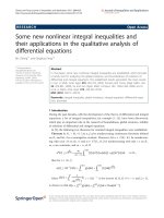
3.1.1.1. Effect of the concentration ratio NaBH4/Ag+
The UV-VIS spectrophotometer (Fig 3.1) showed that the
nanosilver colloid was absorbed at the wavelengths about 400 nm
and the synthesized efficiency of silver nanoparticles was
maximum achieved at a ratio 1:2. TEM images (Figure 3.2)
showed that silver nanoparticle size was less than 20 nm.
M1
M3
Figure 3.1. The UV-VIS spectra
of nanosilver colloid depends on
the NaBH4/Ag+ concentration
ratios
M2
M4
M5
Figure 3.2. The TEM images of
nanosilver colloid depends on
the BH4-/Ag+ concentration ratio
4
3.1.1.2. Effect of stabilizer concentration chitosan
The UV-VIS measurements in Figure 3.4 showed that the
nanosilver colloid is absorbed at the wavelengths 402-411 nm. The
TEM image of the silver nanoparticles depends on the
concentration of chitosan shown in Figure 3.5. The optimum
chitosan concentration of nanosilver colloid fabricating was chosen
as 300 mg/L.
M6
M7
M8
M9
M10
Figure 3.4. The UV-VIS spectra
Figure 3.5. The TEM images
of nanosilver colloid depends on
of nanosilver colloid depends
chitosan concentrations
on the chitosan concentrations
3.1.1.3. Effect of citric acid concentration
The UV-VIS measurements in Figure 3.7 showed that the
nanosilver colloid is absorbed at the wavelengths 402-411 nm. At
the rate of [Citric]/[Ag+] = 3.0 the silver nanoparticles obtained
were of the most uniform, small size and less than 20 nm, the TEM
measurement is shown in Figure 3.8.
M11
M12
M13
M14
M15
M16
Figure 3.7. The UV-VIS
spectra of nanosilver colloid
depends on acid concentration
Figure 3.8. The TEM images of
nanosilver colloid depends on
the [Citric]/[Ag+] concentration
5
Figure 3.9. The HR-TEM of nanosilver colloid was tested at
optimal ratio
The structure of silver nanoparticle at the optimum ratio indicates
that they have a typical hexagon crystal structure of metallic
nanoparticles. The HR-TEM images in Figure 3.9 showed that the
crystals has got Fcc (Face-centered cubic) structure. The silver
nanomaterial at the conditions such as: the ratio of NaBH4/Ag+ is
1/4, the [Citric]/[Ag +] is 3.0 and a concentration of chitosan
stabilizer is 300 mg/L were synthesized to experimented the effect of
material on the growth of the studied subjects in the thesis.
3.1.2. Synthesis of copper nanomaterial by chemical reduction
method
3.1.2.1. Effect of the concentration ratio NaBH4/Cu2+
The results in Figure 3.10 show that, in the XRD spectrum
appears the three peak with the intensity match for the standard
spectra of the copper metal at the side (111), (200), (220)
corresponding to angle 2θ = 43.3; 50.4 and 74.00 belong to the
Bravais network in the fcc structure of the copper metal.
M2
M1
M3
M4
M5
Figure 3.10. The XRD pattern
Figure 3.11. The SEM images
of CuNPs were tested in
of CuNPs in NaBH4/Cu2+ ratio
NaBH4/Cu2+ concentration
The SEM measurements (Fig 3.11) of the material were
performed to determine the distribution of the copper particles and
6
the TEM measurement for determine the size of copper
nanoparticles (Fig 3.12).
M1
M3
M2
M4
M5
Figure 3.13. The XRD
spectrum of CuNPs was tested
by Cu0 concentration
The TEM image results showed that, when the NaBH4/Cu2+
concentration ratio is 1: 1 and 1.5: 1, the size of synthesized copper
nanoparticles are bigger than 50 nm. The nanoparticles are
distributed rather uniformly with a size about 20-50 nm when the
NaBH4/Cu2+ ratio is 2 : 1. The nanoparticles are clumped together,
unevenly distributed with the size nanoparticle > 50 nm when the
NaBH4/Cu2+ ratio is 3: 1 and 4: 1 and match with the SEM results.
To respone the objective of this thesis,
the M3 sample
2+
(NaBH4/Cu ratio is 2: 1) was chosen as the representative sample.
3.1.2.2. Effect of Cu0 concentration
XRD spectrum in Figure 3.13 showed that the of copper
nanoparticles presents the characteristic peaks of copper
nanomaterial. The characteristic peaks on the schematic have the
sharpness intensity and the wide range of the absorption peak
relatively narrow. In addition, the XRD spectrum of the material
also shows the characteristic peaks of CuO, Cu2O crystals.
The SEM (Fig 3.14) measurement results showed that, the
copper nanoparticles form of the unequal size distribution when the
concentration of Cu0 increases. At concentrations of Cu0 is 2g/L,
the copper nanoparticles are distributed rather uniformly with the
size at 20-40 nm. When the concentration of Cu0 increases to 3;
4g/L, the synthesized copper particles will clump together and
form of the particle sizes >50 nm; at Cu0 concentration is 6, 7 g/L,
Figure 3.12. The TEM images
of CuNPs in NaBH4/Cu2+ ratio
7
the nanoparticles distributed unevenly and match for the TEM
measurement (Fig 3.15).
N2
N1
N3
N4
N3
N5
Figure 3.14. The SEM image of
copper nanomaterial was tested at
Cu0 concentration
a)
3000
N2
N1
N5
N4
Figure 3.15. The TEM image
of copper nanomaterial was
tested at Cu0 concentration
b)
Faculty of Chemistry, HUS, VNU, D8 ADVANCE-Bruker - Cu-51
2900
2800
2700
2600
2500
2400
2300
d=2.089
2200
2100
2000
1900
Lin (Cps)
1800
1700
1600
1500
1400
1300
1200
d=1.808
1100
1000
900
800
d=1.278
700
600
500
400
300
200
100
0
c)
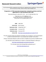
Figure 3.16. The detail characteristics of the N1 copper
nanomaterials sample: (a) SEM image, (b) TEM image, (c) XRD
spectrum
The structure of copper nanomaterial at selected ratio showed
that, the formed copper nanoparticles have
the rather
homogeneous surface (SEM image, Fig 3.16a), the uniformly size
in the range of 30 - 40 nm (TEM image, Fig 3.16b) and have the
Fcc structure with diffraction peaks of the netface (111), (200) and
(220) corresponding to angle 2θ = 43.3; 50.4 and 74.00 with high
intensity (XRD spectrum, Fig 3.16c). This material sample is
suitable with the objective of the thesis and were choosen for
further experiment.
1
10
20
30
40
50
60
70
2-Theta - Scale
File: ThuyVCNMT Cu-51.raw - Type: 2Th/Th locked - Start: 1.000 ° - End: 79.990 ° - Step: 0.030 ° - Step time: 0.3 s - Anode: Cu - WL1: 1.5406 - Generator kV: 40 kV - Generator mA: 40 mA - Creation: 06/10/2016 3:54:39 P
Left Angle: 42.490 ° - Right Angle: 44.350 ° - Obs. Max: 43.281 ° - d (Obs. Max): 2.089 - Max Int.: 1890 Cps - Net Height: 1668 Cps - FWHM: 0.231 ° - Raw Area: 852.6 Cps x deg. - Net Area: 440.4 Cps x deg.
01-085-1326 (C) - Copper - Cu - Y: 16.13 % - d x by: 1. - WL: 1.5406 - Cubic - a 3.61500 - b 3.61500 - c 3.61500 - alpha 90.000 - beta 90.000 - gamma 90.000 - Face-centered - Fm-3m (225) - 4 - 47.2416 - I/Ic PDF 8.9 - F4
1)
80
8
3.1.3. Synthesis of magnetic solution nanomaterial by coprecipitation method
3.1.3.1. Effect of the CMC stabilizer concentration
The tested result of morphological, size and the dispersion of
material in the ratio of CMC stabilizer and precursor (Fe3O4)
respectively were 1/1; 2/1; 3/1; 4/1 and 1/2 by the SEM and
methods shown in Figure 3.17 and 3.18. The SEM result showed
that the concentration of CMC in the solution is high, the
ferromagnetic nanoparticles are unevenly and the particle size is
big, the accumulation of nanoparticles is easy to occur. At the rate
of CMC/Fe3O4 is 2/1, the obtained ferromagnetic nanoparticles are
uniformly sized and less 20 nm.
Figure 3.17. The SEM image of Figure 3.18. The TEM image of
magnetic solution nanostructure magnetic solution nanostructure
tested in ratios of CMC/Fe3O4
tested in ratios of CMC/Fe3O4
The TEM results showed that the nanoparticle size varies
considerably when the CMC concentrations changed. When the
Fe3O4/CMC is 2:1, the obtained nanoparticles were the smallest,
most uniform and less than 20nm within the superparamagnetic size
range. Therefore, the material sample has a Fe3O4/CMC ratio of 2:1
(encoded sample is FC21) selected to tested for the further factors.
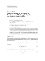
3.1.3.2. The result of infrared measurement of the material
Figure 3.19. The infrared spectrum
of Fe3O4 (a), CMC (b), FC21 (c) and
spectrum of three samples (d)
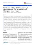
Figure 3.20. The
magnetization hysteresis
result of material FC21
9
The observation in Figure 3.19 showed that the IR spectrum of
ferromagnetic nanoparticles have peaks similar with CMC and
Fe3O4, this proves that the structure of CMC is not broken by the
material synthesis conditions. Therefore, the co-precipitation
method for synthesis of material is suitable for purity as well as
efficiency.
3.1.3.3. The magnetization hysteresis result of material
The result of saturate magnetization hysteresis measurement in
Figure 3.20 showed that ferromagnetic nanoparticles are in the
form of superparamagnetic. The saturate magnetization of Fe3O4
and FC21 is 68 emu/g and 49 emu/g, corresponding to the content
of magnetic phase of the material. The result proves that the
surface interaction of the magnetic phase with the polymer
decreased the saturate magnetization and suitable with the results
of the TEM analysis.
3.2. Evaluating the ability of growth inhibition and prevent
microalgae by synthesized nanomaterials
3.2.1. Study on the selection of concentrations of three types of
nanomaterials
Table 3.1. The screening results of removal M. aeruginosa KG
cyanobacteria of fabricated nanomaterials
No.
Samples
Experimental
concentration (mg/L)
The growth
inhibition of
cyanobacteria
1
3
5
6
Ag nano
Cu nano
Fe3O4 nano
Control
3, 5 and 10
3, 5 and 10
5, 10, 100, 150 and 200
0
+++
+++
-
Notes: +++: Very strong inhibitory effect, ++: Strong inhibitory effect, +:
Normal inhibitory effect, -: Non inhibitory effect.
Figure 3.21. Effect of nanomaterials on growth of cyanobacteria M.
aeruginosa KG after for 7 days.
10
The concentration screening tests were conducted to rapidly
assess inhibition effect to M. aeruginosa KG for 7 days. The
results in Table 3.1 and Figure 3.21 showed that the two silver and
copper nanomaterials inhibited the growth and development of
cyanobacteria M. aeruginosa KG after 6 days (Table 3.1 and Fig
3.21a, b), whereas that the ferromagnetic nanomaterial were not
effective against M. aeruginosa KG (Table 3.1 and Fig 3.21c).
3.2.2. Effect of silver nanoparticles on growth and development
of cyanobacteria M. aeruginosa KG and green algae C. vulgaris
3.2.2.1. Effect of silver nanoparticles on growth and development
of cyanobacteria M. aeruginosa KG
The experiments were conducted with the concentrations of
silver nanoparticles increasing from 0; 0.001; 0.005; 0.01; 0.05; 0.1
to 1 ppm in 10 days. The evaluation parameters include: optical
density (OD), chlorophyll a and cell density at 0, 2, 6 and 10 days
(Fig 3.22a, b). The toxicity of silver nanoparticles on growth of the
cyanobacteria M. aeruginosa KG as measured by the concentration
of supplementary material into the culture medium that affected
50% of the individuals (EC50) was 0.0075 mg/L.
Figure 3.22. Effect of silver
Figure 3.23. Effect of silver
nanomaterial on growth of the nanomaterial was measured by
cyanobacteria M. aeruginosa
the cell density (a) and the
KG after 10 days was
growth inhibition efficiency on
measured by (OD) (a),
cyanobacteria M. aeruginosa
chlorophyll a (b)
KG (b)
The cell density and chlorophyll a showed that, the cell density
and biomass in the control sample increased from the first day (D0)
(110,741 ± 6,317 cells/mL and 1.98 ± 0.06 μg/L, respectively) to the
end of experiment (D10) (5,475, 556 ± 541,274 cells/mL and 23.4 ±
2.96 μg/L, respectively) (Fig 3.23a). All five tested concentration
ranges are toxic to cyanobacteria M. aeruginosa KG. The growth
11
inhibition efficiency (Fig 3.23b) > 75% appears in only 4 tested
concentrations from 0.01; 0.05; 0.1 and 1 ppm.
The SEM image result of cell surface structure after 48h exposed
to silver nanoparticles at the concentration of 1 ppm is shown in
Figures 3.24a (the control sample) and 3.24b (the sample exposed to
the concentration of 1ppm silver nanoparticles). In the control sample,
the morphological of cyanobacteria M. aeruginosa KG cells
maintained a round and had a spherical shape with a smooth exterior
surface (Fig 3.24a). In the experimental sample, the cells were
changed to with a distorted and shrunk cell after exposure to silver
nanoparticles (Fig 3.24b). It is said that the silver nanoparticles have
significantly altered the morphology of the cell.
a)
b)
a)
b)
Figure 3.24. Scanning Electron
Figure 3.26. Transmission
Microscopy (SEM) micrograph of
Electron Microscopy (TEM)
M. aeruginosa KG
micrograph of M. aeruginosa KG
The SEM combined with EDX analysis was used to
characterize the chemical composition and the location of AgNPs
on the cell surface of M. aeruginosa KG. The EDX result in Figure
3.25 showed that the silver nanoparticles appear on the surface of
the cyanobacteria M. aeruginosa KG with 0.37% Ag by weight.
The TEM image in the control sample (Fig 3.26a), the M.
aeruginosa KG ultrastructure image had clearly cell wall and the
organelle lie neatly in the cell. When exposed to silver
nanoparticles at a concentration of 1ppm after 48 hours, the
cyanobacteria cells were destroyed (Fig 3.26b). It is proved that the
silver nanoparticles was affected to structure of the cyanobacteria
M. aeruginosa KG cell.
Elements
% Weight
% Element
CK
38.69
55.90
OK
30.59
33.18
Na K
1.95
1.47
Al K
6.02
3.87
Cu L
11.82
3.23
Ag L
0.37
0.06
12
Totals
100.00
Figure 3.25. The EDX spectrum and the element composition
appear on the cell surface of M. aeruginosa KG after 48 h of
exposure with AgNPs (1ppm)
3.2.2.2. Effect of silver nanoparticles on growth and development
of green algae Chlorella vulgaris
The experiments were conducted with the concentrations of
silver nanoparticles increasing from 0.005; 0.01; 0.05; 0.1; 1 to 5
ppm in 10 days. The evaluation parameters include: optical density
(OD), chlorophyll a and cell density at 0, 2, 6 and 10 days (Fig
3.27 b). The toxicity of silver nanoparticles on growth of the green
algae C. vulgaris as measured by the concentration of
supplementary material into the culture medium that affected 50%
of the individuals (EC50) was 0.017 mg/L.
Figure 3.28. Effect of silver
nanomaterial to the green algae
C. vulgaris was measured by
and the growth inhibition
efficiency (a) and the cell
density (b)
After 48h exposure to silver nanoparticles, the cell density
decreased from 195,925 ± 18,770 (D0) to 82,778 ± 41,384 (D10)
cells/mL (Fig 3.27a). At concentrations of 0.005 and 0.01 ppm,
AgNPs did not affect the growth of the green algae C. vulgaris, the
cell density after 2, 6 and 10 days increased linearly with control
samples. Figure 3.28b shows the analysis results of the chlorophyll
a, in the control sample and the experimental samples
supplemented with 0.005 and 0.01 ppm silver nanoparticles, the
content of chlorophyll a increased from 2.0604 ± 0.3505 μg/L (D0)
and reached to the highest value at the end of the testing period
27.285 ± 4.6893 µg/L (D10). The growth inhibition efficiency of
silver nanomaterial concentrations after 10 days is shown in Figure
Figure 3.27. Effect of
silver nanomaterial on growth
of the green algae C. vulgaris
a) OD and b) cell density
13
3.28a. At the tested concentrations from 0.05 to 1 ppm, the
inhibition efficiency was achieved > 90%.
a)
b)
a)
b)
Figure 3.31. TEM
micrograph of the green
algae C. vulgaris
The SEM image result of cell surface structure after 48h
exposed to silver nanoparticles at the concentration of 1 ppm is
shown in Figures 3.29a (the control sample) and 3.29b (the sample
exposed to the concentration of 1ppm silver nanoparticles). In the
control sample, the green algae cells had spherical or elliptical
shape with a smooth exterior and the organelles were seen clearly
(Fig 3.29a). The cell was distorted with a rough and clumpy
exterior surface after exposure with AgNPs (Fig 3.29b). This
suggests that silver nanoparticles have significantly altered the
morphology of the cell.
The SEM-EDX results in Figure 3.30 confirm that silver
nanoparticles appeared and attached to the surface of green algae
with 5.76% Ag by weight. The ultrastructure TEM image of C.
vulgaris cell (Fig 3.31 a) showed that, in the control sample, the
cells had spherical or elliptical, smooth and the organelle in cells
can be seen clearly. When exposed to silver nanoparticles at a
concentration of 1ppm after 48 hours, the cyanobacteria cells were
slightly distorted, rough and clustered with other (Fig 3.31b). It is
proved that the silver nanoparticles was affected to structure of the
green algae C. vulgaris.
Elements % Weight % Elements
CK
41.56
50.84
OK
52.68
48.38
Ag L
5.76
0.78
Totals
100.00
Figure 3.30. The EDX spectrum and the element composition
appear on the cell surface of the green algae C. vulgaris after 48 h
of exposure with AgNPs (1ppm)
Figure 3.29. SEM micrograph of
the green algae C. vulgaris
14
3.2.3. Effect of copper nanoparticles on growth and development
of cyanobacteria Microcystis aeruginosa KG and green algae
Chlorella vulgaris.
3.2.3.1. Effect of copper nanoparticles on growth and development
of cyanobacteria Microcystis aeruginosa KG
The similar experiments were conducted with copper
nanomaterial to test the effect of materials on the growth and
development of the cyanobacteria M. aeruginosa KG. The results
are shown in Figure 3.32.
Figure 3.32. The growth of cyanobacteria M. aeruginosa KG at
different concentrations CuNPs (0.01; 0.05; 0.1; 1 and 5 ppm):
(OD) (a); chlorophyll a (b); cell density (c)
During the first two days of testing, the results showed that no
significantly difference in growth between the control and five
samples in which supplemented with CuNPs. At the tenth day
(D10), in the experimental samples were recorded the biomass
content of cyanobacteria M. aeruginosa KG larger than the control
sample (Fig 3.32a, b).
a)
b)
Figure 3.33. The growth
Figure 3.34. SEM image of the
inhibition efficiency of
cyanobacteria M. aeruginosa
cyanobacteria M. aeruginosa
KG: a) control sample and b)
KG after 10 days
the sample with 1 ppm after 48h
The chlorophyll a (D0) in the experimental samples in which
supplemented with 1 and 5 ppm CuNPs were achieved 1.845 ±
0.1569 μg/L and 2.295 ± 0.1155 μg/L. At the last day (D10), this
value was only 1.068 ± 1.001 μg/L and 0.11168 ± 0.0501 μg/L,
respectively. In contrast, the chlorophyll a content in the control
sample increased from 2.485 ± 0.135 μg/L (D0) to 7.1501 ± 0.9766
15
μg/L (D10). This result showed that CuNPs do not affect the growth
of cyanobacteria M. aeruginosa KG at concentrations from 0.01 to
0.1 ppm. The inhibition effect of the copper nanomaterial on the
growth of cyanobacteria M. aeruginosa KG after 10 days (Fig 3.33)
at the concentration 1 and 5 ppm were 90.1% 93.7%, respectively.
The calculation results of the optical density (OD) recorded the
efficiency concentration of 50% (EC50) of CuNPs on growth of
cyanobacteria M. aeruginosa KG were 0.7159 mg/L.
The SEM image in Figure 3.34 showed that, when exposed to 1
ppm CuNPs after 48 hours, the cyanobacteria M. aeruginosa KG
cells are slightly distorted and clustered. The SEM-EDX result was
used to characterize the chemical composition and the location of
CuNPs on the cell surface of the cyanobacteria M. aeruginosa KG
cells. The results confirm that copper nanoparticles appeared and
attached to the surface of green algae with 11.63% Cu by weight.
Elements % Weight % Elements
CK
57.97
69.85
OK
30.40
27.50
Cu L
11.63
2.65
Totals
100.00
Figure 3.35. The EDX spectrum and the element composition
appear on the cell surface of the cyanobacteria M. aeruginosa KG
after 48 h of exposure with CuNPs
The result of TEM image (Fig 3.36) showed that the cell wall of
the M. aeruginosa KG in which exposed to copper nanoparticles
was broken, the organelle were destroyed. The membrane and cell
wall are not intact compared to the cells in the control sample.
a)
b) Figure 3.36. TEM micrograph
of the cyanobacteria M.
aeruginosa KG: (a) control
sample and (b) the sample with
1 ppm CuNPs after 48h
3.2.3.2. Effect of copper nanoparticles on growth and development
of the green algae C. vulgaris
The similar experiments were conducted with copper
nanomaterial to test the effect of materials on the growth and
development of the green algae C. vulgaris. Three parameters:
16
optical density (OD) at 680 nm, chlorophyll a and cell density were
analyzed at 0, 2, 6 and 10 days. The results are shown in Figure 3.37.
Figure 3.37. The growth of the green algae C. vulgaris at different
CuNPs concentrations: OD (a); chlorophyll a (b); cell density (c)
The results of the three tested parameters are similar each other.
At all test concentrations, the biomass increased linearly with the
CuNPs concentration by the time and reached the maximum value
at the end of the experiment period (D10). The average value of
optical density (OD) was 0.012 ± 0.002 at the first day (D0) and
0.514 ± 0.117 at the last day (D10) (Fig 3.37a). The content of
chlorophyll a increased in all experimental samples, the biomass
density after 10 days increased from 0.0121 ± 0.0019 μg/L (D0) to
0.5137 ± 0.17171 μg/L (D10) (Fig 3.38b). The cell density also
shows the same result (Fig 3.37c).
Figure 3.38a shows that, in the control sample, the cells had
clearly cell wall and the organelle lie neatly in the cell. When
exposed to silver nanoparticles at a concentration of 1ppm after 48
hours, the cell wall of the green algae C. vulgaris was shrunk but
the cell was not broken (Fig 3.38b).
The results of TEM (Fig 3.40) showed that the cells in the control
sample are spherical or elliptical, smooth and the organelle in cells
such as chloroplasts, thylakoid, granules and the cell wall can be
seen clearly by TEM technique (Fig 3.40a). When exposed to copper
nanoparticles, the cell wall of the green algae C. vulgaris was
slightly distorted, the cell surface is rough but the cell remains intact,
unbroken (Fig 3.40b).
a) b
a)
b)
)
Figure 3.38. SEM image of the
green algae C. vulgaris: a)
control sample and b) the sample
Figure 3.40. TEM of the green
algae C. vulgaris: (a control
sample and (b) the sample with
17
with 1 ppm CuNPs after 48h
1 ppm CuNPs after 48h
The SEM-EDX result was used to characterize the chemical
composition and the location of CuNPs on the cell surface of the
green algae C. vulgaris cells. The results confirm that copper
nanoparticles appeared and attached to the surface of green algae
with 0% Cu by weight.
Elements % Weight % Elements
CK
51.48
58.56
OK
48.52
41.44
Cu L
0.00
0.00
Totals
100.00
Figure 3.39. The EDX spectrum and the element composition
appear on the cell surface of the green algae C. vulgaris after 48 h
of exposure with CuNPs (1ppm)
The EC50 results of the two materials (Table 3.2) showed that,
both AgNPs and CuNPs have effected on the growth inhibition of
microalgae. However, the copper nanomaterial have the potential
to prevent algae more selectively than silver nanomaterial. This
material is toxic to the cyanobacteria M. aeruginosa KG but has
negligible effect on the development of the useful C. vulgaris
(Table 3.2). Therefore, copper nanomaterial was selected for
further studies.
Table 3.2. The toxicity of silver and copper nanomaterials on
growth of the cyanobacteria M. aeruginosa KG and the green algae
C. vulgaris
EC 50
Ag nano (mg/L) Cu nano (mg/L)
0.017
C. vulgaris
0.0075
0.7159
M. aeruginosa
3.2.3.3. Size effect of copper nanoparticles on growth and
development of the cyanobacteria M. aeruginosa
The experimental results of the growth inhibition of M.
aeruginosa KG cyanobacteria strain under the affection of copper
nanoparticle solution concentrations (0; 0.01, 0.05, 0.1; 1 and 5
ppm) with three forms of different particle sizes (<10 nm, 25-40
nm and > 50 nm) on D0, D1, D3, D6 and D10 days are shown in
Figure 3.41.
18
In all three types of particle size, the highest inhibition ability
was observed at concentrations 1 and 5 ppm, the growth of
cyanobacteria was recorded as time-dependent and as the
nanomaterial concentration were added to the medium. The optical
density (OD) increased insignificantly and reached 13÷18% (at the
concentration 1ppm) or decreased many times than the initially
value -42%÷-66% (at the concentration 5 ppm). In addition, there
was no difference in growth and biomass of cyanobacteria in the
experimental samples in which supplemented with nanoparticles
size of 25-40 and > 50 nm. In experiments to test the growth
inhibition ability on the cyanobacteria of CuSO4 material, the
results showed that the cyanobacteria cells die immediately after
exposure to copper sulphate solution, the cell biomass decreases
with time compared to the first day D0 (0.63 0.21g/L) and
reached the lowest value at D10 (0.48 0.075 g/L).
Figure 3.41. The growth of the cyanobacteria M. aeruginosa KG
under the impact of solution concentrations and different copper
particle sizes (nm) a) size <10; b) size 25-40 and c) size > 50
In the experiments with the big size nanomaterials (30 nm ÷ 40
nm and ≥ 50 nm), the optical density and the content of chlorophyll
a were increased over time with the measured values at the end of
the experiment. This value increased approximately 5 ÷ 6 times
compared with the original value and 20% to 30% higher than the
control sample, respectively. Meanwhile, at particle size ≤10 nm,
these values have the same trend in both sizes, but the inhibition
ability of the M. aeruginosa KG is more clearly showed when the
parameters of OD and chlorophyll a are lower and achieved only
15% at the same time (Fig 3.42). This value is still lower than the
biomass of the cyanobacteria cells on D10 in samples that
supplemented nanomaterial with the sizes of 25 ÷ 40 and > 50 nm.
With copper particle size <10 nm, the effect of copper
nanoparticles on the growth of the cyanobacteria M. aeruginosa
19
KG cells was significantly different from that of the two 25-40 nm
particle size and> 50 nm.
Figure 3.42. Changes in
Figure 3.43. The growth
chlorophyll a (A) and OD (B) of
inhibition efficiency of the M.
the M. aeruginosa KG strain
aeruginosa KG strain at the
over time under the effect of at
different sizes of CuNPs
different sizes of CuNPs
The results shown in Figure 3.43 showed that the growth inhibition
efficiency was recorded only at the concentration of CuNPs 1 and 5
ppm (> 85%) with the sizes of 25 ÷ 40 and> 50 nm. Meanwhile, the
growth inhibition efficiency of CuNPs with the size < 10 nm was
recorded even at the CuNPs concentration of 0.01 to 0.1 ppm (with the
growth inhibition efficiency varied from 22.1% to 55%).
3.3. The evaluation results of the safety of nanomaterials (effect
of copper nanomaterial to some other organisms)
3.3.1. Effect of copper nanomaterial on crustacean Daphnia
magna
Figure 3.44. The
survival/mortality ratios of D.
magna after 24h and 48h
The results in Figure 3.44 showed that the different copper
nanoparticle concentrations will be affected different to D. magna.
The percentage of death individuals after 24 hours exposure in the
control sample ( the sample without CuNPs) was 2.5% and in the
20
sample with CuNPs was 100%. At 48 hours, 100% the Daphnia
individuals died at the concentrations from 1 ppm to 5 ppm
compared to only 10% in the control sample. For the remaining
concentrations (0.01; 0.05 and 0.1 ppm) the survival rates were
quite high, at concentrations of 0.05 and 0.1ppm after 24h, these
rates ranged from 75 to 97 % and after 48h is 50 to 90%. The
concentration of 0.01 ppm did not recorded the death of D. magna
individual at the two exposure time (24 and 48h), the survival rate
of the experimental crustaceans was 97.5 and 90% at two exposure
time compared to the control samples, respectively.
The LC50 (Lethal Concentration 50%) value of copper
nanomaterial for D. magna populations was recorded at 24 and 48
hour exposure times, respectively, 0.298 and 0.1 ppm (Table 3.3).
3.3.2. Effect of copper nanomaterial on duckweed Lemna sp.
Effects of different copper nanomaterial concentrations on
growth of duckweed Lemna sp. between the first tested day (D0)
and the seventh tested day (D7) were shown in Figure 3.45.
Figure 3.45. The biomass difference of Lemna sp. biomass
between the first tested day (D0) and the last tested day (D7) under
the different copper nanomaterial concentrations
At the initial time (D0), the weight of the duckweed Lemna sp.
in the control sample was 0.028 ± 0.0006 g. In the samples that
supplemented of copper nanoparticle solution with concentrations:
0.01; 0.05; 0.1; 1 and 5 ppm, the biomass of Lemna sp. recorded
as: 0.0363 ± 0.0163 g; 0.0286 ± 0.0013 g; 0.0306 ± 0.004 g;
0.0272 ± 0.0035 g and 0.0288 ± 0.0023 g, respectively. After the
experimental 7 days, in the control sample and the experimental
sample that supplemented of copper nanoparticle solution with
concentrations: 0.01; 0.05; 0.1; 1 and 5 ppm varied respectively
as: 0.0363 ± 0.004 g; 0.0343 ± 0.004 g; 0.0393 ± 0.0069 g; 0.0366
± 0.0027 g; 0.0226 ± 0.0006 g and 0.0208 ± 0.0021 g.
The results in Fig 3.46 showed that in the samples in which
supplemented with copper nanoparticles concentration of 1 and 5
21
ppm, the growth of duckweed was affected and compared to the
first day (D0), the biomass of Lemna sp. decreased on the seventh
day (D7) at these concentrations. However, when observing the
duckweed’s leaves in these concentrations, from the initial six
duckweed individuals (24 leaves, the root length: 2cm) to the end
of the experimental day (D7), we recorded that the duckweed leaf
has increased to 35 leaves with a root length of 0.1 cm. Therefore,
it can be seen that the roots are affected after exposure to copper
nanomaterial.
Figure 3.46. The growth
inhibition efficiency of the
copper nanomaterial to Lemna
sp. after 7 days.
The study results of Figure 3.46 showed that in the two samples
with nanocopper concentrations 1 and 5 ppm, the inhibition
efficiency was low, only > 40%. This shows that copper
nanomaterial is capable of growth inhibition to Lemna sp. at the
certain concentrations.
3.4. The experimental results with the lake water samples (Tien
Lake)
The biomass fluctuation of the phytoplankton community in the
Tien Lake under the effected of the 1 ppm nanocopper solution are
shown in Figure 3.47. The initial biomass was 11.42 ± 0.17 g/L
(D0) and increased slightly until the end of the experiment (D8)
12.6 ± 1.18 g/L. In contrast, in the experimental sample that
supplemented 1 ppm nanocopper solution, the biomass at the initial
time (D0) was 12.03 ± 0.21 g/L and then reduced to 6.46 ± 0.89
g/L at the last day (D8).
Figure 3.47. Variation of
chla between the control
and the sample were
exposured with 1ppm
Figure 3.48. Variation of the cell
density of phytoplankton (a) and
Microcystis cyanobacteria genus (b)
between the control sample and the
22
CuNPs
sample were exposured with 1ppm
CuNPs
Figure 3.48a, b shows the variation in the phytoplankton and the
Microcystis cyanobacteria genus cell density in the control sample
and the experimental sample. In the control sample, the cell density
of phytoplankton and the Microcystis cyanobacteria genus did not
differ significantly between the first day (D0) and the last day (D8).
In contrast, after exposure to copper nanoparticles with
concentration 1 ppm, in the experimental sample the total cell
density decreased compared to the control samples, between the first
day (D0) and the last day (D8) there was a significantly difference,
the lowest value received at the end of the experiment (D8).
The experimental results showed that the inhibition efficiency
of nanocopper solution by the content of chlorophyll a was 48%;
the cell density of the phytoplankton and Microcystis cyanobacteria
were 44.7% and 52%, respectively. This study results may confirm
that nanocopper solution are capable of controlling the growth of
Microcystis cyanobacteria.
To overall assess the effect of nanomaterials on the environment
when applied, in addition to biological indicators, chemical and
physical parameters such as pH, temperature, dissolved oxygen,
turbidity... is also determined to assess the quality of the
environment before and after treatment with nanomaterials (Table
3.4). The results in Table 3.4 showed that the content of ammonium
varied from 0.309-1.45 mg N/L and the content of phosphorus is
0.01 mg P/L. The parameter values such as: electrical conductivity,
total dissolved solids, the content of salt are quite stable during the
period and varied from 19.4 to 19.6 and 0.11, respectively. The
values of the pH and dissolved oxygen (DO) varied from 8.1 to 8.8
and 1.4 to 1.7 mg/L. The water temperature in the experimental
sample ranged from 18-230C. In the experimental sample, the
content of nitrogen salt was higher than in the control sample, but
the content of nitrogen salt and phosphorus salt in the experimental
samples were below the limit of Vietnam Standard
08:2015/MONRE for surface water resource quality.
23
Table 3.4. Variation of the chemical and physical parameters in
experimental samples (exposure with 1 ppm CuNPs) and control
samples (Tien Lake water sample without CuNPs)
Parameters
Control
The sample (add
1mg/L of
nanocopper)
pH
8.8 (8.4-9)
8.1 (7.1-9)
0
Temperature ( C)
21.4 (18.8-23)
21.3 (18-23.2)
Conductivity (µS/cm)
19.4 (18.6-19.1)
19.6 (18.1-20)
DO (mg/L)
1.61 (1.4-1.7)
1.56 (1.4-1.7)
TDS (mg/L)
0.11
0.11
NH4+-N (mg/L)
0.309 (0.17-0.57)
1.45 (0.36-1.02)
PO43--P (mg/L)
0.01 (0.0025-0.03)
0.014 (0.0020.056)
Cu (mg/L)
0
0.6
CONCLUSIONS
Based on the results of the nanomaterial synthesis and the
experiments to evaluate the preventing and inhibition of
cyanobacteria, there are some main conclusions following:
1. To synthesized and identify the characteristic of three types of
nanomaterials: silver, copper and ferromagnetic. The silver and
copper nanomaterials are synthesized by chemical reduction method,
the ferromagnetic nanomaterial is synthesized by co-precipitation
method. The SEM and TEM results showed that the nanoparticles
were evenly distributed, the average size of the silver nanoparticles
is 15 nm, the copper nanoparticles are 30nm and the ferromagnetic
nanoparticles are 15-20 nm with superparamagnetic properties. The
two types of nanomaterials (silver and copper nanoparticles) have
capable of growth inhibition on M. aeruginosa KG cyanobacteria.
2. To evaluated the toxicity of silver nanomaterial to the M.
aeruginosa KG cyanobacteria and the C. vulgaris green algae was
higher than the copper nanomaterial, the EC50 (Ag) value is 0.0075
mg/L with the M. aeruginosa KG cyanobacteria and 0.07 mg/L with
the C. vulgaris green algae; EC50 (Cu) is 0.7159 mg/L with the M.
aeruginosa KG cyanobacteria. The growth inhibition efficiency is
>75% that recorded at 4 supplemental silver nanoparticles (0.01;
0.05; 0.1 and 1 ppm) and reached >90% at the concentration of nano