Adhesion prevention efficacy of composite meshes parietex®, proceed® and 4Dryfield® PH covered polypropylene meshes in an IPOM rat model
Bạn đang xem bản rút gọn của tài liệu. Xem và tải ngay bản đầy đủ của tài liệu tại đây (1.01 MB, 6 trang )
936
Int. J. Med. Sci. 2016, Vol. 13
Ivyspring
International Publisher
International Journal of Medical Sciences
Research Paper
2016; 13(12): 936-941. doi: 10.7150/ijms.16215
Adhesion Prevention Efficacy of Composite Meshes
Parietex®, Proceed® and 4DryField® PH Covered
Polypropylene Meshes in an IPOM Rat Model
Markus Winny1, Lavinia Maegel2, Leonie Grethe1, Torsten Lippmann2, Danny Jonigk2, Harald Schrem1, 3,
Alexander Kaltenborn3,4, Juergen Klempnauer1, Daniel Poehnert1
1.
2.
3.
4.
Department of General, Abdominal and Transplantation Surgery, Hannover Medical School, Germany.
Institute of Pathology, Hannover Medical School, Germany.
Core Facility Quality Management & Health Technology Assessment in Transplantation, Integrated Research and Treatment Center-Transplantation
(IFB-Tx), Hannover Medical School, Germany.
Department of Trauma and Orthopaedic Surgery, Federal Armed Forces Hospital Westerstede, Westerstede, Germany.
Corresponding author: Dr. Daniel Poehnert, PhD, Carl-Neuberg-Strasse 1, D-30625 Hannover (Germany). Tel. +49 511 5326534 Fax +49 511 5324010 E-Mail
© Ivyspring International Publisher. Reproduction is permitted for personal, noncommercial use, provided that the article is in whole, unmodified, and properly cited. See
for terms and conditions.
Received: 2016.05.18; Accepted: 2016.09.27; Published: 2016.11.23
Abstract
Background: Adhesions to intraperitoneally implanted meshes (IPOM) are a common problem following
hernia surgery and may cause severe complications. Recently, we showed that missing peritoneal
coverage of the intestine is a decisive factor for adhesion formation and 4DryField® PH (4DF) gel
significantly prevents intestine-to-mesh adhesions even with use of uncoated Ultrapro® polypropylene
mesh (UPM). The present study investigates adhesion prevention capability of coated Parietex® mesh
(PTM) and Proceed® mesh (PCM) in comparison to 4DF treated UPM.
Methods: 20 rats were randomized into two groups. A 1.5 x 2 cm patch of PTM or PCM was attached to
the abdominal wall and the cecum was depleted from peritoneum by abrasion. After seven days
incidence of intestine-to-mesh adhesions was evaluated using Lauder and Hoffmann adhesion scores.
Histological specimens were evaluated; statistics were performed using student’s t-test. The data were
compared with recently published data of 4DF treated uncoated UPM.
Results: Use of PTM or PCM did not significantly diminish development of intestine-to-mesh adhesions
(adhesion reduction rate PTM: 29%, p = 0.069 and PCM: 25%, p = 0.078). Histological results confirmed
macroscopic finding of agglutination of intestine and abdominal wall with the mesh in between.
Compared to these data, the use of UPM combined with 4DF gel reveals significantly better adhesion
prevention capability (p < 0.0001) as shown in earlier studies. However, in clinical situation
interindividual differences in adhesion induction mechanisms cannot be excluded by this experimental
approach as healing responses towards the different materials might vary.
Conclusion: This study shows that in case of impaired intestinal peritoneum coated PTM and PCM do not
provide significant adhesion prevention. In contrast, use of UPM combined with 4DF gel achieved a
significant reduction of adhesions. Hence, in case of injury of the visceral peritoneum, application of a
polysaccharide barrier device such as 4DF gel might be considered more effective in reducing
intestine-to-mesh adhesions than coated mesh devices.
Key words: hernia mesh rat model, adhesion prevention, Parietex®, Proceed®, 4DryField® PH.
Introduction
Adhesion formation can be a severe problem
following hernia repair with intraperitoneally
implanted meshes (IPOM). Depending on the
operation technique and the type of mesh,
postoperative adhesion formation is reported in up to
80% or more of patients 1, 2. Adhesions of
intraabdominal organs to the mesh may lead to
complications such as chronic abdominal pain 3, 4,
937
Int. J. Med. Sci. 2016, Vol. 13
4-7,
bowel
obstruction
or
incarceration
enterocutaneous fistulas 5, 8, 9 or female infertility 10, 11.
To reduce the incidence of adhesion formation,
IPOM with coatings for adhesion prevention have
been introduced 12-16. Two of the most commonly used
coated meshes are Parietex® (PTM; Medtronic,
Ireland) and Proceed® (PCM; Ethicon, USA). PTM is a
polyester mesh with a collagen coating, PCM a
polydioxanone polymer–encapsulated polypropylene
mesh with an oxidized cellulose coating 17. Both
meshes are implanted with the coated side directed
towards the intestine to prevent intestine-to-mesh
adhesions, while the uncoated side is directed
towards the abdominal wall to allow mesh ingrowth.
Recently, we introduced a new rat model
mimicking clinical IPOM situation with a direct
contact of the mesh to impaired intestinal peritoneum,
as found in the center of a hernia sac in terms of a
hernia operation 18. Using this challenging model it
could be shown that the application of a barrier gel
basing on the polysaccharide 4DryField® PH (4DF;
PlantTec Medical GmbH, Germany), even with use of
uncoated Ultrapro® polypropylene mesh (UPM),
significantly reduced adhesion formation 19. The
present study uses this model to analyse whether the
composite approach of coating as exemplified by PTM
and PCM is equally efficient in preventing adhesion
formation in the presence of impaired intestinal
peritoneum.
Materials and Methods
This study was approved by The Lower Saxony
State Office for Consumer Protection and Food Safety
(LAVES, Hannover, Germany; approval code
13/1095). All experiments were performed at the
Zentrales Tierlabor of Hanover Medical School
(MHH, Hanover, Germany). In order to provide and
assure adequate life quality of the laboratory animals
all protocols were conducted in accordance with
national and European animal protection laws.
A total of 20 male Lewis rats, weighing 351–392 g
(mean 370 g ± 13 g) were included in this study. Rats
had continuous access to fresh water and ad libitum
food availability. Animals’ welfare was assessed by
monitoring of weight and behavioural changes with a
standard observation chart (body condition scoring,
GV-SOLAS, Charité - Universitätsmedizin Berlin,
Germany).
Surgical procedure
In the present study our recently described rat
model was used 18. General anaesthesia was achieved
by intraperitoneal injection of 80 mg/kg body weight
ketamine and 5 mg/kg xylazine. The required level of
narcosis for surgery was stated when flexor reflexes
failed to appear. For laparotomic access to the
abdominal cavity the abdomen was shaved and
sanitized before a 3 cm long median laparotomy was
performed. Prior to the surgical intervention, rats
were separated into two groups: in one group a 1.5 x 2
cm PTM patch and in the other group a 1.5 x 2 cm
PCM patch was implanted at the lateral side of the
abdominal wall. Implantation was performed
according to the instructions for use of the respective
product, i.e. the coated side of the corresponding
mesh was implanted towards the intestinal
peritoneum. Additionally, the cecal visceral
peritoneum was removed by abrasion with dry gauze
until petechial hemorrhages over a 1 x 2 cm area were
visible. Furthermore, a meso-suture between the
cecum and the abdominal side wall with the
implanted mesh assured approximation of mesh and
abraded cecum. Subsequently, the abdomen was
closed using a two-layer closure technique and a
consecutive suture.
To treat postoperative pain animals received
metamizole subcutaneously after surgery with 200
mg/kg body weight and subsequently during the
experiment by mixing 40 droplets (= 1 g) to 500 mL
drinking water. All animals were sacrificed on day 7
after mesh implantation by carbon dioxide narcosis
followed by cervical dislocation. Immediately
afterwards, the peritoneal cavity was re-entered via
an incision remote to the former laparotomy scar for
evaluation of mesh adhesions. Cecum and the part of
the abdominal wall with mesh integrated were
harvested for histopathological assessment following
a standard protocol.
Evaluation parameters
On day 7 after initial surgery the abdomen was
re-entered and the mesh surface was evaluated for
adhesion formation by two independent observers
according to scoring schemes by Lauder et al. 20 and
Hoffmann et al. 21. The Lauder scoring scheme takes
into account number, strength, and distribution of
adhesions with the following adhesion scoring: 0; no
adhesions, 1; thin filmy adhesions, 2; more than one
thin adhesion, 3; thick adhesions with focal point, 4;
thick adhesions with planar attachment, 5; very thick
vascularised adhesions or more than one planar
adhesion. The Hoffmann scoring scheme covers three
different aspects: 1) the area of adhesion formation,
graded 0 to 4 (0; no adhesions, 1; cecum to bowl
adhesion, 2; cecum to sidewall adhesion over less than
25% of the abraded surface area, 3; cecum to sidewall
adhesion between 25% and 50% of the abraded
surface area, 4; cecum to sidewall adhesion over 50%
of the abraded surface area); 2) the strength, graded 0
to 3 (0; no adhesion, 1; gentle traction required to
938
Int. J. Med. Sci. 2016, Vol. 13
break adhesion, 2; traction required to break
adhesion); 3) the extent, also graded 0 to 3 (0; no
adhesion, 1; filmy adhesion, 2; vascularized adhesion,
3; opaque or cohesive adhesion). These three
subscores were summed for a total Hoffmann
adhesion score. Hoffmann gross and Lauder scores
were set against the corresponding score of controls
expressed in percentages. Values were averaged and
then subtracted from 100 to allow expression as
reduction rate. That means if there was no adhesion
formation the adhesion reduction rate was 100%.
Photographs of the affected areas were taken
from each animal for documentation purposes (20.0
megapixel digital camera, Cyber-shot DSC-RX100,
Sony, Germany).
Recently published data of 10 rats using the
same testing conditions but UPM without
anti-adhesive treatment were used as a control (CT)
group 18, data of 20 rats using the same testing
conditions but UPM combined with 4DF gel for
adhesion prevention were used for further
comparison 19.
Histology
Samples were excised en bloc, rinsed and
immersed in 4% buffered formalin. Specimens were
embedded in paraffin blocks. Serial sections were
stained with haematoxylin and eosin or with a PAS
staining kit and evaluated by light microscopy in a
blinded fashion.
Statistical analysis
Statistical analyses were performed using
GraphPad PRISM software (Version 6 for Mac OS,
GraphPad Software, Inc., La Jolly, USA). Differences
in mean adhesion scores were evaluated using
student’s t-test. Significance levels were set to
p < 0.05.
Results
A total of 20 animals completed the study, none
had to be sacrificed during the experiment. All
animals showed equitable viability and course of
body weight (mean loss of body weight on day 7 was
30.6 ± 11.9 g). Table 1 shows the adhesion scores of all
groups following Lauder and Hoffmann scoring
schemes expressed as a percentage. Original and
mean adhesion scores of all animals of the present
study can be found in Table 2. Due to the lack of
significant differences between both scoring systems,
a mean Lauder-Hoffmann score was calculated. This
score was expressed as an adhesion reduction rate,
based on the results of the control group (UPM
without any anti-adhesive treatment).
Table 1: Mean Lauder and total Hoffmann scores, combined
mean of both scores, adhesion reduction rate and p-value as
compared to control (CT).
mean
Lauder
score
Parietex® coated mesh
(PTM)
Proceed® coated mesh
(PCM)
uncoated mesh without
anti-adhesive treatment;
control (CT) 18
uncoated mesh combined
with 4DryField® PH
premixed gel 19
uncoated mesh combined
with 4DryField® PH
in-situ mixed gel 19
combined
mean of
both
scores
65%
adhesion p-value
reduction to CT
rate
56%
mean
total
Hoffmann
score
73%
29%
0.069
58%
79%
69%
25%
0.078
92%
90%
91%
0%
-
30%
29%
30%
68%
<
0.0001
16%
21%
19%
80%
<
0.0001
Table 2: Original Lauder and Hoffmann, as well as mean
Lauder-Hoffmann scores of all animals. Numbering of animals
continued from 18 and 19.
animal
Parietex®
E1
E2
E3
E4
E5
E6
E7
E8
E9
E10
Proceed®
F1
F2
F3
F4
F5
F6
F7
F8
F9
F10
Lauder score
total Hoffmann
score
score
percentage
score
mean
Lauder-Hoffmann
score
percentage percentage
0
4
0
4
2
2
4
4
4
4
0%
80%
0%
80%
40%
40%
80%
80%
80%
80%
0
4
0
4
2
2
4
4
4
4
20%
100%
20%
100%
40%
60%
100%
100%
90%
100%
10%
90%
10%
90%
40%
50%
90%
90%
85%
90%
4
4
2
0
4
3
4
2
4
2
80%
80%
40%
0%
80%
60%
80%
40%
80%
40%
4
4
2
0
4
3
4
2
4
2
100%
100%
70%
20%
100%
70%
100%
70%
100%
60%
90%
90%
55%
10%
90%
65%
90%
55%
90%
50%
In comparison with controls (UPM without
anti-adhesive treatment), animals with PTM or PCM
showed no significantly reduced adhesion scores.
Animals in the PTM group had an overall mean
adhesion score of 65% (p = 0.069 as compared to
controls), equivalent to an adhesion reduction rate of
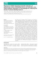
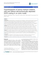
29%. In this group dense agglutinations were detected
in 6 rats (85%; 90%; 90%; 90%; 90%; 90%, Fig. 1B),
medium adhesions developed in 2 rats (40%; 50%)
and minor adhesions also in 2 rats (10% each). In
animals with PCM implantation, an overall mean
adhesion score of 69% (p = 0.078 as compared to
Int. J. Med. Sci. 2016, Vol. 13
controls), equivalent to an adhesion reduction rate of
25%, was observed. In this group dense
agglutinations between cecum and PCM were visible
in 5 rats (90% each, Fig. 1D), medium adhesions in 4
rats (50%; 55%; 55%; 65%), and minor adhesions in 1
rat (10%).
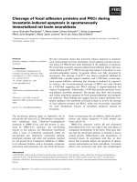
As described recently 19, animals with UPM
implantation and anti-adhesive treatment with 4DF
gel had significantly lower adhesion scores as
compared to controls (p < 0.0001) (Fig. 2). A
comparison with the adhesion scores of already
coated devices used in the present study revealed a
939
significantly better adhesion prevention capability of
UPM combined with 4DF gel (Table 1 and Fig. 2).
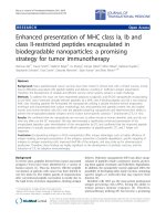
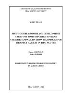
Evaluation of histological findings showed that
PTM (Fig. 3A) and PCM (Fig. 3B) fibers were
surrounded by granulating tissue connecting the
abdominal wall, the implanted mesh and the cecum.
Remnants of the collagen coating of PTM were visible
as a thin layer within the granulating tissue (Fig. 3A).
The
histologic
examination
confirmed
the
macroscopic adhesion scoring in extent and severity
of adhesion formation between the abraded cecum
and the mesh.
Figure 1: Representative photographs of implanted meshes, cecal abrasion and meso-stich approximation on day 0 (A and C). Dense agglutinations on postoperative
day 7 (B and D). (A and B) Parietex®, (C and D) Proceed®.
Figure 2: Comparison of mean adhesion scores of intestine-to-mesh adhesions: control (CT; uncoated mesh without anti-adhesive treatment), Parietex® mesh
(PTM), Proceed® mesh (PCM), uncoated Ultrapro® mesh combined with 4DryField® PH premixed gel (UPM/4DF premixed gel), and uncoated Ultrapro® mesh
combined with 4DryField® PH in-situ gel (UPM/4DF in-situ gel). Original data of CT published in 18, original data of UPM/4DF premixed gel and UPM/4DF in-situ gel
in 19.
Int. J. Med. Sci. 2016, Vol. 13
940
Figure 3: Representative histological findings one week after mesh implantation in rats with cecal abrasion with (A) Parietex® and (B) Proceed® meshes. Black arrows
= mesh fibers, hollow arrowhead = remnants of collagen coating of Parietex® mesh visible as a thin layer within granulating tissue.
Discussion
Intestinal
adhesions
to
intraperitoneally
implanted mesh materials (IPOM) are still a problem
and may cause chronic pain, bowel obstruction or
enterocutaneous fistulas, possibly followed by
infection 22. Furthermore, adhesions can be associated
with secondary female infertility 4, 10, 11 and
complicated re-operations 5, 8, 23, 24. In the long run,
these adhesions can lead to an impairment of the
stability and flexibility of the mesh 15.
Recent studies showed that not only the foreign
material or making of mesh but also the impairment
of visceral peritoneum is a co-decisive factor for the
development of intestine-to-mesh adhesions 18, 25, 26.
This especially accounts for areas where intestine has
been dissected from the hernia sac. Here, unavoidably
an intact peritoneal coverage is missing. Our recently
introduced rat model, comprising the placement of
mesh on intact peritoneum and the secure exposition
of mesh towards injured intestine, combines two
challenges of IPOM surgery: (1) integration of the
implanted mesh into the abdominal wall, (2) coping
with the trigger for adhesion formation originating
from injury of intestinal peritoneum.
In Parietex® (PTM) and Proceed® (PCM) the
synthetic mesh material is coated with an
anti-adhesive layer on the visceral side, while on the
parietal side plastic is directly exposed to the
abdominal wall. The purpose is to allow unhindered
integration into the abdominal wall, while the
temporary coating is supposed to prevent formation
of adhesions. Coating of PTM consists of collagen,
PCM uses oxidized cellulose for coverage. The results
of the present study indicate that integration into the
abdominal wall occurred unhindered and was not
negatively affected by the anti-adhesive coverage,
even though after one week still remnants of the
anti-adhesive coating were visible. With respect to
adhesion prevention, a significant efficiency could not
be found. The presence of remnants of the
anti-adhesive coverage in PTM animals possibly
suggests limited efficiency of the coating material.
Agglutination and absence of coating material in PCM
animals may indicate that the coating material might
have been degraded before a neo-mesothelial layer on
the cecum could develop. The more favourable
experimental results of PTM and PCM found in the
literature 27-30 can be explained by a different
experimental approach, in which the visceral
peritoneum was left intact. However, the model of the
present study with injury of intestinal peritoneum
mimics clinical practice, as found commonly, where
effective adhesion prevention is desired.
The results of our recent study, in which a
barrier gel was applied between injured intestine and
mesh 19, deserve special consideration. In this
approach the adhesion prevention device is not
limited as a coating layer to singular mesh fibers,
leaving unprotected gaps between the fibers. Instead,
Int. J. Med. Sci. 2016, Vol. 13
the 4DF gel barrier covers the complete area at high
risk for adhesion formation so that no gaps are left, in
which de-peritonealized bowel could get in contact to
the parietal peritoneum. Transferred to clinical IPOM
surgery, it could be useful to administer the barrier
gel in a selective way, i.e. especially to areas where an
intact peritoneal coverage is missing. However, in
clinical situation interindividual differences in
adhesion induction mechanisms cannot be excluded
by this experimental approach as healing responses
towards the different materials might vary.
Conclusion
Our results indicate that in case of impaired
intestinal peritoneum coated Parietex® and Proceed®
meshes do not provide significant adhesion
prevention. Considering the superior outcome of
4DryField® PH gel applied between injured intestine
and uncoated polypropylene Ultrapro® mesh, the use
of a polysaccharide barrier device as a prevention for
adhesions can be acknowledged a promising
alternative to coated mesh technology in IPOM
surgery.
Competing Interests
The authors have no conflicts of interest to
disclose.
References
1.
2.
3.
4.
5.
6.
7.
8.
9.
10.
11.
12.
13.
14.
Chelala E, Debardemaeker Y, Elias B, Charara F, Dessily M, Allé L. 85 Redo
surgeries after 733 laparoscopic treatments for ventral and incisional hernia:
Adhesion and recurrence analysis. Hernia 2010;14(2): 123-129.
Lamber B, Grossi JV, Manna BB, Montes JH, Bigolin AV, Cavazzola LT. May
polyester with collagen coating mesh decrease the rate of intraperitoneal
adhesions in incisional hernia repair? Arq Bras Cir Dig 2013;26(1): 13-17.
Holmdahl L, Risberg B, Beck DE, Burns JW, Chegini N, diZerega GS, Ellis H.
Adhesions: pathogenesis and prevention-panel discussion and summary. Eur J
Surg Suppl 1997;577: 56-62.
Leber GE, Garb JL, Alexander AI, Reed WP. Long-term Complications
Associated With Prosthetic Repair of Incisional Hernias. Arch Surg 1998;133(4):
378-382.
Béllon JM, Rodríguez M, Grarcía-Honduvilla N, Pascula G, Gómez V, Buján J.
Peritoneal Effects of Prosthetic Meshes Used to Repair Abdominal Wall
Defects. Journal of Laparoscopic & Advanced Surgical Techniques 2007;17(2).
Cassar K, Munro A. Surgical treatment of incisional hernia. British journal of
surgery 2002;89: 534-545.
Sauerland S, Walgenbach M, habermalz B, Seiler CM, Miserez M.
Laparoscopic versus open hernia repair. The Cochrane Libraty 2011(3).
Ellis H. The clinical significance of adhesions: focus on intestinal obstruction.
Eur J Surg Suppl 1997;577: 5-9.
Parker MC, Wilson MS, van Goor H, Moran BJ, Jeekel J, Duron J-J, Menzies D,
Wexner SD, Ellis H. Adhesions and Colorectal Surgery - Call for Action. The
Associantion of Coloproctology of Great Britain and Ireland 2007;2: 66-72.
Bageacu S, Blanc P, Breton C, Gonzales M, Porcheron J, Chabert M, Balique JG.
Laparoscopic repair of incisional hernia: a retrospective study of 159 patients.
Surg Endosc 2002;16(2): 345-348.
Halm JA, de Wall LL, Steyerberg EW, Jeekel J, Lange JF. Intraperitoneal
polypropylene mesh hernia repair complicates subsequent abdominal
surgery. World J Surg 2007;31(2): 423-429; discussion 430.
Schreinemacher MH, Emans PJ, Gijbels MJ, Greve JW, Beets GL, Bouvy ND.
Degradation of mesh coatings and intraperitoneal adhesion formation in an
experimental model. Br J Surg 2009;96(3): 305-313.
Schreinemacher MH, van Barneveld KW, Dikmans RE, Gijbels MJ, Greve JW,
Bouvy ND. Coated meshes for hernia repair provide comparable
intraperitoneal adhesion prevention. Surg Endosc 2013;27(11): 4202-4209.
van't Riet M, Burger JW, Bonthuis F, Jeekel J, Bonjer HJ. Prevention of
adhesion formation to polypropylene mesh by collagen coating: a randomized
controlled study in a rat model of ventral hernia repair. Surg Endosc 2004;18(4):
681-685.
941
15. Schug-Pass C, Sommerer F, Tannapfel A, Lippert H, Kockerling F. The use of
composite meshes in laparoscopic repair of abdominal wall hernias: are there
differences in biocompatibily?: experimental results obtained in a laparoscopic
porcine model. Surg Endosc 2009;23(3): 487-495.
16. Judge TW, Parker DM, Dinsmore RC. Abdominal wall hernia repair: a
comparison of sepramesh and parietex composite mesh in a rabbit hernia
model. J Am Coll Surg 2007;204(2): 276-281.
17. Jacob BP, Hogle NJ, Durak E, Kim T, Fowler DL. Tissue ingrowth and bowel
adhesion formation in an animal comparative study: polypropylene versus
Proceed versus Parietex Composite. Surg Endosc 2007;21(4): 629-633.
18. Winny M, Grethe L, Maegel L, Jonigk D, Lippmann T, Klempnauer J, Poehnert
D. Impairment of the Peritoneal Surface as a Decisive Factor for Intestinal
Adhesions in Intraperitoneal Onlay Mesh Surgery - Introducing a New Rat
Model. Int J Med Sci 2016;13(2): 108-112.
19. Winny M, Maegel L, Grethe L, Jonigk D, Borchert P, Kaltenborn A, Schrem H,
Klempnauer J, Poehnert D. Treatment of de-peritonealized intestine with
4DryField® prevents adhesions between non-resorbable intra-peritoneal
hernia mesh and bowel. American Journal of Translational Research, in press.
20. Lauder CI, Garcea G, Strickland A, Maddern GJ. Use of a modified
chitosan-dextran gel to prevent peritoneal adhesions in a rat model. The
Journal of surgical research 2011;171(2): 877-882.
21. Hoffmann NE, Siddiqui SA, Agarwal S, McKellar SH, Kurtz HJ, Gettman MT,
Ereth MH. Choice of hemostatic agent influences adhesion formation in a rat
cecal adhesion model. The Journal of surgical research 2009;155(1): 77-81.
22. Fortelny RH, Petter-Puchner AH, Glaser KS, Offner F, Benesch T, Rohr M.
Adverse effects of polyvinylidene fluoride-coated polypropylene mesh used
for laparoscopic intraperitoneal onlay repair of incisional hernia. Br J Surg
2010;97(7): 1140-1145.
23. Chuback JA, Singh RS, Sills C, Dick LS. Small bowel obstruction resulting from
mesh plug migration after open inguinal hernia repair. Surgery 2000;127(4):
475-476.
24. Losanoff JE, Richman BW, Jones JW. Entero-colocutaneous fistula: a late
consequence of polypropylene mesh abdominal wall repair: case report and
review of the literature. Hernia : the journal of hernias and abdominal wall surgery
2002;6(3): 144-147.
25. Hooker GD, Taylor BM, Driman DK. Prevention of adhesion formation with
use of sodium hyaluronate-based bioresorbable membrane in a rat model of
ventral hernia repair with polypropylene mesh--a randomized, controlled
study. Surgery 1999;125(2): 211-216.
26. Dinsmore RC, Calton WC, Jr., Harvey SB, Blaney MW. Prevention of
adhesions to polypropylene mesh in a traumatized bowel model. J Am Coll
Surg 2000;191(2): 131-136.
27. Burger JW, Halm JA, Wijsmuller AR, ten Raa S, Jeekel J. Evaluation of new
prosthetic meshes for ventral hernia repair. Surg Endosc 2006;20(8): 1320-1325.
28. Brown CN, Finch JG. Which mesh for hernia repair? Ann R Coll Surg Engl
2010;92(4): 272-278.
29. Gaertner WB, Bonsack ME, Delaney JP. Visceral adhesions to hernia
prostheses. Hernia 2010;14(4): 375-381.
30. van 't Riet M, de Vos van Steenwijk PJ, Bonthuis F, Marquet RL, Steyerberg
EW, Jeekel J, Bonjer HJ. Prevention of adhesion to prosthetic mesh:
comparison of different barriers using an incisional hernia model. Ann Surg
2003;237(1): 123-128.