Specific antiproliferative activity against several human cancer cells with metabolites from Onygena corvina
Bạn đang xem bản rút gọn của tài liệu. Xem và tải ngay bản đầy đủ của tài liệu tại đây (401.54 KB, 10 trang )
Int.J.Curr.Microbiol.App.Sci (2019) 8(1): 1659-1668
International Journal of Current Microbiology and Applied Sciences
ISSN: 2319-7706 Volume 8 Number 01 (2019)
Journal homepage:
Original Research Article
/>
Specific Antiproliferative Activity against Several Human Cancer Cells with
Metabolites from Onygena corvina
Yukiko Ogawa1*, Fumihide Takano2, Nobuo Yahagi3, Marie Yahagi3,
Yuki Kobayashi3 and Hidemitsu Kobayashi4
1
Divisions of Infection Control and Prevention, 4Divisions of Microbiology, Department of
Pharmacy, Faculty of Pharmaceutical Science, Nagasaki International University, 2825-7
Huis Ten Bosch, Sasebo, Nagasaki 859-3298, Japan
2
Division of Kampo Pharmaceutical Sciences, Nihon Pharmaceutical University, 10281
Komuro, Ina-machi, Kitaadachi-gun, Saitama 362-0806, Japan
3
Yahagi Bio Institute, Mamurogawa-machi, Mogami-gun, Yamagata, 999-5604, Japan
*Corresponding author
ABSTRACT
Keywords
Onygena corvina,
Antitumor activity,
Cell specificity,
Medicinal plants,
Lung carcinoma
Article Info
Accepted:
12 December 2018
Available Online:
10 January 2019
We succeeded in large volume artificial cultivation of Onygena corvina. The appearance
(shape, color, and size) of fruiting bodies of cultured bacteria was almost the same as that
of wild one. We screened their metabolites for antiproliferative activities against nine
human cancer cells in vitro. Culture filtrates (metabolite-containing media) of O. corvina
were lyophilized and resuspended in phosphate buffer to investigate them in vitro. As a
result, these metabolites effectively inhibited the growth of some cancer cells in a
concentration-dependent manner. In particular, the effect on lung carcinoma Hara cells
was remarkable, but there was almost no effect on hepatoma HepG2 cells. In addition,
almost no effect was exerted on three normal cells [human hepatocyte (NHH), human
mammary epithelial cells (NHME), and human epidermal melanocytes (NHEM)] at the
same concentration. Therefore, it was revealed that the human cell proliferation inhibitory
effect of the O. corvina metabolites greatly differs in susceptibility depending on the type
of cancer cell under the concentration range not affecting normal cells. In vivo experiments
demonstrated that the oral administration of the O. corvina metabolites (OC-FD) decreased
the tumor growth compared with untreated mice. The OCT extract obtained by extracting
ethanol had the same anti-tumor activity as the OC-FD, but this activity was weaker than
with OC-FD. On the other hand, when the same experiment was performed on OCT with
2.O.C.11 and 1.O.C.5, respectively, for the EtOAc soluble fraction and water layer, this
demonstrated stronger inhibitory activity than when only the OC-FD or OCT was
administered. These results revealed these active components may be both low-molecular
weight with low polarity (EtOAc soluble) and high-molecular weight, such as a
polysaccharide or protein (water soluble). These findings suggest that the unknown
compounds in the metabolite-containing media from cultures of O. corvina are potential
lead compounds for developing anticancer drugs with extremely mild side effects.
1659
Int.J.Curr.Microbiol.App.Sci (2019) 8(1): 1659-1668
Treatment
strategies
have
undergone
st
significant changes in the 21 Century.
Surgical treatment is, even now, an essential
part of solid cancer treatment, but treatment
policy has shifted from the extended
operations used thus far to functional
preservation strategies that focus on the
patient QOL, and treatment methods that
highly value the informed consent of the
patient (Tian et al., 2018). With
chemotherapy,
there
is
high-dose
chemotherapy through targeting therapy that
uses DDS (Nakamura et al., 2018), as well as
bone
marrow
transplantation.
For
radiotherapy, treatment takes place using new
methods such as the X knife, etc. (Alster et al.,
2018), and a new method for which there are
high expectations in the 21st Century is cancer
gene therapy. Under particular focus is
“precision therapy,” which is starting to be
introduced for lung cancer therapy to provide
individual treatment for different types of
cancer on an individual level (Kehl et al.,
2018, Lu et al., 2018).
been shown to have a number of benefits, such
as cancer cell inhibitory effects (Bok et al.,
1999, Chen et al., 2013, Ogawa et al., 2014),
anti-oxidant effects, and immunostimulatory
effects (Choi et al., 2004, Paterson et al.,
2008, Bhatti et al., 2013, Cheng et al., 2013).
The caterpillar fungus in silkworms promises
to have a repairing effect on the hippocampus
for Alzheimer’s-type dementia (Tsushima et
al., 2010). Caterpillar fungus works to
strengthen the lungs and kidneys and is used
as a core treatment for emphysema and asthma
in China, in combination with other herbal
medicines (Wang et al., 2016). β-glucan is
included in extremely high quantities in
caterpillar fungus (Smiderle et al., 2014), 17
times the amount contained in agaricus
mushrooms and 170 times that of normal
fungi. It has a rich composition rate of
nutritional elements, including minerals such
as zinc or selenium, as well as natural amino
acids and proteins. It also includes cordycepin
and antioxidant enzymes (S.O.D), and is
gaining attention for its anticancer effects
(Choi et al., 2012, Choi et al., 2013, Shao et
al., 2016).
Genus Cordyceps is a type of natural
caterpillar fungus, in which caterpillar fungus
infests living insects (Sato et al., 2002), forms
hypha nuclei within the body, and seasonally
extends and develops from the head section
and joints of the insect to the fruiting body
(Takano et al., 1996, Yahagi et al., 1999,
Yahagi et al., 2004). Approximately 350 or
more types of genus cordyceps have been
discovered throughout the world and, since
ancient times in China (Zhu et al., 1998), it
has been known to be a nourishing tonic, and
considered effective in increasing longevity,
providing anti-oxidant effect, and increasing
immunological effects (Jeong et al., 2013,
Xiao et al., 2017). In Japan, 300–400 types of
caterpillar fungus infesting a large number of
insects, such as cicada and silkworms (Chen et
al., 2002), have been discovered. These have
On the other hand, Onygena corvina (feather
stalkball) and Onygena equinea (horn
stalkball), both species of the fungal genus
Onygena in Onygenaceae family, can live as
saprophytes on feathers, hooves, horn, and
hair (Lange et al., 1975) which is a rare
bacterium in the world, is a fungus that infests
the bones of animals. Whereas Japanese
caterpillar fungus is hosted by insects,
Onygena is hosted in the bones, vomit, and
excreta of mammals, and therefore promises
to
provide
more
powerful
enzyme
“physiologically active substances.” However,
examples of its occurrence are extremely rare,
and there are virtually no reports of it being
picked in the wild. This, in turn, means that
there are few examples of academic papers or
experiments, and there are many fields which
are yet unknown. On this occasion, we have
Introduction
1660
Int.J.Curr.Microbiol.App.Sci (2019) 8(1): 1659-1668
been successful in artificially cultivating it for
the first time in Japan. We have evaluated the
anti-tumor effects of the picked and cultivated
Onygena and metabolites from Onygena
corvina by administering four types of
Onygena extract using Sarcoma-180 solid
sarcoma tumor model mice with the aim of
exploring its anti-tumor activity elements.
Additionally, we performed screening in
relation to anti-growth inhibitory activities in
several types of human cancer cells within the
culture metabolite.
Materials and Methods
Genus Onygena fungal mycelial cultibation
Parasitic mushroom, Onygena corvina was
harvested from skull of a weasel at Sakekawamura (Mogami-Gun, Yamagata Prefecture,
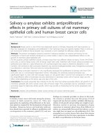
Japan). The photograph of O.carvina in the
natural field was shown in figure 1A. Onygena
picked in the wild and its conidium was first
injected into agar. After cultivation, the
cultivated mushroom bed was moved to a
liquid culture including yeast and cultured for
a fixed period of time.
The bacteria were removed when a fixed
number of conidophore bundles formed in the
liquid medium, and the liquid medium was
filtered and freeze-dried (OC culture medium
extract: OC-FD). In addition, the OC extract
was infiltrated with ethanol and, after
concentrating the ethanol-soluble fraction, an
extract was obtained (OCT). 150 mL of water
was added to the OCT extract and a separating
funnel was used to distribute 150 mL of ethyl
acetate (EtOAc).
The distribution operation was repeated three
times with 150 mL of the EtOAc and water,
respectively. The EtOAc soluble fraction and
water fraction obtained from this were
vacuum-concentrated
or
freeze-dried,
respectively, to make 2.O.C.11 (EtOAc
soluble) and 1.O.C.5 (water soluble).
Chemicals
Cell Counting Kit-8 including 2-(2-methoxy4-nitrophenyl)-3-(4-nitrophenyl)-5-(2,4disulfophhenyl)-2H-tetrazolium, monosodium
salt (WST-8) was from Dojindo Co.,
Kumamoto, Japan. RPMI 1640 medium was
from Nissui Pharmaceutical Co., Tokyo,
Japan. Fetal calf serum (FCS) was from Life
Technologies Co., Carlsbad, CA, USA.
Penicillin- streptomycin was from Roche
Diagnostics K. K., Tokyo, Japan. Trypan blue
solution was from Nacalai Tesque, Inc.,
Kyoto, Japan. Aphidicolin was purchased
from Wako Pure Chemical Industries, Osaka,
Japan.
Cells
Human breast cancer MCF-7 and MDA-MB231 cells, human gastric cancer KatoⅢ cells,
human colon adenocarcnoma Colo201 and
Caco2 cells, human hepatoma HepG2 cells,
human lung squamous cell carcinoma Hara
cells, human promonocytic leukemia U937
cells and human malignant melanoma GAK
cells were obtained from the JCRB Cell Bank
(National Institute of Biomedical Innovation,
Health and Nutrition) and ATCC Cell Biology
Collection, respectively.
Normal human hepatocytes (NHH), normal
human mammary epithelial cells (NHME),
Normal human epidermal melanocytes
(NHEM) were purchased from Promo Cell
Co. (Heidelberg, Germany). MCF-7, Caco-2
and HepG2 were maintained in E-MEM and
D-MEM, respectively. KATO III, Colo201,
U937 and Hara cells were grown in
RPMI1640 medium. All cell cultures were
supplemented with heat-inactivated fetal calf
serum 10% (v/v), penicillin (100 IU/ml), and
streptomycin (100 µg/mL) at 37 °C in an
atmosphere of 95% air/ 5% CO2. MDA-MB231 and GAK were maintained in L-15
medium and Ham’s F12 medium were
supplemented with heat-inactivated fetal calf
1661
Int.J.Curr.Microbiol.App.Sci (2019) 8(1): 1659-1668
serum 20% (v/v), penicillin (100 IU/ml), and
streptomycin (100 g/ml) at 37 °C in an
atmosphere of 95% air/ 5% CO2.
t-test with P values <0.05 considered to be
statistically significant.
Results and Discussion
Creation of a cancer bearing model
Solid sarcoma cell Sarcoma-180 (S-180) was
transplanted at 0.05 mL/animal to the right
lower abdomen limb root of ICR mice
acclimatized and bred over one week to make
a concentration of 1×10⁶ cells/mL.
After transplantation, the body weight and
tumor volume were periodically measured, the
tumor mass was excised, and its weight was
measured at the end of the experiment. The
tumor volume was calculated from the product
of the major and minor axis of the tumor,
under the presumption that it is the same as
the density of the cancel cells.
The four types of Onygena prepared to make
concentrations of 300 mg/mL (0.01 mL/g
body weight in saline) were administered
orally from the start date of injecting S-180
once per day for 30 days.
Cell viability and cytotoxic assay
Cells (2×10⁴ cels/well) were seeded into a 96well flat-bottom plate and treated with various
concentrations (0-2500 µg/mL) of OC-FD for
48 h at 37 °C in an atmosphere of 95% air and
5% CO2. Cytotoxic activity and cell viability
and cell growth were evaluated by trypan blue
(0.5% (w/v)) exclusion and by the WST-8
assay, respectively. The reduction in
proportion of living cells was assayed by
measurement of absorbance at 450 nm
(reference, 600 nm) using the GloMax Multi
Detection System.
Statistical analysis
The results of experiments are presented as
mean ± standard error (SE). Differences in
means were evaluated by two-tailed Student’s
Culture of O. corvina and preparation of
secondary metabolites
The Onygena fungus is in the Ascomycota
group and is often broken down in a
saprophytic manner in animal carcasses, but
there are those that infest and become
pathogens in animals, and those that form
symbiosis and form mycorrhiza in the
Ericaceaee family of plants (Doveri et al.,
2012).
Ecologically, it is a mixture of a wide variety
of groups. Additionally, because some of these
have strong keratin decomposition abilities,
even organisms that are suited to processing
industrial waste such as bird feathers have
gained attention recently (Gupta et al., 2013,
Sharma et al., 2011). However, there are very
few examples demonstrating a scientific basis
for its effectiveness.
We successfully developed a stable Chu-Soh
culture system that has a high level of safety
(Yahagi et al., 1999). It also permits the
efficient formation of sexual fruiting bodies on
the culture medium that are comparable with
those that develop in the wild, and allow the
production of different lots with the same
efficacy.
Using this artificial liquid medium, we were
able to culture samples in large quantities
(Yahagi et al., 2004). Figure 1B shows the
fruiting bodies developed from O. corvina on
agar. The fruiting body had similar form, size,
and color compared to wild O. corvina. The
metabolite-containing media obtained from
this culture system were filtered through a
membrane filter and freeze-dried for use in
subsequent experiments.
1662
Int.J.Curr.Microbiol.App.Sci (2019) 8(1): 1659-1668
Table.1 Influence of Onygena corvina on mouse body weight changes
Body weight (g)
Day 0
Day 5
Day 10
Day 15
Day 20
Day 325
Day 30
Control
20.1
20.7
21.0
21.7
23.5
25.2
26.9
OC-FD i.p.
20.2
20.6
21.1
21.6
23.8
25.4
26.3
OC-FD p.o
19.6
20.3
20.9
21.3
22.6
24.6
26.8
OC-FD i.v.
20.1
20.8
31.1
21.8
23.3
25.0
27.1
OC-FD were orally, intraperitoneally and intravenously administered to mice, respectively, and the changes in
weight of the mice over 4-weeks were measured.
Table.2 Antitumor effect of Onygena corvina on tumor bearing model
Body weight (g)
Day 0
Day 16
Day 35
Av.tumor
weight (g)
Inhibition
ratio (%)
Control
25.6
29.6
42.1
7.43
OC-FD
25.1
29
39.7
5.84
21.3
OCT
25.1
29.1
36.7
4.7
36.7
2.O.C.11
25.5
28.7
36.5
3.24
56.4
1.O.C.5
25.3
29.2
37.6
2.75
62.9
S-180 was transplanted to the ICR mice. After transplantation, the body weight and tumor volume were periodically
measured, the tumor mass was excised, and its weight was measured at the end of the experiment. The tumor
volume was calculated from the product of the major and minor axis of the tumor, under the presumption that it is
the same as the density of the cancel cells. The four types of Onygena prepared to make concentrations of 300
mg/mL (0.01 mL/g body weight in saline) were administered orally from the start date of injecting S-180 once per
day for 30 days. Data are the means of triplicate assay mean ± SE.
Fig.1 Artificial culture and natural products of Onygena corvina
A
B
(A) Specimens of Onygena corvina. in natural field
(B) Fruiting bodies produced by Onygena corvina. in culture medium.
1663
Int.J.Curr.Microbiol.App.Sci (2019) 8(1): 1659-1668
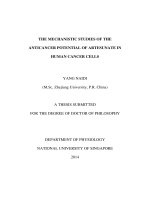
Fig.2 Inhibitory effect of secondary metabolites of Onygena corvine (OC-FD) on the growth of
human tumor cells
(A)
Relative Cell Viability (%)
100
90
80
70
60
50
40
30
20
10
0
2500
1250
630
310
160
80
160
80
Concentration (m g/mL)
(B)
Relative Cell Viability (%)
100
90
80
70
60
50
40
30
20
10
0
2500
1250
630
310
Concentration (m g/mL)
Cells were treated with OC-FD 〔1250-80µg/mL〕 for 48h. Then cell viability was determined by WST-8 assay and
trypan blue dye exclusion assay. Error bars: SE from three different cell preparations assayed individually. (A)
MCF7 (●), MDA (■), KATOIII (▲), Colo201 (○), Caco2 (□) and HepG2 (△), (B) HARA (●), U937 (■), GAK (▲),
NHH (○), NHME (□) and NHEM (△)
Antiproliferative activity of O. corvina
secondary metabolites in human cancer
cells
We succeeded in large volume artificial
cultivation of O. corvina. To investigate the
effect of O. corvina secondary metabolites
(OC-FD) on cell proliferation in various types
of human cancer cells [human breast cancer
(MCF7, MDA), gastric cancer (Kato III),
1664
Int.J.Curr.Microbiol.App.Sci (2019) 8(1): 1659-1668
colon adenocarcinoma (Colo201, Caco2),
leukemia (U937), lung carcinoma (Hara),
hepatoma (HepG2), human malignant
melanoma (GAK)], and three normal cells
[human hepatocyte (NHH), human mammary
epithelial cells (NHME), human epidermal
melanocytes (NHEM)]. OC-FD significantly
suppressed cell growth in MCF7, Caco2 and
Hara cells and extremely strong growth
inhibition activity was especially seen for
lung carcinoma Hara cells. At this
concentration, there was hardly any effect on
normal cells (Fig. 2). The antiproliferative
metabolites obtained from OC-FD were
highly specific to human cancer cell lines.
These findings suggest that the unknown
compounds in the metabolite-containing
media from cultures of these mushrooms are
potential lead compounds for developing
anticancer drugs with extremely mild side
effects.
Influence of O. corvina on mouse body
weight changes
Antiproliferative activities of culture filtrates
from O. corvina are rarely observed in normal
human skin fibroblasts at a range of
concentration where the effects are observed
in cancer cells. Moreover, in our experiments
on acute and chronic toxicities in mice, there
was not a large difference in weight gain
between the control and test groups (Table 1),
and no abnormalities in isolated organs were
observed in the test group (data not shown).
Taking into consideration data from the
above, some ingredients in metabolitecontaining media of O. corvina are expected
to be potential novel lead compounds for
developing anticancer drugs with extremely
mild side effects.
Antitumor effect of O. corvina on tumor
bearing model
As shown in Table 2, in control mice for
which the S-180 intake sample was not
administered, solid cancer was formed and, as
time passed, the mass and volume of the
tumor increased. In contrast, in the mice for
which OC extract (OC-FD) as administered at
doses of 300 mg/kg/day, the volume and mass
of the tumors increasing over time was
controlled. The OCT extract obtained by
extracting ethanol had the same anti-tumor
activity as the OC-FD, but this activity was
weaker than with OC-FD. On the other hand,
when the same experiment was performed on
OCT with 2.O.C.11 and 1.O.C.5, respectively,
for the EtOAc soluble fraction and water
layer, this demonstrated stronger inhibitory
activity than when only the OC-FD or OCT
was administered. In the diagram comparing
the weight of the solid cancer excised at the
end of the experiment, the strongest activity
was shown in the OC fraction (2.O.C.11
[EtOAc soluble] and 1.O.C.5 [water soluble]).
From the above results, oral administration of
the O. corvina cultured metabolite solution
OC clearly demonstrated anti-tumor activity,
and it was shown that this active element was
not an element that was soluble in ethanol and
had comparatively high water solubility.
Additionally, in the ethyl acetate (EtOAc)
soluble fraction 2.O.C.11 and water layer
1.O.C.5
obtained
from
liquid-liquid
distribution from OCT, stronger activity was
recognized than in single administrations of
OC, which suggests that this active
components may be both low-molecular
weight with low polarity (EtOAc soluble) or
high-molecular
weight,
such
as
a
polysaccharide or protein (water soluble).
Moving forward, investigating what kind of
elements are contained in 1.O.C.5 fractions
that demonstrate particularly strong activity
and reevaluating the anti-tumor activity of
model animals will provide evidence of the
effectiveness of O. corvina in battling cancer.
To the best of our knowledge, the present
study was the first to demonstrate that culture
1665
Int.J.Curr.Microbiol.App.Sci (2019) 8(1): 1659-1668
filtrates (metabolite-containing media) of O.
corvina may inhibit the viability of breast
cancer cells, colon carcinoma cells and lung
carcinoma cells in vitro, and sarcoma solid
tumor cells in vivo. The metabolite-containing
medium harvested from O. corvina is
considered to contain many substances
including polysaccharides. In order to identify
the bioactive forms of O. corvina, we are
currently
working
towards
isolated
purification of the active fractions using highperformance liquid chromatography after
fractioning by various organic solvents and
determining the chemical structures by
focusing on instrumental analysis such as
NMR method, and mass spectrometry. On the
other hand, experiments to clarify anti-cancer
activity mechanisms of the active fractions
which have been purified by chromatography,
and toxicity tests on mice have carried out
accurately, and publication of the details are
planned for the next thesis. In the near future,
O. corvina is expected to become a valuable
resource for combined therapy with an anticancer drug, at a dosage which does not show
side effects, and a leading compound for
novel anticancer agents.
References
Alster, P., Koziorowski, D.M., Za Bek, M.,
Dzierzȩcki, S., Ma Dry, J., DuszyńskaWa, S. K., Grygarowicz, H., Zielonko,
J., Królicki, L., Friedman, A., 2018.
Making a Difference-Positive Effect of
Unilateral
VIM
Gamma
Knife
Thalamotomy in the Therapy of Tremor
in Fragile X-Associated Tremor/Ataxia
Syndrome (FXTAS). Front. Neurol., 9,
512. doi:10.3389/fneur.2018.00512.
Bok, J.W., Lermer, L., Chilton, J.,
Klingeman, H.G., Towers, G.H., 1999.
Antitumor sterols from the mycelia of
Cordyceps sinensis. Phytochemistry, 51,
891-898.
Bhatti, M.T., Freedman, S.M., Mahmoud,
T.H., 2013. Fingolimod Therapy and
Mocular
Hemorrhage.
J.
neuroophthalmol., 33(4), 370-372.
Chen, R., Ichida, M., 2002. Infection of the
silkworm,
Bombyx
mori,
with
Cordyceps militaris. J. Inect Biotech.
Sericol., 71, 61-63.
Chen, Y.H., Wang, J.Y., Pan, B.S., Mu, Y.F.,
Lai, M.S., So, E.C., Wong, T.S., Huang,
B.M., 2013. Cordycepin enhances
cisplatin apoptotic effect through
caspase/MAPK pathways in human
head and neck tumor cells. Onco.
Tagets Ther., 25, 983-998.
Choi, G.Y., Choi, B.T., Jeog, Y.H., Jeong,
Y.K, 2013. Apoptosis induction of
human prostate carcinoma cells by
cordycepin through reactive oxygen
species-mediated mitochondrial death
pathway. J. Oncology, 42, 1036-1044.
Gupta, R., Sharma, R., Beg, Q.K., 2013.
Revisiting microbial keratinases: next
generation proteases for sustainable
biotechnology. Crit., Rev., Biotechnol.,
33, 216-228.
Cheng, Y.W., Chen, Y.I., Tzeng, C.Y.,
Chang, C.H., Lee, Y.C., Chen, H.C.,
Tsai, C.C., Hsu, T.H., Lai, Y.K., Chang,
S.L., 2013. Aqueous extracts of
Cordyceps militaris (Ascomycetes)
lower the levels of plasma glucose by
activating the cholinergic nerve in
streptozotocin-induced diabetic rats. Int
J Med Mushrooms, 15, 277-286.
Choi, S.B., Park, C.H., Choi, M.K., Jun,
D.W., Park, S., 2004. Improvement of
insulin resistance and insulin secretion
by water extracts of Cordyceps
militaris, Phellinus linteus, and
Paecilomyces
tenuipes
in
90%
pancreatectomized
rats.
Biosci.
Biotechnol. Biochem., 68, 2257-2264.
Doveri, F., Pecchia, S., Vergara, M.,
Sarrocco, S., Vannacci, G., 2012. A
comparative study of Neogymnomyces
virgineus, a new keratinolytic species
1666
Int.J.Curr.Microbiol.App.Sci (2019) 8(1): 1659-1668
from dung, and its relationships with the
Onygenales. Fungal Divers, 52, 13-34.
Jeong, M.H., Lee, C.M., Lee, S.W., Seo, S.Y.,
Kang, B.W., Jeong, Y.K., Choi, Y.J.,
Yang, K.M., Jo, W.S., 2013.
Cordycepin-enriched
Cordyceps
militaris induces immunomodulation
and tumor growth delay in mousederived breast cancer. Oncol. Rep., 30,
1996-2002.
Jeong, J.W., Jin, C.Y., Park, C., Han, M.H.,
Kim, G.Y., Moon, S.K., Kim, C.G.,
Jeong, Y.K., Kim, W.J., Lee, J.D., Choi,
Y.H., 2012. Inhibition of migration and
invasion of LNCaP human prostate
carcinoma cells by cordycepin through
inactivation of Akt. Int. J.Oncol., 40,
1697-1704.
Kehl, K.L., Lathan, C.S., Johnson, B.E.,
Schraq, D., 2018. Race, Poverty, and
Initial Implementation of Precision
Medicine for Lung Cancer. J. Natl.
Cancer Inst., doi:10.1093/jnci/djy202.
Lange, M., Hora, F.B.(eds), 1975. Collins
guide to mushrooms & toadstool.
Collins, London.
Lu, J., Han, B., 2018. Liquid Biopsy
Promotes Non-Small Cell Lung Cancer
Precision Therapy, Technol Cancer
Res.Treat., 17, 1533033818801809
Nakamura, T., 2018. Development of a Nano
DDS for Cancer Immunotherapy Based
on Llipid Nanoparticles. Yakugaku
Zasshi, 138, 1443-1449.
Ogawa, Y., Yahagi, N., Yahagi, R.,
Kobayashi,
H.,
2014.
Specific
antiproliferative activity against human
cancer cells with metabolites from
several species related to the genus
Cordyceps, Int. J. Curr. Microbiol. App.
Sci., 3(5), 607-617.
Paterson, R.R.M., 2008. Cordyceps-A
traditional Chinese medicine and
another fungal therapeutic biofactory.
Phytochemistry. 69, 1469-1495.
Sharma, R., Murty, N. A., Gupta, R., 2011.
Molecular characterization of Nterminal pro-sequence of keratinase ker
P from Pseudomonas aeruginosa:
identification of region with chaperone
activity. Appl. Biochem. Biotechnol.,
165, 892-901.
Sato, H., Shimizu, M., 2002. Storomata
production for Cordyceps militaris
(Clavicipitales: Clavicipitaceae) by
infection of hyphal bodies to alternative
host insects. Appl. Entomol. Zool., 37,
85-92.
Shao, L.W., Huang, L.H., Yan, S., Jin, J.D.,
Ren, S.Y., 2016. Cordycepin induces
apoptosis in human liver cancer HepG2
cells through extrinsic and intrinsic
signaling pathways. Oncol. Lett., 12(2),
995-1000.
Smiderle, F.R., Baggio, C.H., Borato,
D.G., Santana-Filho, A.P., Sassaki,
G.L., Iacomini, M., Van Griensven
Leo,
J.L.D.,
2014.
Antiinflammatory properties of the
medicinal
mushroom
Cordyceps
militaris might be related to its linear
(1→3)-β-D-glucan.
PLOS
ONE,
9(10),
doi.org/10.1371/journal.pone.01102
66
Takano, F., Kikuchi, Y., Fushiya, S., Hojo,
H., Nozoe, S., Yahagi, N., Kondo, Y.,
1996. The culture fluid of Isaira
japonica yasuda augments anti-sheep
red blood cell antibody response in
mice. Biol. Pharm. Bull. 19, 641-643.
Tian, J.Y., Ying, Y.L., Xin, H., Gen,
H.Di., 2018. Survival following breastconserving therapy is equal to that
following mastectomy in young women
with early-stage invasive lobular
carcinoma. Eur. J. Sur. Oncol.,
44(11), 1703-1707.
Tsushima, M., Yamamoto, K., Goryou, M.,
Suzuki, F. and Suzuki, K., 2010. Hotwater extract of Paecilomyces tenuipes
from the silkworm pupae improves D-
1667
Int.J.Curr.Microbiol.App.Sci (2019) 8(1): 1659-1668
galactose-induced brain aging in mice.
J. Insect Biotec. Sericol., 79, 45-51.
Wang, N., Li J., Huanq, X., Chen, W., Chen,
Y., 2016. Herbal Medicine Cordyceps
sinensis
Improves
Health-Related
Quality of Life in Moderate-to-Severe
Asthma. Evid. Based Complement.
Alternat. Med., 2016, Article ID
6134593, 8 pages.
Yahagi, N., Komatsu, M., Hiramatsu, M.,
SHI, H., Yahagi, R., Kobayashi, H.,
Kamada, H., Takano, F., Fushiya, S.,
1999. Radical Scavenging Activities of
Condensed Culture Medium of Isaria
japonica Yasuda and Hot water Extract
of Fomes fomentarius (L.:Fr) Kicrx.
Nat. Med., 53, 319-323.
Yahagi, N., Yahagi, R., Takano, F., Fushiya,
S., Tanaka, T., Tanaka, K., Ohta, T.,
2004. Growth of ascocarps from
cultured Cordyceps militaris (L.:Fr.) Fr.
and Cordyceps formicarum Kobayasi in
an agar medium, Kingakukai kaiho, 45,
15-19.
Xiao, Y., Huanq, Q., Zhenq, Z., Quan, H.,
Liu, S., 2017. Construction of a
Cordyceps sinensis exopolysaccharideconjugated selenium nanoparticles and
enhancement of their antioxidant
activities. Int. J. Biol. Macromol., 99,
483-491.
Zhu, J.S., Halpern, G.M., Jones, K., 1998.
The Scientific Rediscovery of an
Ancient Chinese Herbal Medicine:
Cordyceps sinensis Part I, J. Altern.
Complement. Med., 4: 289-303.
How to cite this article:
Yukiko Ogawa, Fumihide Takano, Nobuo Yahagi, Marie Yahagi, Yuki Kobayashi and
Hidemitsu Kobayashi. 2019. Specific Antiproliferative Activity against Several Human Cancer
Cells with Metabolites from Onygena corvine. Int.J.Curr.Microbiol.App.Sci. 8(01): 1659-1668.
doi: />
1668