Nuclear expression of GS28 protein: A novel biomarker that predicts prognosis in colorectal cancers
Bạn đang xem bản rút gọn của tài liệu. Xem và tải ngay bản đầy đủ của tài liệu tại đây (1.46 MB, 8 trang )
Int. J. Med. Sci. 2017, Vol. 14
Ivyspring
International Publisher
515
International Journal of Medical Sciences
2017; 14(6): 515-522. doi: 10.7150/ijms.19368
Research Paper
Nuclear Expression of GS28 Protein: A Novel
Biomarker that Predicts Prognosis in Colorectal
Cancers
Sung Hak Lee1, Hyung Jae Yoo2, Do Eun Rim2, Yinji Cui3, Ahwon Lee1, Eun Sun Jung1, Seung Taek Oh4, Jun
Gi Kim4, Oh-Joo Kwon2, Su Young Kim3, Seong-Whan Jeong2
1.
2.
3.
4.
Department of Hospital Pathology, Seoul St. Mary’s Hospital, College of Medicine, The Catholic University of Korea, Seoul, Republic of Korea;
Department of Biochemistry, College of Medicine, The Catholic University of Korea, Seoul, Republic of Korea;
Department of Pathology, College of Medicine, The Catholic University of Korea, Seoul, Republic of Korea;
Department of Surgery, Seoul St. Mary’s Hospital, College of Medicine, The Catholic University of Korea, Seoul, Republic of Korea.
Corresponding authors: Seong-Whan Jeong MD, PhD, Department of Biochemistry, College of Medicine, The Catholic University of Korea, 222,
Banpo-daero, Seocho-gu, Seoul, 06591, Republic of Korea Tel: +82 2 2258 7291; FAX: +82 2 596 4435; E-mail: Su Young Kim MD, PhD,
Department of Pathology, College of Medicine, The Catholic University of Korea, 222, Banpo-daero, Seocho-gu, Seoul, 06591, Republic of Korea Tel: +82 2 2258
7315; FAX: +82 2 537-6586; E-mail:
© Ivyspring International Publisher. This is an open access article distributed under the terms of the Creative Commons Attribution (CC BY-NC) license
( See for full terms and conditions.
Received: 2017.01.26; Accepted: 2017.03.23; Published: 2017.04.09
Abstract
Aims: GS28 (Golgi SNARE protein, 28 kDa), a member of the soluble N-ethylmaleimide-sensitive
factor attachment protein receptors (SNARE) protein family, plays a critical role in mammalian
endoplasmic reticulum (ER)-Golgi or intra-Golgi vesicle transport. To date, few researches on the
GS28 protein in human cancer tissues have been reported. In this study, we assessed the
prognostic value of GS28 in patients with colorectal cancer (CRC).
Methods and results: We screened for GS28 expression using immunohistochemistry in 230
surgical CRC specimens. The CRCs were right-sided and left-sided in 28.3% (65/230) and 71.3%
(164/230) of patients, respectively. GS28 staining results were available in 214 cases. Among these,
there were 26 nuclear predominant cases and 188 non-nuclear predominant cases. Stromal GS28
expression was noted in 152 cases of CRC. GS28 nuclear predominant immunoreactivity was
significantly associated with advanced tumour stage (p = 0.045) and marginally associated with
perineural invasion (p = 0.064). Decreased GS28 expression in the stromal cells was significantly
associated with lymph node metastasis (N stage; p = 0.036). GS28 expression was not associated
with epidermal growth factor receptor (EGFR) immunohistochemical positivity or KRAS mutation
status. Investigation of the prognostic value of GS28 with Kaplan-Meier analysis revealed a
correlation with overall survival (p = 0.004). Cases with GS28 nuclear predominant expression had
significantly poorer overall survival than those with a non-nuclear predominant pattern.
Conclusions: Taken together, these results indicate that GS28 nuclear predominant expression
could serve as a prognostic marker for CRC and may help in identifying aggressive forms of CRC.
Key words: GS28 protein, Biologic Marker, Colorectal Carcinoma, Prognosis, Golgi Complex, SNARE proteins.
Introduction
Colorectal cancer (CRC) is the third most
common cancer, and an important contributor to
cancer mortality and morbidity worldwide [1].
According to the cancer statistics data of the Ministry
of Health and Welfare in Korea, CRC incidence rates
in 2012 were 69.3 and 45.9 per 100,000 among men
and women, respectively, with rapidly increasing
incidence rates in both sexes [2]. Although diagnosis
and treatment of CRC have significantly improved
over the past two decades, the survival rates in
individuals with advanced CRC remain suboptimal,
owing to recurrence and metastasis [3]. CRC
Int. J. Med. Sci. 2017, Vol. 14
progression is an intricate process associated with
cumulative genomic changes [4]. However,
underlying mechanisms that control CRC progression
and metastasis remain poorly understood. Thus, it is
essential to identify proteins regulating CRC
progression and metastasis, which will assist in the
discrimination of prognostic biomarkers to provide
information regarding clinical outcomes of CRC
patients, as well as in the development of novel
therapeutic targets.
The Golgi apparatus is a polarized organelle,
comprising three distinct cisternae: cis, medial, and
trans. The Golgi complex functions as a factory in
which membrane transport intermediates received
from the endoplasmic reticulum (ER) are further
processed and sorted for delivery to their eventual
destinations: lysosomes, plasma membrane, or
secretion [5]. Soluble N-ethylmaleimide-sensitive
factor attachment protein receptors (SNAREs) are a
group of tail-anchored membrane proteins that play
important roles in these membrane trafficking steps.
SNAREs on transport vesicles (v-SNAREs) interact
with SNAREs on the target membrane (t-SNAREs) in
membrane docking and fusion [6]. In mammalian
cells, at least 12 different proteins classified as
SNAREs were identified in the Golgi [7].
The Golgi apparatus is a platform for molecular
signalling between the Golgi and other organelles [8].
Through the organelle networking, the Golgi is
involved in crucial roles in cellular activities,
including stress sensing, cell death, mitosis
checkpoints, and malignant transformation [8].
Numerous proapoptotic/autophagic factors and
mitosis-related molecules are localized in the Golgi
[9]. Therefore, the Golgi apparatus is becoming
increasingly important as an anti-cancer target.
GS28 (Golgi SNARE protein, 28 kDa) has been
described as a member of the SNARE protein family
that plays a critical role in mammalian ER-Golgi or
intra-Golgi vesicle transport [10, 11]. To date, all
reports have focused on the roles of GS28 in vesicular
transport, and little is known about the possible roles
of this protein in pathological conditions. A recent
study demonstrated that deletion mutants of GS28 in
C. elegans demonstrated reduced seam cell numbers
and a missing ray phenotype during development,
suggesting that GS28 has roles in cell proliferation
and differentiation [12]. Another report showed that
mutations in GS28 lead to retinal degeneration in
Drosophila [13]. However, few researches on the GS28
protein in human cancer tissues have yet been
reported. We reported very recently that High nuclear
expression of GS28 is associated with poor prognosis
in cervical cancer patients [14]. The observation
suggests the GS28 as a novel prognostic marker in
516
cervical cancers.
Here, we evaluated GS28 expression in CRC in
Korean patients. To our knowledge, this is the first
study to assess the prognostic value of GS28 in CRC.
Materials and Methods
Patients and tumour tissues
A total of 230 patients (140 men and 90 women)
with CRC who had undergone surgical procedures at
Seoul St. Mary’s Hospital, The Catholic University of
Korea, between 2008 and 2011 were enrolled in the
study. Clinicopathological data were obtained
retrospectively from medical records and pathology
reports. Patients ranged in age from 32 to 93 (mean,
62.3) years. Mean tumour size was 4.85 cm (range,
0.7–17.0). The study was approved by the Institutional
Review Board of the Catholic University of Korea,
College of Medicine (MC14SNSI0093, Oct. 6, 2014).
Tissue microarray construction and
immunohistochemistry
Following review of histologic sections from the
230 cases of CRC, tissue microarrays (TMAs) were
constructed from paraffin-embedded blocks with a
Manual Tissue Arrayer (Beecher Instruments, Inc.,
Sun Prairie, WI, USA) with a 2.0-mm tip. The TMA
blocks were sectioned at a thickness of 4 µm, and the
sections were transferred to ProbeOn Plus slides
(Fisher Scientific, Pittsburgh, PA, USA) and baked for
2 hours in a dry oven at 56°C (Agilent Technologies,
Santa Clara, CA, USA). Immunohistochemistry using
diluted (1:500) anti-GS28 antibody (BD Biosciences,
Franklin Lakes, NJ, USA) was performed according to
a previously reported protocol [15]. GS28 expression
was categorized into 4 grades according to the
intensity of nuclear, cytoplasmic, and stromal
staining, respectively (0, no stain; 1, weak; 2,
moderate; 3, strong). Additionally, the authors
evaluated CRC according to the differences between
nuclear and cytoplasmic staining. Cases in which the
nuclear staining score exceeded the cytoplasmic
staining
score
were
considered
“nuclear
predominant”, and cases in which the cytoplasmic
staining score exceeded the nuclear staining score, or
cases with equal scores for nuclear and cytoplasmic
staining,
were
considered
“non-nuclear
predominant”. Positivity for EGFR expression was
defined as > 10% of tumour cells with any membrane
staining above the background level. Cytoplasmic
staining without associated membrane staining was
considered negative, as in our previous study [16].
Immunohistochemical staining was independently
examined by 2 pathologists (S. H. Lee and E. S. Jung).
Int. J. Med. Sci. 2017, Vol. 14
KRAS mutation test
Genomic
DNA
was
isolated
from
formalin-fixed, paraffin-embedded tissue sections at a
thickness of 10 μm, containing a representative
tumour-rich area, with the QIAamp DNA Mini Kit
(Qiagen, Hilden, Germany). Tumour areas were
manually microdissected from glass slides with a
scalpel under a dissecting microscope in a subset of
samples. We performed mutational analysis of exons
2 and 3 of KRAS genes using a previously described
extraction method [17].
Statistical analysis
The chi-square or Fisher’s exact test was used to
assess the association between GS28 expression and
various clinicopathological parameters and molecular
markers. The survival rate was calculated with the
Kaplan-Meier method and differences were evaluated
using the log-rank test. In all tests, two-sided P values
< 0.05 were considered statistically significant. Data
were analysed using the SPSS statistical software
version 21.0 (IBM Corp., Armonk, NY, USA) for
Windows.
Results
Patient characteristics
In the 230 patients who underwent operation,
masses were right-sided and left-sided in 28.3%
(65/230) and 71.3% (164/230) of patients,
respectively. In one case, no information was
available regarding the tumour site. Histologic
examinations revealed 216 (93.9%) adenocarcinomas,
10 (4.3%) mucinous adenocarcinomas, and 4 other
tumours.
Patient
characteristics
and
clinicopathological features are summarized in
Table 1.
Association of GS28 expression with
clinicopathological features and molecular
markers
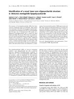
In the normal colorectal mucosa, GS28 is
expressed in the cytoplasm of the crypt epithelium
with weak to moderate intensity (Figure 1A). In the
CRC tissues, GS28 staining results were available in
214 cases. GS28 immunoreactivity was revealed in 213
cases (99.5%) of CRC. Among these, 28 cases showed
weak immunopositivity, and 92 and 93 cases showed
moderate and strong staining, respectively (Figure
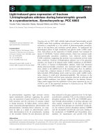
1B–1D). There were 26 nuclear predominant cases and
188 non-nuclear predominant cases (Figure 2A and
2B). Stromal GS28 expression was demonstrated in
152 cases of CRC.
GS28 nuclear predominant immunoreactivity
was significantly associated with advanced tumour
517
stage (T stage; p = 0.045) and marginally associated
with perineural invasion (p = 0.064) (Table 2). Other
clinicopathological features were not associated with
GS28 expression. As stromal cells of tumour tissues
are important in the progression of CRC, we
evaluated the association of GS28 expression with
clinicopathological parameters. Decreased GS28
expression in the stromal cells was significantly
associated with lymph nodes metastasis (N stage; p =
0.036) (Table 3). EGFR expression and KRAS
mutations are important well-known molecular
markers in CRC. However, GS28 expression was not
associated
with
EGFR
immunohistochemical
positivity or KRAS mutation status in the current
study (Tables 4 and 5).
Table 1. Clinicopathological data and molecular marker
expression in 230 CRC patients
Characteristics
Sex
Male
Female
Age
≤ 55 years
> 55 years
Tumour stagea
T1
T2
T3
T4
Nodal stageb
N0
N1
N2
Metastasis
M0
M1
Sitec
Right colon
Left colon
Rectum
N (%)
140 (60.9)
90 (39.1)
60 (26.1)
170 (73.9)
2 (0.9)
14 (6.1)
157 (68.3)
51 (22.2)
86 (37.4)
76 (33.0)
64 (27.8)
211 (91.7)
19 (8.3)
65 (28.3)
86 (37.4)
78 (33.9)
Data regarding tumour stage were unavailable in 6 cases.
Data regarding nodal stage were unavailable in 4 cases.
cData regarding tumour location were unavailable in 1 case.
CRC: colorectal cancer
a
b
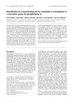
Prognostic values of GS28 expression in CRC
Thirty-one patients expired during the study
period. We investigated the prognostic value of GS28
with Kaplan-Meier analysis, and revealed a
correlation with overall survival (p = 0.004) (Table 6
and Figure 3). Our results showed that the cases with
GS28
nuclear
predominant
expression
had
significantly poorer overall survival than those with a
non-nuclear predominant pattern. Additionally, there
were no significant survival differences between
CRCs with GS28 stromal expression and
non-expression (data not shown). Taken together,
these results indicate that GS28 nuclear predominant
expression could serve as a prognostic marker for
CRC.
Int. J. Med. Sci. 2017, Vol. 14
518
Figure 1. Representative GS28 immunohistochemical staining in (A) normal colonic mucosa and CRC with (B) weak cytoplasmic staining, (C) moderate cytoplasmic
staining and (D) strong cytoplasmic staining results (× 400). Stromal immunoreactivity is also shown in myofibroblastic cells of the lamina propria (C and D).
Figure 2. Representative GS28 immunohistochemical staining in CRC with (A) nuclear predominant pattern (nuclear staining: 3, cytoplasmic staining: 1) (B)
non-nuclear predominant pattern (nuclear staining: 0, cytoplasmic staining: 2) (× 400).
Int. J. Med. Sci. 2017, Vol. 14
519
Table 2. Relationship between GS28 expression
clinicopathological parameters in CRC patients
Parameter
Sex
Male
Female
Age
≤ 55 years
> 55 years
Tumour stage
T1, T2 or T3
T4
Nodal stage
N0 or N1
N2
Metastasis
M0
M1
Lymphatic
invasion
Absent
Present
Vascular
invasion
Absent
Present
Perineural
invasion
Absent
Present
Differentiation
Well-to-moderate
Poor
Site
Right colon
Left colon or
rectum
GS28 expression (n = 214a)
nuclear
non-nuclear
predominant
predominant
and
P value
0.987
16
10
116
72
5
21
49
139
16
10
148
40
18
8
138
50
22
4
176
12
0.452
0.045*
0.654
0.102
0.738
10
16
66
122
0.822
23
3
169
19
0.064
14
12
136
52
24
2
178
10
7
19
56
132
0.644
0.764
aOne case with GS28 non-immunoreactivity is included in the non-nuclear
predominant subgroup.
* Statistically significant
CRC: colorectal cancer
Table 3. Relationship between stromal GS28 expression and the
clinicopathological parameters in CRC patients
Parameter
Sex
Male
Female
Age
≤ 55 years
> 55 years
Tumour stage
T1, T2 or T3
T4
Nodal stage
N0 or N1
N2
Metastasis
M0
M1
Lymphatic
invasion
Absent
Present
Vascular
invasion
Absent
Present
Perineural
invasion
Absent
Present
Differentiation
Well-to-moderate
Poor
Site
Right colon
Left colon or
rectum
Marker
KRAS mutation
Positive
Negative
EGFR expression
Positive
Negative
GS28 expression (n = 214)
nuclear
non-nuclear
predominant
predominant
P value
0.937
11
15
78
110
17
9
142
46
EGFR: epidermal growth factor receptor; CRC: colorectal cancer
0.487
96
56
36
26
35
117
19
43
116
36
48
14
117
35
39
23
144
8
54
8
0.244
0.863
0.036*
0.054
0.995
54
98
22
40
0.853
136
16
56
6
0.631
108
44
42
20
143
9
59
3
43
109
20
42
1.000
0.563
Table 5. Relationship between stromal GS28 expression and
epidermal growth factor receptor expression, and KRAS mutation
status in CRC patients
KRAS mutation
Positive
Negative
EGFR expression
Positive
Negative
Stromal GS28 expression (n = 214)
Positive
Negative
P value
0.247
67
85
22
40
115
37
44
18
0.476
EGFR: epidermal growth factor receptor; CRC: colorectal cancer
Table 6. Kaplan-Meier analysis of overall survival in CRC patients
Variable
0.267
P value
* Statistically significant
CRC: colorectal cancer
Marker
Table 4. Relationship between GS28 expression and epidermal
growth factor receptor expression, and KRAS mutation status in
CRC patients
Stromal GS28 expression (n = 214)
Positive
Negative
Kaplan-Meier analysis
M ± SE (Days)
95% CI
1881.42 ± 189.38
1510.24 - 2252.61
GS28 nuclear
predominant
GS28 non-nuclear 2300.14 ± 43.42
predominant
P value
0.004*
2215.04 - 2385.23
* Statistically significant
CRC: colorectal cancer; M: mean; SE: standard error; CI: confidence interval
Int. J. Med. Sci. 2017, Vol. 14
520
Figure 3. Association of the overall survival of CRC patients with GS28 tumour cell expression. Cases with GS28 nuclear predominant expression showed
significantly poorer overall survival.
Discussion
We found that increased nuclear expression of
GS28 in primary CRC tissues significantly correlated
with advanced T stage tumours (p = 0.045), and
decreased stromal expression of GS28 significantly
correlated with advanced N stage tumours (p = 0.036).
Additionally, increased nuclear GS28 expression was
marginally associated with perineural invasion (p =
0.064). We were unable to find an association of
cytosolic or nuclear GS28 expression with other
clinicopathological parameters, such as sex, M stage,
tumour differentiation, EGFR expression, or KRAS
mutation. Therefore, a larger-scale study might be
necessary to further evaluate the clinicopathological
values in CRC tissues. This study is the first to
examine the correlation between GS28 expression and
clinicopathological parameters in CRC tissues.
The ER and Golgi apparatus are two major
organelles that play important roles in the processing,
sorting, and transport of newly synthesized secretory
and transmembrane proteins [9]. The ER-Golgi
network is a hub for various signalling pathways
involved in crucial cellular activities, including cell
death and malignant transformation [8]. The
localization of caspase 2, Polo-like kinase 3 (Plk3), and
GD3 synthase to Golgi suggested that the Golgi may
be active in the crucial cellular activities [18-20].
GS28 is a 28 kDa membrane protein that appears
to play an essential role in intra-Golgi or ER-Golgi
vesicle transport [10]. Mammalian SNAREs known to
participate in vesicular transport include GS28, Bet1,
Sec22b, and syntaxin 5 [21, 22]. Very few studies have
focused on the possible roles of these proteins in
pathological conditions, however. Studies in C. elegans
and Drosophila GS28 mutants have suggested that
GS28 plays important roles in proliferation and
differentiation of seam cells and in maintenance of
retinal neurons [12, 13]. We reported previously that
GS28 plays a protective role in hydrogen
peroxide-induced cell death via inhibition of p38
MAPK in glutathione-depleted neuronal cells [23].
However, few researches to examine GS28 expression
in human pathological tissues have yet been reported.
Recent studies have shown that the ER
stress-related signalling pathways and malfunction of
the Golgi apparatus are involved in cancer
development [9, 24]. The present study demonstrated
that increased nuclear expression of GS28 in CRC is
significantly correlated with advanced T stage
tumours. Considering that GS28 is a protein located in
the Golgi apparatus, it can be speculated that
translocation of GS28 into the nuclear compartment
may be related to increases in tumour cell migration
and invasion, possibly via interactions between
GS28-induced nuclear functions and the Golgi
apparatus. Syntaxin 17, another SNARE protein, was
found to be localized in the cytoplasm, nucleus, and
both in several types of cells [25]. Furthermore, its
localization was altered in tumour cells compared
with their normal counterparts, suggesting that
syntaxin 17 may possess additional novel roles in cell
proliferation and transformation. We observed
nuclear GS28 expression in TMAs of cervical cancer,
Int. J. Med. Sci. 2017, Vol. 14
and a significant association between the high nuclear
expression of GS28 and the advanced T stage tumors
[14]. We, furthermore, demonstrated that patients
with high nuclear expression of GS28 showed
significantly
worse
overall
survival
and
progression-free survival, compared to those with low
or no nuclear expression. These suggest that the
nuclear expression of GS28 protein plays important
roles in the progression of CRC. However, molecular
mechanisms of protein translocation and its roles
remain unknown. Sun et al. [26] revealed that GS28
forms a complex with p53 and its ubiquitin ligase
MDM2. They showed that overexpression of GS28
promotes cisplatin-induced apoptosis by reducing the
ubiquitination and degradation of p53. In contrast,
knockdown of GS28 using shRNA (short hairpin
RNA) demonstrated the opposite result in response to
cisplatin. These findings offer the first evidence that
SNARE proteins can be involved in chemosensitivity,
although these results have only been observed in
vitro. It has not yet been confirmed that interactions
among p53, MDM2, and GS28 proteins occur in the
cytosolic or nuclear compartments.
We predicted conserved motifs in the GS28
protein (250 amino acids) using web-based software
PROSITE and PredictProtein. Only one hit displayed
in the prediction is coiled-coil helices (called SNARE
motifs), which mediate the interactions between
SNARE proteins. A nuclear localization signal motif is
not contained in the GS28 protein. Motifs with high
probability of occurrence are glycosylation sites and
target sites of phosphorylation for casein kinase II
(CKII), protein kinase C (PKC), and cAMP- and
cGMP-dependent protein kinases. Involvement of
CKII and the tumour promoter PKC as poor
prognostic factors in CRC has been reported [27].
However, GS28 phosphorylation and its nuclear
localization have not yet been reported. Further
studies should be performed to confirm the molecular
mechanisms of the protein kinases and the
phosphorylation of GS28 in CRC.
It has been shown that 30% of patients with
node-negative CRC on conventional histopathological
analysis die from metastatic disease [28]. However,
there is no standard method to identify lymphatic and
blood vessel invasion, which are reliable independent
prognostic factors in patients with node-negative CRC
[28]. We identified a reverse relationship between N
stage of CRC and GS28 expression in the stromal
fibroblasts. Stromal cells contribute to CRC
development and progression via secreting regulatory
molecules [29]. Thus, low GS28 expression in stromal
fibroblasts might be a prognostic factor for patients
with node-negative CRC.
We observed an association trend of increased
521
nuclear GS28 protein with perineural invasion in
CRC. The presence of perineural invasion was
suggested as an independent prognostic factor for a
more aggressive phenotype and poor prognosis in
CRC [30, 31]. Perineural invasion was strongly
correlated with high tumour stage, poor
differentiation, nodal involvement, infiltrative
growth, lymphatic invasion, and venous invasion.
Adjuvant therapy was suggested particularly for
node-negative CRC patients with perineural invasion
[31]. However, further studies with larger populations
of CRC patients should be performed to confirm a
significant association between GS28 expression and
perineural invasion.
Thus, we assessed for the first time the
prognostic
value
of
GS28
in
colorectal
adenocarcinoma. Our findings indicate that GS28
nuclear predominant expression appears to be an
independent predictor of poorer survival in patients
with CRC. GS28 may be a potential novel candidate
for a prognostic biomarker in the battle against CRC.
Our study results provide a better understanding of
the importance of GS28 in tumour development and
may enable the establishment of clinically useful
therapeutic targets.
Acknowledgments
This research was supported by the Basic Science
Research Program through the National Research
Foundation of Korea (NRF) funded by the Ministry of
Education (2013R1A1A2011752).
Author Contributions
S. H. Lee, S. Y. Kim, and S. W. Jeong designed the
research. H. J. Yoo, Y. Cui, S. H. Lee, S. Y. Kim, D. E.
Rim, E. S. Jung, and A. Lee performed the
experiments. S. T. Oh and J. G. Kim collected the
tissues. S. H. Lee, S. Y. Kim, O. J. Kwon, and S.W.
Jeong analysed the data. S. H. Lee, S. Y. Kim, and S.
W. Jong wrote the paper.
Competing Interests
The authors have declared that no competing
interest exists.
References
1.
2.
3.
4.
5.
6.
Shike M, Winawer SJ, Greenwald PH, Bloch A, Hill MJ, Swaroop SV. Primary
prevention of colorectal cancer. The WHO Collaborating Centre for the
Prevention of Colorectal Cancer. Bull World Health Organ. 1990; 68: 377-85.
Jung KW, Won YJ, Kong HJ, Oh CM, Cho H, Lee DH, et al. Cancer statistics in
Korea: incidence, mortality, survival, and prevalence in 2012. Cancer Res
Treat. 2015; 47: 127-41.
Schmoll HJ, Stein A. Colorectal cancer in 2013: Towards improved drugs,
combinations and patient selection. Nat Rev Clin Oncol. 2014; 11: 79-80.
Cancer Genome Atlas Network. Comprehensive molecular characterization of
human colon and rectal cancer. Nature. 2012; 487: 330-7.
Mellman I, Simons K. The Golgi complex: in vitro veritas? Cell. 1992; 68:
829-40.
Malsam J, Söllner TH. Organization of SNAREs within the Golgi stack. Cold
Spring Harb Perspect Biol. 2011; 3: a005249.
Int. J. Med. Sci. 2017, Vol. 14
7.
8.
9.
10.
11.
12.
13.
14.
15.
16.
17.
18.
19.
20.
21.
22.
23.
24.
25.
26.
27.
28.
29.
30.
31.
522
Tai G, Lu L, Wang TL, Tang BL, Goud B, Johannes L, et al. Participation of the
syntaxin 5/Ykt6/GS28/GS15 SNARE complex in transport from the
early/recycling endosome to the trans-Golgi network. Mol Biol Cell. 2004; 15:
4011-22.
Wilson C, Venditti R, Rega LR, Colanzi A, D'Angelo G, De Matteis MA. The
Golgi apparatus: an organelle with multiple complex functions. Biochem J.
2011; 433: 1-9.
Wlodkowic D, Skommer J, McGuinness D, Hillier C, Darzynkiewicz Z.
ER-Golgi network--a future target for anti-cancer therapy. Leuk Res. 2009; 33:
1440-7.
Subramaniam VN, Peter F, Philp R, Wong SH, Hong W. GS28, a 28-kilodalton
Golgi SNARE that participates in ER-Golgi transport. Science. 1996; 272:
1161-3.
Nagahama M, Orci L, Ravazzola M, Amherdt M, Lacomis L, Tempst P, et al. A
v-SNARE implicated in intra-Golgi transport. J Cell Biol. 1996; 133: 507-16.
Maekawa M, Inoue T, Kobuna H, Nishimura T, Gengyo-Ando K, Mitani S, et
al. Functional analysis of GS28, an intra-Golgi SNARE, in Caenorhabditis
elegans. Genes Cells. 2009; 14: 1003-13.
Rosenbaum EE, Vasiljevic E, Cleland SC, Flores C, Colley NJ. The Gos28
SNARE protein mediates intra-Golgi transport of rhodopsin and is required
for photoreceptor survival. J Biol Chem. 2014; 289: 32392-409.
Cho U, Kim HM, Park HS, Kwon OJ, Lee A, Jeong SW. Nuclear Expression of
GS28 Protein: A Novel Biomarker that Predicts Worse Prognosis in Cervical
Cancers. PLoS One. 2016; 11: e0162623.
Kim YI, Lee A, Lee BH, Kim SY. Prognostic significance of syndecan-1
expression in cervical cancers. J Gynecol Oncol. 2011; 22: 161-7.
Lee SH, Lee YS, Hong YG, Kang CS. Significance of COX-2 and VEGF
expression in histopathologic grading and invasiveness of meningiomas.
APMIS. 2014; 122: 16-24.
Kim SY, Choi EJ, Yun JA, Jung ES, Oh ST, Kim JG, et al. Syndecan-1 expression
is associated with tumor size and EGFR expression in colorectal carcinoma: a
clinicopathological study of 230 cases. Int J Med Sci. 2015; 12: 92-9.
Mancini M, Machamer CE, Roy S, Nicholson DW, Thornberry NA,
Casciola-Rosen LA, et al. Caspase-2 is localized at the Golgi complex and
cleaves golgin-160 during apoptosis. J Cell Biol. 2000; 149: 603-12.
Ruan Q, Wang Q, Xie S, Fang Y, Darzynkiewicz Z, Guan K, et al. Polo-like
kinase 3 is Golgi localized and involved in regulating Golgi fragmentation
during the cell cycle. Exp Cell Res. 2004; 294: 51-9.
Rippo MR, Malisan F, Ravagnan L, Tomassini B, Condo I, Costantini P, et al.
GD3 ganglioside directly targets mitochondria in a bcl-2-controlled fashion.
FASEB J. 2000; 14: 2047-54.
Zhang T, Hong W. Ykt6 forms a SNARE complex with syntaxin 5, GS28, and
Bet1 and participates in a late stage in endoplasmic reticulum-Golgi transport.
J Biol Chem. 2001; 276: 27480-7.
Glick BS, Nakano A. Membrane traffic within the Golgi apparatus. Annu Rev
Cell Dev Biol. 2009; 25: 113-32.
Lee HO, Byun YJ, Cho KO, Kim SY, Lee SB, Kim HS, et al. GS28 Protects
Neuronal Cell Death Induced by Hydrogen Peroxide under
Glutathione-Depleted Condition. Korean J Physiol Pharmacol. 2011; 15:
149-56.
Gong H, Feng L. Computational analysis of the roles of ER-Golgi network in
the cell cycle. BMC Syst Biol. 2014; 8 Suppl 4: S3.
Zhang Q, Li J, Deavers M, Abbruzzese JL, Ho L. The subcellular localization of
syntaxin 17 varies among different cell types and is altered in some malignant
cells. J Histochem Cytochem. 2005; 53: 1371-82.
Sun NK, Huang SL, Chien KY, Chao CC. Golgi-SNARE GS28 potentiates
cisplatin-induced apoptosis by forming GS28-MDM2-p53 complexes and by
preventing the ubiquitination and degradation of p53. Biochem J. 2012; 444:
303-14.
Lin KY, Tai C, Hsu JC, Li CF, Fang CL, Lai HC, et al. Overexpression of nuclear
protein kinase CK2 alpha catalytic subunit (CK2alpha) as a poor
prognosticator in human colorectal cancer. PLoS One. 2011; 6: e17193.
van Wyk HC, Roxburgh CS, Horgan PG, Foulis AF, McMillan DC. The
detection and role of lymphatic and blood vessel invasion in predicting
survival in patients with node negative operable primary colorectal cancer.
Crit Rev Oncol Hematol. 2014; 90: 77-90.
Sun XF, Zhang H. Clinicopathological significance of stromal variables:
angiogenesis, lymphangiogenesis, inflammatory infiltration, MMP and
PINCH in colorectal carcinomas. Mol Cancer. 2006; 5: 43.
Poeschl EM, Pollheimer MJ, Kornprat P, Lindtner RA, Schlemmer A, Rehak P,
et al. Perineural invasion: correlation with aggressive phenotype and
independent prognostic variable in both colon and rectum cancer. J Clin
Oncol. 2010; 28: e358-60.
Liebig C, Ayala G, Wilks J, Verstovsek G, Liu H, Agarwal N, et al. Perineural
invasion is an independent predictor of outcome in colorectal cancer. J Clin
Oncol. 2009; 27: 5131-7.