Hyperglycemia inhibits osteoblastogenesis of rat bone marrow stromal cells via activation of the Notch2 signaling pathway
Bạn đang xem bản rút gọn của tài liệu. Xem và tải ngay bản đầy đủ của tài liệu tại đây (858.27 KB, 8 trang )
Int. J. Med. Sci. 2019, Vol. 16
Ivyspring
International Publisher
696
International Journal of Medical Sciences
2019; 16(5): 696-703. doi: 10.7150/ijms.32707
Research Paper
Hyperglycemia inhibits osteoblastogenesis of rat bone
marrow stromal cells via activation of the Notch2
signaling pathway
Kuo-Chin Huang 1,2,, Po-Yao Chuang2, Tien-Yu Yang2, Tsan-Wen Huang2, Shun-Fu Chang3
1.
2.
3.
School of Traditional Chinese Medicine, Chang Gung University College of Medicine, Taoyuan City 33302, Taiwan
Department of Orthopaedic Surgery, Chiayi Chang Gung Memorial Hospital, Chiayi County 61363, Taiwan
Department of Medical Research and Development, Chiayi Chang Gung Memorial Hospital, Chiayi County 61363, Taiwan
Corresponding author: Kuo-Chin Huang, MD, PhD and Shun-Fu Chang, PhD. Department of Orthopaedic Surgery, Chiayi Chang Gung Memorial Hospital,
No. 6, West Sec., Chiapu Rd., Putz City, Chiayi County 61363, Taiwan. Tel: (+886)-5-362-1000 ext. 2855. E-mail: and
© Ivyspring International Publisher. This is an open access article distributed under the terms of the Creative Commons Attribution (CC BY-NC) license
( See for full terms and conditions.
Received: 2019.01.01; Accepted: 2019.03.23; Published: 2019.05.10
Abstract
Background: Bone fragility and related fractures are increasingly being recognized as an important
diabetic complication. Mesenchymal progenitors often serve as an important source of bone
formation and regeneration. In the present study, we have evaluated the effects of diabetes on
osteoblastogenesis of mesenchymal progenitors.
Methods: Primary bone marrow stromal cells (BMSCs) were isolated from control and
streptozotocin-induced diabetic rats. These cells were evaluated for the effects of in vivo
hyperglycemia on the survival and function of mesenchymal progenitors. We concomitantly
investigated the effects of different concentrations of glucose, osmolality, and advanced glycation
end product (AGE) on osteogenic differentiation and matrix mineralization of rat bone marrow
mesenchymal stem cells (RMSC-bm). The relationship between the expression levels of Notch
proteins and the corresponding ALP levels was also examined.
Results: Our results revealed that in vivo hyperglycemia increased cell proliferation rate but
decreased osteogenic differentiation and matrix mineralization of primary rat BMSCs. In vitro high
glucose treatment, instead of high AGE treatment, induced a dose-dependent inhibition of
osteoblastogenesis of RMSC-bm cells. Activation of the Notch2 signaling pathway, instead of the
Notch1 or osmotic response pathways, was associated with these diabetic effects on
osteoblastogenesis of mesenchymal progenitors.
Conclusions: Hyperglycemia might inhibit osteoblastogenesis of mesenchymal progenitors via
activation of the Notch2 signaling pathway.
Key words: Diabetes mellitus, Hyperglycemia, Osteoblastogenesis, Mesenchymal Progenitor, Notch signaling
pathway
Introduction
Fragility fractures are being increasingly
recognized as an important complication of both type
1 diabetes mellitus (T1DM) and type 2 diabetes
mellitus (T2DM), and are associated with excessive
morbidity, mortality and health-care costs [1]. The
extensive prospective Nurses’ Health Study revealed
that the incidence of hip fractures in T1DM patients is
2.5-fold higher than that in the T2DM population, and
6-fold higher than that in the general population [2].
There is also a moderate increase in the number of
fractures of the spine, distal forearm and proximal
humerus in diabetic populations [3]. The
pathophysiological mechanisms underlying bone
fragility and increased risk of fragility fractures in
Int. J. Med. Sci. 2019, Vol. 16
diabetes mellitus (DM) are complex and not
completely understood [4]. Some of these mechanisms
can potentially alter the fate of mesenchymal
progenitors, the initial precursor and major source of
osteoblasts, leading to impaired osteogenic
differentiation, compromised matrix mineralization,
and thus reduced bone strength [5]. Therefore,
understanding the diabetes-bone association and
studying the process of cell fate determination of
mesenchymal
progenitors
in
the
diabetic
microenvironment is critical for improving bone
fragility and decreasing the risk of fragility fractures
in patients with DM.
Various signaling factors have been implicated
in the regulation of maintenance and expansion of
mesenchymal progenitors, including those from the
Notch signaling pathway [6]. Notch signaling is an
important pathway in that it regulates cell-to-cell
signal transduction, which plays an essential role in
skeletal remodeling [7]. Notch signaling requires
cell-cell contact, and is initiated when the Notch
ligands bind to the Notch receptors expressed on the
surface of a neighboring cell, then the Notch
intracellular domain (NICD) is cleaved and released,
followed by its translocation from the cell membrane
to the nucleus. In the nucleus, the NICD interacts with
the transcriptional regulator of the CSL family to
regulate downstream target genes [8]. A study, shows
that, NICD overexpression prevents the biological
effects of BMP-2 and Wnt by suppressing Wnt but not
BMP signaling [9], however, others showed that
Notch signaling promotes osteogenic differentiation
of mesenchymal progenitors by enhancing BMP
signaling [10], whereas, inhibition of Notch signaling
impaired BMP2-induced osteoblast differentiation
[11]. Besides, Notch1 and Notch2 proteins may
function differently and even in reverse manners in
the same cell type [12]. Therefore, further studies are
necessary to delineate the relationship between Notch
and the other signaling pathways under physiological
or pathological conditions.
In the present study, we evaluated the effects of
diabetes on osteoblastogenesis of mesenchymal
progenitors. To address this issue, we investigated the
effects of in vivo hyperglycemia, increased glucose or
normal glucose levels in vitro, and high osmotic
treatments on cellular proliferation, osteogenic
differentiation, and matrix mineralization of
mesenchymal progenitors. We also assessed the
relationship between the Notch expression and
osteogenic differentiation in response to treatment
with different concentrations of glucose and advanced
glycation end product (AGE).
697
Materials and methods
Animals and streptozotocin (STZ)-induced
diabetic models
Seven-week-old male Sprague-Dawley (SD) rats
were purchased from BioLASCO Taiwan Co., Ltd.
(Taipei, Taiwan), and were housed under
environmentally controlled conditions with ad
libitum access to standard laboratory chow. The
Institutional Animal Care and Use Committee
(IACUC) of Chang Gung Memorial Hospital
approved the animal use protocol, and all animal
experiments followed the Animal Protection Law by
the Council of Agriculture, Executive Yuan, ROC, and
were performed according to the guidelines for the
Care and Use of Laboratory Animals as promulgated
by the Institute of Laboratory Animal Resources,
National Research Council, USA.
Diabetes was induced in SD rats with a single
intraperitoneal (IP) administration of STZ (65 mg/kg
of body weight) diluted in citrate buffer (0.01 M, pH =
4.3). Control animals received the buffer alone.
Animals were given food and water ad libitum and
their body weights were continuously monitored.
Blood glucose levels were evaluated at regular
intervals using a glucometer (Accu-Check, Roche
Diagnostics, Basel, Switzerland), and rats with blood
glucose level > 250 mg/dL were considered to be
diabetic. Glycated hemoglobin (HbA1c) levels were
concomitantly evaluated using the ARKRAY
Automatic Glycohemoglobin Analyzer ADAMS A1c
(ARKRAY Factory Inc., Shiga, Japan).
Isolation of primary rat bone marrow MSCs
(BMSCs)
We isolated primary rat bone marrow by cutting
both ends of the femur and tibia, and flushing the
bone marrow with Dulbecco’s modified Eagle’s
medium
(DMEM,
Life
Technologies
Inc.,
Gaithersburg, MD, USA). After flushing the bone
marrow, we filtered the cell suspension through a 70
µm filter to obtain a single cell suspension. Then, we
lysed the RBCs using ammonium chloride (at a 9:1
ratio) for 10 min on ice. After lysing RBCs, we
removed ammonium chloride by centrifugation and
washing the cell pellet once with culture medium. We
then plated the BMSCs isolated from the rats into a
T75 flask. The first change of medium was performed
less than 72 h after plating the cells in the flask.
Cell lines and cell cultures
Rat bone marrow mesenchymal stem cells
(RMSC-bm) were obtained from ScienCell Research
Laboratory (Carlsbad, CA, USA) and grown in
DMEM supplemented with 10% fetal bovine serum
Int. J. Med. Sci. 2019, Vol. 16
(FBS;
Life
Technologies
Inc.)
and
1%
penicillin/streptomycin at 37 °C in a 5% CO2
humidified incubator. The culture medium was
exchanged with a medium that was identical except
that it contained only 0.5% FBS, and the cells were
further incubated for 24 h before treatment with high
glucose or normal glucose but high osmotic
conditioned media.
Preparation of high glucose and normal
glucose but high osmotic conditioned media
To prepare high glucose conditioned media, we
supplemented DMEM (5.5 mM of glucose) with
additional glucose at 4 final concentrations of 5.5, 15,
25, and 35 mM of glucose. To prepare normal glucose
but high osmotic conditioned media, we
supplemented DMEM (5.5 mM of glucose) with 0, 9.5,
19.5, and 29.5 mM of mannitol, respectively. AGE was
purchased from BioVision (Glucose AGE-BSA-II,
BioVision Inc., Milpitas, CA, USA). For osteogenic
differentiation, cells were cultivated in 6-well plates
until 80% confluence and then incubated in the
medium containing ascorbate, ß-glycerophosphoate,
and
dexamethasone
(Sigma-Aldrich
GmbH,
Schnelldorf, Germany).
Analysis of cell proliferation
Cell proliferation was analyzed using XTT
assays (Biological Industries, Kibbutz Beit-Haemek,
Israel), for which the cells were plated in 96-well
plates and allowed to adhere for 24 h. Culture
medium was changed every 3 days. An ELISA plate
reader (Thermo Labsystems Multiskan RC, Vantaa,
Finland) was used to measure absorbance of samples
at a wavelength of 490 nm.
Western blotting
Cultured/treated cells were lysed with the lysis
buffer containing protease and phosphatase inhibitors
(phenylmethylsulfonylfluoride,
aprotinin,
and
sodium orthovanadate). Total protein from the cell
lysate (100 µg of protein) was separated using
SDS-PAGE (using a 10% running, and 4% stacking
polyacrylamide gel) and separated proteins were
transferred onto a nitrocellulose membrane
(Immobilon P; 0.45-µm pore size). The blot was then
treated
with
the
indicated
antibodies.
Chemiluminescent bands were detected by using the
Western-Light chemiluminescent detection system
(Applied Biosystems). COL1 and ALP expression
levels were normalized with those of ß-actin internal
control from the same sample.
ARS quantification assay
We used Alizarin Red-S (ARS) staining to study
the effects of high glucose treatment on matrix
698
mineralization. After treatment for 14 days, culture
medium was removed and the cells were fixed with
70% ice-cold ethanol (v/v) for 10 min and rinsed
thoroughly with distilled water. Cultures were then
stained with 40 mM ARS in deionized water (pH 4.2)
for 10 min at room temperature. After removing the
ARS solution by aspiration, cells were rinsed with
fresh PBS and dried at room temperature. ARS
concentrations were then calculated by comparison
using an ARS dye standard curve and expressed as
nM/mL.
Statistical analysis
Results were expressed as means ± SEM.
One-way analysis of variance (ANOVA) with
Scheffe’s post hoc test was performed and Spearman’s
rank correlation coefficient (rs) was calculated using
SPSS v13.0 (SPSS, Chicago, IL, USA). P < 0.05 was
considered as statistically significant.
Results
Effects of in vivo hyperglycemia on cell
proliferation, osteogenic differentiation and
matrix mineralization of primary rat BMSCs
Administration of STZ successfully induced
diabetes in all rats, as revealed by increased mean
blood glucose at the time of sacrifice (349.3 ± 16.1
mg/dL vs. 101.8 ± 3.9 mg/dL in control rats, P < 0.001)
(Fig. 1A). HbA1c levels were elevated accordingly
(9.18 ± 0.12% vs. 4.20 ± 0.09%, respectively, P < 0.001)
(Fig. 1B).
To determine whether in vivo hyperglycemia
impairs survival and functions of mesenchymal
progenitors, we harvested primary BMSCs from
control and STZ-induced diabetic rats. As determined
using the XTT assay, our data revealed that primary
BMSCs from the STZ-induced diabetic rats had a
higher proliferation rate than that of BMSCs from the
control rats (1.54 fold on day 7, P < 0.05) (Fig. 1C). On
the other hand, these cells showed decreased
osteogenic differentiation and matrix mineralization,
as seen using western blotting (31% and 42% decrease
in COL1 and ALP expression, respectively, P < 0.05
for both) (Fig. 1D) and ARS staining quantification
assay (0.35 ± 0.03 vs. 0.41 ± 0.01, P = 0.121) (Fig. 1E).
These results suggest that in vivo hyperglycemia in
STZ-induced diabetic rats is associated with an
increased proliferation rate but decreased osteogenic
differentiation and matrix mineralization of their
mesenchymal progenitors.
Effects of different concentrations of glucose
on osteogenic differentiation and matrix
mineralization of RMSC-bm cells
To
examine
the
effects
of
different
Int. J. Med. Sci. 2019, Vol. 16
699
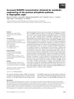
Figure 1. Hyperglycemia in STZ-induced diabetic rats is associated with an increased proliferation rate but decreased osteogenic differentiation and matrix
mineralization of primary bone marrow stromal cells (BMSCs). (A) Blood glucose levels and (B) HbAlc levels of normoglycemic (control) and diabetic (STZ) rats.
Primary BMSCs from control and diabetic rats were harvested, and their cell proliferation rates were determined using the XTT assay (C), COL1 and ALP
expression levels in BMSCs from control and diabetic rats were determined using western blotting (D), and matrix mineralization in BMSCs was determined using
the ARS staining quantification assay (E). Data are represented as mean ± SEM from three to four independent experiments. *, P < 0.05 vs. control cells. **, P < 0.001
vs. control cells.
concentrations
of
glucose
on
osteogenic
differentiation and matrix mineralization, we used
RMSC-bm cells in culture as a model system. As
determined using western blotting, high glucose (25
nM/L) treatment for 7 days inhibited expression of
both COL1 and ALP (27% and 36% decrease,
respectively, P < 0.05 for both) (Fig. 2A). As
determined using the ARS staining quantification
assay, high glucose treatment for 14 days induced
dose-dependent inhibition of calcium deposition (P <
0.05) (Fig. 2B). These results suggested that high
glucose treatment could lead to a deleterious impact
on
osteogenic
differentiation
and
matrix
mineralization of mesenchymal progenitors.
Effects of the osmotic effect of glucose on
osteogenic differentiation of RMSC-bm cells
To determine the role of osmotic effect of
glucose in osteogenic differentiation, we cultured
RMSC-bm cells in different concentrations of high
glucose and normal glucose but high osmotic
conditioned media. As determined using western
blotting, our data revealed that there were no
significant differences in either COL1 or ALP
expression among treatments with different
concentrations of normal glucose but high osmotic
conditioned media. On the contrary, high glucose
treatment for 7 days induced dose-dependent
inhibition of COL1 and ALP expression in RMSC-bm
cells (Fig. 3). These results suggest that the deleterious
effects of high glucose treatment on osteogenic
differentiation could not be ascribed to the osmotic
effect of glucose.
Effects of different concentrations of glucose
and AGE on Notch expression and osteogenic
differentiation of RMSC-bm cells
Because of the recent insights into the role of
Notch signaling in regulating bone physiology and in
causing human bone diseases [7], we examined the
relationship between Notch expression and high
glucose-induced
inhibition
of
osteogenic
differentiation. As determined using western blotting,
our data revealed that high glucose treatment for both
2 and 7 days would induce dose-dependent increase
in Notch2 expression in RMSC-bm cells. However,
there were no significant differences in Notch1
expression among treatments with different
concentrations of glucose. Meanwhile, Notch2
expression levels showed a moderate to strong
negative association with corresponding ALP
expression levels (rs = -0.674, P < 0.05) in response to
Int. J. Med. Sci. 2019, Vol. 16
700
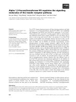
Figure 2. High glucose treatment presents a dose-dependent inhibitory effect on osteogenic differentiation and matrix mineralization of mesenchymal progenitors.
RMSC-bm cells were used as control (5.5 mM), or stimulated with high glucose (15, 25, and 35 mM) for 7 and 14 days. Western blotting was used to determine
COL1 and ALP expression levels in RMSC-bm cells (A), β-actin used as an internal control. The ARS staining quantification assay was used to define the extent of
matrix mineralization in RMSC-bm cells (B). Data are represented as mean ± SEM from three to four independent experiments. *, P < 0.05 vs. control cells.
treatments with different concentrations of glucose
(Fig. 4A). We further investigated the effects of
different concentrations of AGE on Notch expression
and osteogenic differentiation of RMSC-bm cells as
the gradual increase or accumulation of AGE is
considered to be an important cause of diabetic
complications. Our data revealed that AGE treatment
results in non-significant changes of Notch2 and ALP
expression. Besides, Notch2 expression levels did not
indicate an association with the corresponding ALP
expression levels (rs = 0.487, P = 0.11) in response to
treatments with different concentrations of AGE (Fig.
4B). These results suggest that activation of the
Notch2 signaling pathway might play a role in high
glucose-induced inhibition of osteoblastogenesis of
mesenchymal progenitors.
Discussion
This study suggests the potential mechanism
underlying
the
effects
of
diabetes
on
osteoblastogenesis of mesenchymal progenitors. Our
findings revealed that in vivo hyperglycemia in
STZ-induced diabetic rats is associated with an
increased cell proliferation rate; however, shows
decreased osteogenic differentiation of primary
BMSCs. High glucose treatment presents a
dose-dependently inhibitory effect on osteogenic
differentiation and matrix mineralization of
RMSC-bm cells. The deleterious effects of high
glucose treatment on osteogenic differentiation could
not be ascribed to the osmotic effect of glucose. On the
other hand, activation of the Notch2, instead of the
Notch1, signaling pathway might play a critical role
in
high
glucose-induced
suppression
of
osteoblastogenesis and subsequent compromised
osteogenic differentiation and reduced matrix
mineralization. Our findings provide insights into the
molecular mechanisms by which DM affects cell fate
determination of mesenchymal progenitors. Given
that Notch signaling affects cell fate decision of
BMSCs and that the effects are dependent on the
cellular context, it may be critical to develop
therapeutic options that can preferentially target the
osteoblastic lineage cells of diabetic patients with
bone fragility and related fragility fractures.
DM is characterized by hyperglycemia. Some in
vitro studies have indicated that hyperglycemia can be
toxic to osteoblasts [4]. Botolin et al. have shown that
hyperglycemia and its associated hyperosmolality
suppress expression of genes involved in osteoblast
maturation [13]. Cunha et al. further demonstrated
that MC3T3-E1 osteoblasts respond to high
extracellular glucose concentrations through an
osmotic response pathway that results in modulation
of ALP and OCN expression and uptake of calcium by
osteoblasts
in
culture
[14].
Contrastingly,
mesenchymal progenitors are more resistant to the
toxicity caused by hyperglycemia than osteoblasts,
Int. J. Med. Sci. 2019, Vol. 16
depending on the stemness of mesenchymal
progenitors [15]. This is supported by our findings
that primary BMSCs from the STZ-induced diabetic
rats have an increased proliferation rate as compared
to that of BMSCs from control rats. Some studies
showed that hyperglycemia and oxidative stress
might influence differentiation of mesenchymal
progenitors with adipogenesis being favored over
osteoblastogenesis [16]. In agreement with an earlier
observation in mesenchymal progenitors from
STZ-induced diabetic rats [17], we found that both, in
vivo hyperglycemia and in vitro high glucose
treatment, affect osteoblastogenesis of mesenchymal
progenitors. Our data demonstrates that the harmful
effect of hyperglycemia in mesenchymal progenitors
is independent of the extracellular osmolality but is
most likely attributed to the impact on cell fate
decision of mesenchymal progenitors. Indeed and
surprisingly, Notch2 expression levels presented a
moderate to strong negative association with the
corresponding ALP expression levels (rs = -0.674, P <
0.05) in response to treatments with different
concentrations of glucose.
DM is also characterized by accumulation of
AGEs. Levels of AGEs are increased in DM patients as
a result of chronic hyperglycemia and increased levels
of oxidative stress [18], and might play a crucial role
in the development of bone fragility and related
fragility fractures in these patients [5]. Yamamoto et
al. indicated that serum AGE (pentosidine) levels are
701
positively associated with the presence of vertebral
fractures in postmenopausal Japanese women with
T2DM [19]. Saito et al. found that the degree of
mineralization correlates with distinctive patterns of
enzymatic and non-enzymatic cross-links in human
bone [20]. Kume et al. showed that AGEs might lead
to in vivo loss of MSC mass and to the delay of bone
repair by inhibiting the maturation of MSC-derived
cells [21]. Taken together, accumulation of AGEs in
the organic bone matrix by the Maillard reaction
should be negatively associated with biomechanical,
dynamic, and microarchitectural skeletal properties
[22]. These data, although limited, would suggest that
AGE-distorted collagen likely renders the bone more
fragile in DM patients regardless of their bone mineral
density (BMD). In the current study, our data did not
support the idea that AGEs per se directly affect
osteoblastogenesis of mesenchymal progenitors. We
found that the AGE-BSA treatment did not result in
significantly enhanced ALP expression in RMSC-bm
cells, which would metabolize calcium phosphate into
insoluble phosphate salts, thereby mediating matrix
mineralization [23]. As the effects of AGEs
presumably vary according to the source of AGEs, the
type of cells, and culture conditions, further studies
will be required to clarify the details of the mechanism
of AGE-mediated control of MSC osteoblastogenesis,
particularly studies modeling the in vivo
microenvironment for the processes of diabetic
complications.
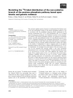
Figure 3. The deleterious effects of high glucose treatment on osteogenic differentiation could not be ascribed to the osmotic effect of glucose. RMSC-bm cells were
used as control (5.5 mM), or stimulated with (A) high glucose (15, 25, and 35 mM), and (B) normal glucose but high osmotic (5.5 mM glucose + 9.5 mM mannitol, 5.5
mM glucose + 19.5 mM mannitol, and 5.5 mM glucose + 29.5 mM mannitol) for 2, 5, and 7 days. Western blotting was used to determine COL1 and ALP expression
levels in RMSC-bm cells while β-actin used as an internal control.
Int. J. Med. Sci. 2019, Vol. 16
702
Figure 4. Activation of the Notch2 signaling pathway might play a role in high glucose-induced inhibition of osteogenic differentiation. RMSC-bm cells were used as
control (5.5 mM), or stimulated with high glucose (15, 25, and 35 mM) for 2 and 7 days. Western blotting was used to determine Notch1, Notch2, and ALP expression
levels in RMSC-bm cells (A), β-actin used as an internal control. RMSC-bm cells were used as control (50 µg/mL), or stimulated with high AGE (100 and 200 µg/mL)
for 2 days. Western blotting was used to determine Notch2 and ALP expression levels (B), β-actin used as an internal control. Data are represented as mean ± SEM
from three to four independent experiments. rs, Spearman’s rank correlation coefficient. *, P < 0.05 vs. control cells. **, P < 0.001 vs. control cells.
Our results revealed that Notch2, instead of
Notch1, plays a role in high glucose-induced
inhibition of osteoblastogenesis of mesenchymal
progenitors. Notch signaling, discovered in
Drosophila, is well known for its role in cell fate
decisions and the development of multiple tissues
including bone [24]. Notch signaling may interact
with other signaling pathways such as BMP and Wnt,
the two critical factors known to enhance
osteoblastogenesis [25,26], to regulate bone
homeostasis [7]. Although many studies reported that
Notch signaling inhibits osteoblastogenesis of
mesenchymal progenitors [9,27,28], some other
studies demonstrated that Notch sensitizes cells of the
osteoblastic lineage to the effects of inducers of
osteoblastogenesis
under
selective
conditions
[10,11,29]. Generally, Notch might be required not for
the function of mature osteoblasts, but for the
differentiation of their mesenchymal precursors [28].
Engin et al. further indicated that the conditional
deletion of notch2 would cause a similar
developmental phenotype to the dual deletion of
notch1 and notch2, which suggests that notch2 might be
the predominant regulator of endochondral bone
formation [27]. Our data contribute to the debate
regarding the role of Notch signaling in
osteoblastogenesis of mesenchymal progenitors,
supporting that the Notch2, instead of the Notch1,
signaling pathway might play a critical role in
diabetic bone fragility and related fragility fractures.
Conclusions
Hyperglycemia, instead of AGE, inhibits
osteoblastogenesis of mesenchymal progenitors,
which might be through activation of the Notch2,
instead of the Notch1 or osmotic response, signaling
pathway. This fact suggests the possibility of a new
therapeutic strategy to treat various diabetic
complications including bone fragility and related
fragility fractures. As a molecule that might
selectively inhibit osteoblastogenesis of mesenchymal
progenitors, Notch2 may be expected to be a unique
target molecule for the treatment of diabetic
complications such as bone fragility and related
fragility fractures.
Acknowledgement
This work was supported by grants from the
Int. J. Med. Sci. 2019, Vol. 16
Chang Gung Memorial Hospital, Taiwan (Grant Nos.
CMRPG 6C0081-3, and CMRPG 6G0061) and from the
Taiwan National Science Council (Grant No.
NSC102-2314-B-182A-033).
Competing Interests
The authors have declared that no competing
interest exists.
References
1.
2.
3.
4.
5.
6.
7.
8.
9.
10.
11.
12.
13.
14.
15.
16.
17.
18.
19.
20.
21.
22.
Janghorbani M, Van Dam RM, Willett WC, Hu FB. Systematic review of type 1
and type 2 diabetes mellitus and risk of fracture. Am J Epidemiol. 2007; 166(5):
495-505.
Janghorbani M, Feskanich D, Willett WC, Hu FB. Prospective study of diabetes
and risk of hip fracture: the Nurses’ Health Study. Diabetes Care. 2006; 29(7):
1573-1578.
Vestergaard P, Rejnmark L, Mosekilde L. Relative fracture risk in patients with
diabetes mellitus, and the impact of insulin and oral antidiabetic medication
on relative fracture risk. Diabetologia. 2005; 48(7): 1292-1299.
Napoli N, Strollo R, Paladini A, Briganti SI, Pozzilli P, Epstein S. The alliance
of mesenchymal stem cells, bone and diabetes. Int J Endocrinol. 2014; 2014:
690783.
Napoli N, Chandran M, Pierroz DD, Abrahamsen B, Schwartz AV, Ferrari SL.
Mechanisms of diabetes mellitus-induced bone fragility. Nat Rev Endocrinol.
2017; 13(4): 208-219.
Dong Y, Long T, Wang C, Mirando AJ, Chen J, O’Keefe RJ, et al.
NOTCH-mediated maintenance and expansion of human bone marrow
stromal/stem cells: a technology designed for orthopedic regenerative
medicine. Stem Cells Transl Med. 2014; 3(12): 1456-1466.
Regan J, Long F. Notch signaling and bone remodeling. Curr Osteoporos Rep.
2013; 11(2): 126-129.
Canalis E. Notch signaling in osteoblasts. Sci Signal. 2008; 1(17): pe17.
Deregowski V, Gazzerro E, Priest L, Rydziel S, Canalis E. Notch 1
overexpression inhibits osteoblastogenesis by suppressing Wnt/ß-catenin but
not bone morphogenetic protein signaling. J Biol Chem. 2006; 281(10):
6203-6210.
Cao J, Wei Y, Lian J, Yang L, Zhang X, Xie J, et al. Notch signaling pathway
promotes osteogenic differentiation of mesenchymal stem cells by enhancing
BMP9/Smad signaling. Int J Mol Med. 2017; 40(2): 378-388.
Nobta M, Tsukazaki T, Shibata Y, Xin C, Moriishi T, Sakano S, et al. Critical
regulation of bone morphogenetic protein-induced osteoblastic differentiation
by Delta1/Jagged1-activated Notch1 signaling. J Biol Chem. 2005; 280(16):
15842-15848.
Sun Y, Gao X, Liu J, Kong QY, Wang XW, Chen XY, et al. Differential Notch1
and Notch2 expression and frequent activation of Notch signaling in gastric
cancers. Arch Pathol Lab Med. 2011; 135(4): 451-458.
Botolin S, Faugere M, Malluche H, Orth M, Meyer R, McCabe LR. Increased
bone adiposity and peroxisomal proliferator-activated receptor-γ2 expression
in type I diabetic mice. Endocrinology. 2005; 146(8): 3622-3631.
Cunha JS, Ferreira VM, Maquigussa E, Naves MA, Boim MA. Effects of high
glucose and high insulin concentrations on osteoblast function in vitro. Cell
Tissue Res. 2014; 358(1): 249-256.
Li YM, Schilling T, Benisch P, Zeck S, Meissner-Weigl J, Schneider D, et al.
Effects of high glucose on mesenchymal stem cell proliferation and
differentiation. Biochem Biophys Res Commun. 2007; 363(1): 209-215.
Aguiari P, Leo S, Zavan B, Vindigni V, Rimessi A, Bianchi K, et al. High
glucose induces adipogenic differentiation of muscle-derived stem cells. Proc
Natl Acad Sci USA. 2008; 105(4): 1226-1231.
Weinberg E, Maymon T, Moses O, Weinreb. Streptozotocin-induced diabetes
in rats diminishes the size of the osteoprogenitor pool in bone marrow.
Diabetes Res Clin Pract. 2014; 103(1): 35-41.
Piperi C, Adamopoulos C, Dalagiorgou G, Diamanti-Kandarakis E,
Papavassiliou AG. Crosstalk between advanced glycation and endoplasmic
reticulum stress: emerging therapeutic targeting for metabolic diseases. J Clin
Endocrinol Metab. 2012; 97(7): 2231-2242.
Yamamoto M, Yamaguchi T, Yamauchi M, Yano S, Sugimoto T. Serum
pentosidine levels are positively associated with the presence of vertebral
fractures in postmenopausal women with type 2 diabetes. J Clin Endocrinol
Metab. 2008; 93(3): 1013-1019.
Saito M, Fujii K, Marumo K. Degree of mineralization-related collagen
crosslinking in the femoral neck cancellous bone in cases of hip fracture and
controls. Calcif Tissue Int. 2006; 79(3): 160-168.
Kume S, Kato S, Yamagishi S, Inagaki Y, Ueda S, Arima N, et al. Advanced
glycation end-products attenuate human mesenchymal stem cells and prevent
cognate differentiation into adipose tissue, cartilage, and bone. J Bone Miner
Res. 2005; 20(9): 1647-1658.
Leslie WD, Rubin MR, Schwartz AN, Kanis JA. Type 2 diabetes and bone. J
Bone Mine Res. 2012; 27(11): 2231-2237.
703
23. Beck GR Jr, Sullivan EC, Moran E, Zerler B. Relationship between alkaline
phosphatase levels, osteopontin expression, and mineralization in
differentiating MC3T3-E1 osteoblasts. J Cell Biochem. 1998; 68(2): 269-280.
24. Bugeon L, Taylor HB, Progatzky F, Lin MI, Ellis CD, Welsh N, et al. The
NOTCH pathway contributes to cell fate decision in myelopoiesis.
Haematologica. 2011; 96(12): 1753-1760.
25. Gazzerro E, Canalis E. Bone morphogenetic proteins and their antagonists.
Rev Endocr Metab Disord. 2006; 7(1-2): 51-65.
26. Krishnan V, Bryant HU, MacDougald OA. Regulation of bone mass by Wnt
signaling. J Clin Invest. 2006; 116(5): 1202-1209.
27. Engin F, Yao Z, Yang T, Zhou G, Bertin T, Jiang MM, et al. Dimorphic effects of
Notch signaling in bone homeostasis. Nat Med. 2008; 14(3): 299-305.
28. Hilton MJ, Tu X, Wu X, Bai S, Zhao H, Kobayashi T, et al. Notch signaling
maintains bone marrow mesenchymal progenitors by suppressing osteoblast
differentiation. Nat Med. 2008; 14(3): 306-314.
29. Tezuka K, Yasuda M, Watanabe N, Morimura N, Kuroda K, Miyatani S, et al.
Stimulation of osteoblastic cell differentiation by Notch. J Bone Miner Res.
2002; 17(2): 231-239.