Efficacy of honeycomb TCP-induced microenvironment on bone tissue regeneration in craniofacial area
Bạn đang xem bản rút gọn của tài liệu. Xem và tải ngay bản đầy đủ của tài liệu tại đây (2.65 MB, 11 trang )
Int. J. Med. Sci. 2016, Vol. 13
Ivyspring
International Publisher
466
International Journal of Medical Sciences
Research Paper
2016; 13(6): 466-476. doi: 10.7150/ijms.15560
Efficacy of Honeycomb TCP-induced Microenvironment
on Bone Tissue Regeneration in Craniofacial Area
Satoko Watanabe,1 Kiyofumi Takabatake,2 Hidetsugu Tsujigiwa,3 Toshiyuki Watanabe,1 Eijiro Tokuyama,1
Satoshi Ito2, Hitoshi Nagatsuka,2 Yoshihiro Kimata1
1.
2.
3.
Department of Plastic and Reconstructive Surgery, Okayama University, Graduate School of Medicine, Dentistry and Pharmaceutical Sciences, Okayama,
Japan.
Department of Oral Pathology and Medicine, Okayama University, Graduate School of Medicine, Dentistry and Pharmaceutical Sciences, Okayama, Japan.
Department of Life Science, Faculty of Science, Okayama University Science, Japan.
Corresponding authors: Kiyofumi Takabatake, e-mail:, TEL: +81-86-235-6652, FAX: +81-86-235-6654. Hidetsugu Tsujigiwa,
e-mail:, TEL/FAX: +81-86-256-9523
© Ivyspring International Publisher. Reproduction is permitted for personal, noncommercial use, provided that the article is in whole, unmodified, and properly cited. See
for terms and conditions.
Received: 2016.03.17; Accepted: 2016.05.18; Published: 2016.06.01
Abstract
Artificial bone materials that exhibit high biocompatibility have been developed and are being
widely used for bone tissue regeneration. However, there are no biomaterials that are minimally
invasive and safe. In a previous study, we succeeded in developing honeycomb β-tricalcium
phosphate (β-TCP) which has through-and-through holes and is able to mimic the bone
microenvironment for bone tissue regeneration. In the present study, we investigated how the
difference in hole-diameter of honeycomb β-TCP (hole-diameter: 75, 300, 500, and 1600 μm)
influences bone tissue regeneration histologically. Its osteoconductivity was also evaluated by
implantation into zygomatic bone defects in rats. The results showed that the maximum bone
formation was observed on the β-TCP with hole-diameter 300μm, included bone marrow-like
tissue and the pattern of bone tissue formation similar to host bone. Therefore, the results
indicated that we could control bone tissue formation by creating a bone microenvironment
provided by β-TCP. Also, in zygomatic bone defect model with honeycomb β-TCP, the result
showed there was osseous union and the continuity was reproduced between the both edges of
resected bone and β-TCP, which indicated the zygomatic bone reproduction fully succeeded. It is
thus thought that honeycomb β-TCP may serve as an excellent biomaterial for bone tissue
regeneration in the head, neck and face regions, expected in clinical applications.
Key words: honeycomb β-TCP, bone tissue regeneration, bone microenvironment, pore size, Bone
morphogenetic protein-2
Introduction
Free bone transplant has been performed for
bone defect reconstruction in areas such as the head
and neck, face and extremities. However, problems
such as sequestration and infection caused by
ischemia of transferred bone tissue tend to occur in
large bone defects. Although a free vascularized bone
graft exhibits good synostosis because of good blood
supply through anastomosis of the vascular pedicle
[1, 2], it requires a long time to harvest the graft and
the volume obtained for harvest is limited because of
donor site morbidity [3-5]. Furthermore, severe
complications like total graft necrosis may occur due
to problems with the vascular pedicle [6]. Recently,
bone tissue reconstruction performed with artificial
bone has received much attention due to its low
invasiveness and shorter surgical time. In addition, it
has the advantage of availability of adequate volume
and shape depending on the required component.
However, some problems remain such as exposure
and infection of material.
Three key factors are essential for the process of
tissue regeneration: cells, extracellular matrix (ECM)
Int. J. Med. Sci. 2016, Vol. 13
and growth factors. In addition, vascularity as a
nutrient source and dynamic elements influence the
factors. In previous studies on ECM which is one of
the
important
elements,
various
synthetic
biomaterials have been developed in order to
reproduce the extracellular microenvironment [7-10].
Several bioceramics having high biocompatibility like
hydroxyapatite (HA), β-tricalcium phosphate (β-TCP)
and calcium are already applied clinically for bone
tissue regeneration [11-14]. These materials function
as a scaffold on which bone cells proliferate and
differentiate, at the same time the scaffold materials
resorb and are replaced with new bone tissue
gradually. HA is hardly or very slowly absorbed in
vivo, but β-TCP is more easily absorbed compared
with HA. The risk of foreign body reaction and
infection of bone prosthetic material can be reduced if
new bone cell infiltration and neovascularization,
serving as a nutrient source, are induced into the
center of the bone material; it will be replaced by bone
tissue almost completely. Recently, the results from
some studies have indicated that the geometric
characteristics of biomaterials play an important role
in neovascularization and osteoinduction, so the pore
shape and size of those materials have been contrived
for optimal osteogenesis [15, 16]. However, most
synthetic bone materials currently on the market have
coecums in those pores acting as barriers that
interrupt osteogenesis and vascular development into
the center of pore, thereby preventing replacement of
most biomaterials by new bone tissue in the center of
pores and remains a foreign body [17]. Therefore, if a
large amount of artificial bone material has to be
transplanted or if vascularity of the recipient site is
poor, a lower graft success can be expected because of
insufficient penetration and proliferation of
osteocytes and vessels to the center.
We have already succeeded in developing new
honeycomb β-TCP which has a through-and-through
hole penetrating the material in order to overcome the
problems mentioned above. It was found that
histologically the honeycomb β-TCP had high
biological activity, when β-TCP at varied sintering
temperatures was embedded into an experimental
animal model [18]. In this study, we reproduced an
extracellular microenvironment using new β-TCP that
contained through-and-through holes and varied
pore size in each material and investigated the effect
of the extracellular microenvironment formed by
honeycomb β-TCP on bone tissue formation. In
addition, we selected the most optimal pore size of
honeycomb β-TCP and evaluated the compatibility as
a material for bone tissue reconstruction when
transplanted into a zygomatic bone defect in rat.
467
Materials and methods
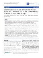
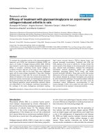
Preparation of honeycomb TCP containing
BMP-2
Honeycomb β-TCP was pressed in a cylindrical
mold with a depth of 5 mm, which contained
through-and-through holes of diameter 75 μm
(75TCP), 300 μm (300TCP), 500 μm (500TCP), 1600 μm
(1600TCP). And each β-TCP was calcinated by heating
to 1200 °C (Fig. 1). The detailed method of β-TCP
manufacture has been described previously [18].
Each β-TCP was sterilized by autoclave, and was
loaded with Bone morphogenetic protein-2 (BMP-2),
which was diluted to a final contained amount of 1000
ng (1000BMP), 500 ng (500BMP), 250 ng (250BMP),
125 ng (125BMP), and 0 ng (0BMP) in Matrigel® (BD
Bioscience). For BMP-2 loading, we centrifuged TCP
and Matrigel® added with BMP-2 (4 °C, 10000 rpm, 5
min). In the control group, we centrifuged TCP and
Matrigel® without BMP-2.
Fig. 1. Images of honeycomb β-TCP used in this experiment.
Animals and implantation procedure
Four-week-old healthy male Wister rats were
used in this experiment. All experiments in this study
were performed in accordance with the Policy on the
Care and Use of the Laboratory Animals, Okayama
University and approved by the Animal Care and Use
Committee, Okayama University, and all surgical
procedures were performed under general anesthesia,
in a pain-free state.
To investigate the osteoconductivity of
honeycomb β-TCP, the animals were randomly
divided into 20 groups: different holes of honeycomb
Int. J. Med. Sci. 2016, Vol. 13
β-TCP (4 types) × different amount of BMP-2 (5
conditions), total 20 groups.
Wistar
male
rats
were
anesthetized
intraperitoneally with ketamine hydrochloride (75
mg/kg body weight), medetomidine hydrochloride
(0.5 mg/kg body weight) and atipamezole
hydrochloride (1 mg/kg body weight) was injected
subcutaneously when awakening. The region of hip
from femoral region was shaved, cleaned with 70%
alcohol and iodine, and cut 10 mm by blunt dissection
to form 8 mm intramuscular pockets. Each sample
was implanted carefully with tweezers in the
intramuscular pockets and sutured. The animals were
killed with an overdose of ether at 3 weeks after
implantation.
For
histological
observations,
implanted β-TCPs were fixed in perfusion fixation by
4% paraformaldehyde (PFA).
Zygomatic bone reproduction of honeycomb
β-TCP
To evaluate the osteoinductive ability of the
β-TCP in a bone defect, we implanted the samples,
which were the most osteoconductive and formed
bone marrow-like tissue in the intramuscular
experiment, into the rat zygomatic bone defect. A
method for preparation of rat zygomatic bone defect
model is described below. At first, skin incision about
8 mm was made just above the zygomatic bone, and
the masseter muscle that adhered to the zygomatic
bone was completely separated from the bone, the
zygomatic bone was exposed entirely. Next, the
zygomatic bone periosteum was incised by a surgical
knife and was completely peeled from the zygomatic
bone, and bone was totally cut in two places using
scissors in front of the arch of zygomatic bone to
create 5 mm bone defect.
Then, β-TCP alone and β-TCP added with BMP
were implanted into this bone defect with the
through-and-through holes of β-TCP and the long
axis of the bone defects was parallel. And completely
zygomatic bone defect without β-TCP were prepared
as a control. For each groups, four to five Wister rats
were used. Each β-TCP was embedded 3 weeks later,
embedded tissues were fixed with 4% PFA reflux
fixation, and we investigated the specimens by micro
CT and histology.
Histological procedure and Immunohistochemical staining of osteopontin
The specimens were decalcified in 10%
ethylenediaminetetraacetic acid for 3 weeks. They
were embedded in paraffin, and sectioned to 5-μm
thickness. Sections were chemically stained with
hematoxylin and eosin (H&E), toluidine blue and
observed histologically.
468
Osteopontin (OPN) is a noncollagenous protein
that is produced in abundance in the bone
extracellular matrix by the osteoblasts responsible for
bone formation. Therefore, the presence of this bone
protein was investigated.
Sections were deparaffinized, rehydrated, and
incubated in proteinase K for 15 min at room
temperature. Endogenous peroxidase was blocked
using a 0.3% hydrogen peroxide solution in methanol
for 20 min. Nonspecific binding sites were blocked
with 10% normal rabbit antiserum (Vector
Laboratories, Burlingame, CA) for 10 min. The
sections were incubated with monoclonal antibodies
against rat OPN (Immuno-Biological Laboratories,
Gunma, Japan) following the Vectastain ABC Mouse
Kit method (Vector Laboratories, Burlingame, CA).
The principal steps were as follows: (1) incubation
with primary antibodies at a dilution of 1:50; (2)
incubation with secondary anti-mouse IgG antibodies
at a dilution of 1:200 for 30 min; (3) incubation with
avidin-biotin-peroxidase complex (ABC; Vector
Laboratories, Burlingame, CA) at a dilution of 1:50 for
30 min; (4) treatment with Diaminobenzidine color
development and nuclear counterstaining with
Mayer's hematoxylin. Staining was visualized using a
light microscope. The control sections were processed
in the same way but in the absence of the primary
antibodies.
Bone and cartilage tissue formation evaluation
by area measurement
HE-stained specimens were taken using a Nikon
Elipse 80i microscope (Teknooptik AB, Huddinge,
Sweden), equipped with an Easy Image 2000 system
(Teknooptik AB) using 103 to 403 lenses. In HE
staining specimens (100× magnification), we
investigated the image taken at a total of 5 fields (5
fields: at the center, both ends, and the center of the
center and both ends) using Image J1.47v [developed
by Wayne Rasband, the National Institute of Health
(NHS)]. In each field, we measured the total area of
bone formation in β-TCP holes and the area of β-TCP
holes and we calculated the ratio of area of bone area
in β-TCP holes to determine the average of the 5
fields. The obtained average value was compared in
each group, the rate of bone formation and cartilage
formation were compared for different pore size and
BMP concentration.
Micro CT
In the zygomatic bone defect model, the head
specimens after fixation were taken with micro CT
(Hitachi Aloka Latheta LCT200), and the resulting
DICOM data was reconstructed three-dimensionally
by using the workstation and software (AZE
Int. J. Med. Sci. 2016, Vol. 13
VirtualPlace Lexus64). Then, we assessed bone tissue
formation in the image.
Results
The effect of honeycomb β-TCP on bone and
cartilage tissue formation
The incidence rate of bone and cartilage tissue
formation depending on the amount of BMP is shown
469
in Table 1. In the control group, bone formation was
observed in all samples with 125BMP, but the
incidence rates of that were not so high. The incidence
rates were getting higher, as the amount of BMP was
increased and all the samples with 1000BMP, except
1600TCP, showed bone formation. 1600TCP seemed
likely to promote less bone formation than others
regardless of the amount of BMP.
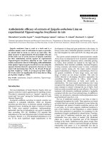
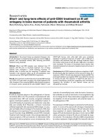
Fig. 2. HE staining images of each honeycomb β-TCP with added 1000 ng BMP-2 3 weeks after implantation. a) Lower-magnification images of 75TCP. b) Higher-magnification
image of corresponding outline area in (a). c) Lower-magnification images of 300TCP. d) Higher-magnification image of corresponding outline area in (c). e) Lower-magnification
images of 500TCP. f) Higher-magnification image of corresponding outline area in (e). g) Lower-magnification images of 1600TCP. h) Higher-magnification image of corresponding
outline area in (g). Bone formation pattern in each pore size TCP was different, bone tissue filled in the holes of 75TCP. Bone formation was observed in 300TCP adding to the
inner wall, and also bone marrow-like tissue was observed in some parts. In 500TCP, cancellous bone-like bone tissue was observed, and in 1600TCP bone tissue formation was
observed in the center of TCP hole. Bone tissue is indicated by arrowheads, and bone marrow-like tissue is indicated by an asterisk.
Int. J. Med. Sci. 2016, Vol. 13
470
There were quite a few samples that showed
cartilage formation except 75TCP. Even though
cartilage formation was observed in some samples,
cartilage tissue extended to just a part of them and the
inner lumen of TCP was not totally replaced by
cartilage tissue. In 1600TCP, with any BMP, cartilage
tissue was not observed. However, the 75TCP group
was different from the others, as there was a high
incidence rate of cartilage tissue formation not only in
samples with large amounts of BMP but also in
samples with small amounts (Table 1).
β-TCP and there were numerous osteoblasts arrayed
in a single line around the bone matrix, which suggest
that bone-forming activity was high. Additionally, a
large amount of capillaries were seen piercing
through the hole surrounded by bone tissue, and also
in the center part of β-TCP. Bone marrow-like tissue
which had many blood cells was observed in part of
the vessel lumen. In 300TCP+1000BMP samples, the
pattern of bone tissue formation was similar
regardless of the amount of BMP. There were some
osteoclasts and some findings showed that β-TCP was
absorbed and replaced by bone tissue (Fig. 2 c,d).
Histological findings as vital reaction on
500TCP with 1000 ng had a similar pattern of
honeycomb β-TCP
bone formation as 300TCP; in addition, there were
In 75TCP with 1000BMP, new bone formation
numerous newly beam-shape cancellous bone tissues.
was seen with fibroblast like cells filling the inner part
However, there were not any kind of tissues that
of hole, but there was a lack of vascularization and
looked like bone marrow tissue in the area
little infiltration of the inflammatory cells. In a part of
surrounded by bone tissue (Fig. 2 e,f).
them, cartilage tissue spread through the hole as if
In 1600TCP, the pattern of bone formation was
they filled the lumen and some findings indicated
different from other pore size β-TCP. For
calcification of cartilage tissue. Although the pattern
1600TCP+1000BMP, isolated spherical new bone
of bone tissue formation was similar to that of
tissue was observed in the center of holes, but the
cartilage regardless of the amount of BMP, cartilage
bone tissue occupancy region was very small. Also
formation with a few bone tissues was remarkably
there were fewer osteoblasts in 1600TCP than in
observed in some samples with a small amount of
300TCP and 500TCP. Although blood vessels and
BMP (Fig. 2 a, b).
fibroblasts were observed in the stroma surrounding
In 300TCP with 1000BMP, bone tissue formation,
new bone tissue, the, number of cells was poorer than
differing from that in 75TCP, was observed on the
in 300TCP or 500TCP. Vascularization and fibroblasts
β-TCP and also on the β-TCP inner wall, but there
were observed in the interstitial tissue around the new
were few cancellous bone-like trabeculae inside the
bone tissue in 1600TCP, but those tissues had poor
hole. Bone formation was present up to the center of
number of cells and consisted of coarse tissue
compared to the other pore size TCP. (Fig. 2
Table 1. The incidence rate of bone and cartilage tissue formation
g,h)
depending on the amount of BMP.
Immunohistochemical and special
staining for biological reaction of
honeycomb β-TCP
For 75TCP, cartilage tissue filled the
holes in the 125BMP group, which was the
lowest concentration, and invasion of blood
vessels in the holes was hardly observed. In
the toluidine blue staining, cartilage matrix
was stained red purple, indicating cartilage
matrix-specific staining. (Fig. 3 a,b)
In 75TCP added with 125 ng BMP-2,
toluidine blue staining positive images
showed cartilage-like tissue filling the holes.
(Fig. 3 c,d)
Immunohistochemical staining of OPN
revealed that an immature bone matrix was
present in the pores of 300TCP+1000BMP and
new bone tissue was observed to be added to
the β-TCP and also lining the β-TCP inner
wall. (Fig. 3 e,f)
Int. J. Med. Sci. 2016, Vol. 13
471
Fig 3. Toluidine blue staining and immunohistochemical staining. a) HE staining images of 75TCP with 125 ng BMP-2 at 3 weeks after implantation. b) Higher-magnification image
of corresponding outline area in (a). c) Toluidine blue staining images of 75TCP with 125 ng BMP-2. d) Higher-magnification image of corresponding outline area in (c). e)
Immunohistochemical staining of osteopontin of 300TCP added with 1000 ng BMP-2. f) Higher-magnification image of corresponding outline area in (e). Toluidine blue staining
positive images were observed to fit the cartilage-like tissue (a-d). The positive images of osteopontin were observed in new bone tissue (e,f), and the positive images of
osteopontin are indicated by arrowheads.
Effect of honeycomb β-TCP pore diameter and
BMP amount on bone and cartilage tissue
formation
We measured cartilage or bone formation area in
honeycomb β-TCP holes, and we analyzed the
relationship between BMP amount and cartilage or
bone tissue formation in β-TCP holes.
Analysis revealed that bone formation was not
observed in honeycomb β-TCP without BMP.
In β-TCP combined with BMP, as the amount of
BMP was increased, bone tissue formation tended to
increase regardless of TCP pore diameter.
Considering the effect of β-TCP pore size on the
amount of bone tissue formation, as the pore size
increased from 75 μm up to 500 μm, bone formation
amount tended to increase regardless of the amount
of BMP. Bone formation in 1600TCP was very little
and was hardly affected although amount of BMP
was increased (Fig. 4 a).
Analysis showed that cartilage formation was
not observed in honeycomb β-TCP without BMP.
In β-TCP combined with BMP, cartilage
formation was observed only in 75TCP+125BMP,
which was the smallest pore size and was the lowest
amount of BMP. And as the amount of BMP increased
Int. J. Med. Sci. 2016, Vol. 13
in 75TCP, it was observed that the area of cartilage
tissue formation was decreased. In 300TCP and
500TCP, only a small amount of cartilage tissue
formation was observed regardless of the BMP
amount, so relationship between BMP amount and
TCP pore size was uncertain. In 1600TCP, cartilage
tissue formation was hardly observed regardless of
the amount of BMP. (Fig. 4 b)
472
β-TCP even 3 weeks after implantation, and the bone
defect area did not change almost immediately after
surgery. (Fig. 5 a,b,c)
In only the β-TCP group, bone defect was
maintained and new bone regeneration was not
observed. There was a marginal gap between the
implanted β-TCP and the bone resection stump,
therefore the osseous union between β-TCP and the
existing bone tissue was not clear. (Fig. 5 d,e,f)
In the β-TCP+BMP group, new bone formation
was observed from the edge of resected bone to
β-TCP, and there was osseous union and the
continuity was reproduced in those areas. In addition,
new bone formation was recognized not only in the
gap between the implanted β-TCP and bone resection
stump, but also covering the β-TCP. (Fig. 5 g,h,i)
Histological analysis on bone tissue
regeneration in zygomatic bone defect model
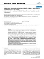
Fig 4. Graphic representation of the experimental result showing the relationship
among the area ratio of bone formation in holes of β-TCP and pore size and amount
of BMP-2. (a) It shows that as the amount of BMP is increased, bone tissue formation
tends to increase regardless of pore size except 1600TCP. Also, it shows that as pore
size becomes bigger, bone tissue formation tends to increase regardless of the
amount of BMP-2 except 1600TCP. In 1600TCP, bone tissue formation decreases
remarkably, which is less affected by increasing amount of BMP-2. (b) It shows that
cartilage formation was observed only in 75TCP+125BMP group, which was the
smallest pore size and was the lowest amount of BMP and as the amount of BMP was
increased in 75TCP, cartilage tissue formation was decreased. In 300TCP and
500TCP, only a small amount of cartilage tissue formation was observed regardless of
the BMP amount.
Micro CT findings of bone tissue regeneration
in zygomatic bone defect model
In the control group, bone tissue regeneration
was not observed from the edge of bone defect to
The pattern of bone tissue formation was similar
to β-TCP implanted intramuscularly, in which bone
was added to the β-TCP inner wall. Rich osteoblasts
existed on the surface of new bone, and bone tissue
formation had reached the center of β-TCP. (Fig. 6 a,b)
In both ends of β-TCP adjacent to existing bone
tissue, new bone regeneration occurred from the bone
resection stump, and new bone formation was
combined with β-TCP. In the holes of β-TCP
surrounded with bone tissue, bone marrow tissue that
had rich blood vessels was observed adjacent to
existing bone tissue. And on the surface of new bone
tissue, osteoblasts were arranged orderly in a
single-layer and osteoblasts showed endosteum-like
structure. In addition, emergence of osteoclast cells
was partially observed and also TCP was replaced by
bone tissue by resorption. (Fig. 6 a,c,d)
Immunohistochemical staining of OPN revealed
that an immature bone matrix was present in the
pores of β-TCP, and new bone had formed adjacent to
TCP in the zygomatic bone defect model experiment.
(Fig. 6 e,f)
Discussion
In tissue regeneration, stem cell, scaffold and
growth factor are important elements [19], so normal
tissue regeneration is disturbed when any one of these
factors is missing. Among these elements, artificial
biomaterial plays a role as scaffold to provide an
environment for proliferation and differentiation of
cells. The characteristics required for ideal artificial
biomaterials are not only cell proliferation and
differentiation but also biocompatibility, a structure
which cells are likely to invade, tissue solubility and
so on.
Int. J. Med. Sci. 2016, Vol. 13
473
Fig 5. Analysis of bone formation on honeycomb β-TCP in micro CT image. (a-c) Micro CT images of facial bone of rat with left zygomatic bone defect. (d-f) Micro CT images
of facial bone of rat with implanted 300TCP in left zygomatic bone defect site. (g-i) Micro CT images of facial bone of rat with implanted 300TCP+BMP-2 in left zygomatic bone
defect site. (a,d,g) axial view of micro CT, (b,c,e,f,h,i) reconstructed 3D images of micro CT. (a-c)New bone tissue formation was not observed at bone defect site (arrow). (d-f)
In TCP alone group, TCP exists at bone defect site with neither resorption nor new bone formation (arrow). (g-i) New bone formation was observed in implanted TCP+BMP-2
group at the boundary between TCP and existing bone (arrow). The osseous union between TCP and existing bone tissue was observed (arrow).
In our study, it was shown that both bone
formation rate and amount of bone formation were
the greatest in 500TCP+BMP-2 in rat thigh muscle.
This was followed by 300TCP+BMP-2, and in these
samples, normal bone tissue-like structure that had
bone marrow tissue formation was observed. Then,
bone formation rate and amount tended to increase in
proportion to the amount of BMP-2. These results are
consistent with our previous experimental result of
the ear canal bone reconstruction [18], and consistent
with a previous report by Tsurug using porous
granular apatite [20]. The bone formation patterns
varied with β-TCP pore size, and bone tissue
formation occurred so as to fill the lumen in 75TCP. In
300TCP, the bone tissue formed on the β-TCP inner
wall. In 500TCP, similar to 300TCP, bone tissue was
formed along the inner wall of the hole, and
furthermore, large amounts of cancellous bone-like
tissue were also formed in β-TCP pores. In 1600TCP,
solitary bone tissue was presented in the center of
TCP hole. In all pore size β-TCP, the pattern of bone
formation did not vary with the concentration of
BMP-2. Our previous study and Kuboki et al [15,21]
suggested that the biological material providing the
microenvironment is not actively involved in bone
formation but provides only space for cell
proliferation when the microenvironment of bone
formation
is
relatively
large.
However,
honeycomb-type hydroxyapatite that had pores of
diameter 300-400 μm directly added to the bone
matrix, and when it was implanted in vivo, the
biomaterial functioned in bone regeneration
effectively [22].
Int. J. Med. Sci. 2016, Vol. 13
474
Fig 6. (a) Histological images of 300TCP+BMP-2 implanted into zygomatic defect. (b,c,d) Higher-magnification image of corresponding outline area in (a). (a-d) New bone tissue
formation was observed from the bone stump, and the bone stump combined with 300TCP. New bone tissue formation was also observed in the center of TCP holes, and bone
marrow-like tissue formation was observed. The positive images of osteopontin were observed in new bone tissue (e,f). Bone tissue is indicated by arrowheads, bone
marrow-like tissue is indicated by asterisk, the positive images of osteopontin are indicated by arrow.
In HE images, rich blood vessels that penetrated
into β-TCP holes were observed only in 300TCP and
500TCP. Many reports indicated that blood vessel
formation plays an important role in regeneration of
not only bone tissue but also various tissues [23-26].
Also, our study suggested that angiogenesis had a
great influence on bone tissue formation.
Furthermore, marked infiltration of inflammatory
cells was not observed in all β-TCP, and this proved
that the β-TCP used in our study had extremely high
biocompatibility.
The results suggest that β-TCP in this
experiment has high biocompatibility, and pore size
of about 300 to 500 μm β-TCP provides an
environment for proliferation and differentiation of
cells in vivo and is the most suitable material for
inducing bone tissue.
On the other hand, in this study an interesting
finding was that strong chondrogenesis was shown
only when using a small amount of BMP-2 in 75TCP
although a little cartilage formation was observed in
large amount of BMP-2 in other pore size TCP. BMP-2
is a well-known growth factor which induces bone
tissue specifically. But when using both low
concentration of BMP-2 and TGF known as
cartilage-induce factor, the cartilage-inducing ability
is higher than the case of using only TGF [27,28]. It is
known that the BMP family is involved in normal
Int. J. Med. Sci. 2016, Vol. 13
cartilage tissue development [29]. Also in ectopic bone
formation experiments using BMP, it has been
reported that endochondral ossification-mediated
cartilage formation occurs, and thus involvement of
BMP-2 in cartilage formation is consistent with this
experimental result. But although little cartilage
formation was observed in 300TCP and 500TCP,
which were recognized for strong bone formation, the
most amount of cartilage tissue formation was shown
in 75TCP. Therefore, it is thought that a specific
microenvironment provided by 75TCP is involved in
cartilage tissue formation. Further investigations are
required as to what kind of environmental factors
provided by 75TCP induce formation of cartilage
tissue.
Bone tissue regeneration in zygomatic bone
defect model
Generally, when a bone defect occurs due to a
bone injury, cells are supplied from the periosteum
and surrounding connective tissue, and bone tissue
regeneration occurs. However, complete regeneration
becomes more difficult the wider the bone defect.
Therefore, various artificial biomaterials made from
hydroxyapatite, calcium phosphate ceramics (TCP),
polylactic acid, and titanium and so on have been
developed and used clinically. In addition, many
studies have reported bone tissue regeneration when
using these biomaterials combined with mesenchymal
stem cell [30].
But when these materials are used in bone tissue
regeneration, there are still many problems such as
early stage strength, efficiency of osteoinductive
activity, and replacement property of bone tissue in
vivo. These materials have already been used as bone
substitutes in clinical practice, but in the current
situation, it is difficult to obtain the regeneration on
such a large total bone defect.
For the bone tissue reconstruction experiment in
zygomatic bone defect model, we used 300TCP which
formed bone tissue structurally similar to biological
bone tissue in an ectopic experiment in which
honeycomb β-TCP of each hole diameter was
embedded into thigh muscle. In micro CT, the
continuity of the zyomatic bone was not observed in
the zygomatic bone resection group and in only the
TCP group 3 weeks after implantation. On the other
hand, new bone formation was observed in the
300TCP+BMP-2, and the continuity of the zygomatic
bone tissue was recovered. In the histological
observation, the pattern of bone tissue formation was
almost the same as the 300TCP that was implanted
into thigh muscle, and new bone formation in the
inner wall of β-TCP was observed. The new bone
tissue in 300TCP with BMP-2, which was
475
accompanied with bone marrow-like tissue having
rich hematopoietic cells, had continuity with the
existing bone tissue and β-TCP was completely
connected with the existing bone tissue. Therefore, the
recovery of bone tissue continuity between β-TCP and
existing bone was confirmed at the tissue level.
Many bone tissue regeneration studies using
various cells have been attempted with the
development of new biomaterials. However, when
using a cell it is difficult to exclude the risk of
tumorigenesis completely, thus bone tissue
regeneration without using cells and by a simple
technique is considered much more ideal.
Honeycomb β-TCP has the characteristic features of
excellent biocompatibility, osteoconductive ability,
and bioabsorbable ability along with bone remodeling
[31,32]. It is reported that the bioabsorbable ability of
TCP is higher than hydroxyapatite which is widely
used clinically [31,32]. It is thought that TCP replaces
the existing bone tissue by absorption [33,34].
Our study indicates that honeycomb β-TCP is an
excellent artificial biomaterial because honeycomb
β-TCP regenerates bone tissue that is similar to
normal bone with bone marrow-like tissue and
endosteum-like tissue in completely transected bone
tissue. Therefore, TCP is expected to serve as a new
biological material in the head and neck region.
Acknowledgement
This study was supported by a Grant-in-Aid for
Scientific Research(C), 15K20309 provided by the
Japan Society for Promotion of Science (JSPS).
Competing Interests
The authors have declared that no competing
interest exists.
References
1.
Lee KS, Park JW. Free vascularized osteocutaneous fibular graft to the tibia.
Microsurgery. 1999; 19:141-147.
2. Muramatsu K, Hashimoto T, Tominaga Y, et al. Vascularized Bone Graft for
Oncological Reconstruction of the Extremities: Review of the Biological
Advantages. Anticancer Res. 2014; 34:2701-2707.
3. Dimitriou R, Mataliotakis GI, Angoules AG, et al. Complications following
autologous bone graft harvesting from the iliac crest and using the RIA: a
systematic review. Injury. 2011; 42 Suppl 2:S3-15.
4. Banwart JC, Asher MA, Hassanein RS. Iliac crest bone graft harvest donor site
morbidity. A statistical evaluation. Spine (Phila Pa 1976). 1995;
20(9):1055-1060.
5. Schaaf H, Lendeckel S, Howaldt HP, et al. Donor site morbidity after bone
harvesting from the anterior iliac crest. Oral Surg Oral Med Oral Pathol Oral
Radiol Endod. 2010; 109(1):52-58.
6. Arce K, Bell RB, Potter JK, et al. Vascularized free tissue transfer for
reconstruction of ablative defects in oral and oropharyngeal cancer patients
undergoing salvage surgery following concomitant chemoradiation. Int J Oral
Maxillofac Surg. 2012; 41(6):733-738.
7. Karageorgiou V, Kaplan D. Porosity of 3D biomaterial scaffolds and
osteogenesis. Biomaterials. 2005; 26(27): 5474-5491.
8. Burg KJ, Porter S, Kellam JF. Biomaterial developments for bone tissue
engineering. Biomaterials. 2000; 21(23): 2347-2359.
9. Stevens M. Biomaterials for bone tissue engineering. Materials Today. 2008;
11(5):18-25.
10. Yoshikawa H, Myoui A. Bone tissue engineering with porous hydroxyapatite
ceramics. J Artif Organs. 2005; 8(3):131-136.
Int. J. Med. Sci. 2016, Vol. 13
476
11. Kaltreider SA, Newman SA. Prevention and management of complications
associated with the hydroxyapatite implant. Ophthal Plast Reconstr Surg.
1996; 12(1):18-31.
12. Walsh WR, Vizesi F, Michael D, et al. Beta-TCP bone graft substitutes in a
bilateral rabbit tibial defect model. Biomaterials. 2008; 29(3):266-271.
13. Asaoka T, Ohtake S, Furukawa KS, et al. Development of bioactive porous
alpha-TCP/HAp beads for bone tissue engineering. J Biomed Mater Res A.
2013; 101(11):3295-3300.
14. Ono I, Tateshita T, Satou M, et al. Treatment of large complex cranial bone
defects by using hydroxyapatite ceramic implants. Plast Reconstr Surg. 1999;
104(2):339-349.
15. Kuboki Y, Jin Q, Takita H. Geometry of carriers controlling phenotypic
expression in BMP-induced osteogenesis and chondrogenesis. J Bone Joint
Surg Am. 2001; 83A(Suppl 1):S105-115.
16. Kuboki Y, Jin Q, Kikuchi M, et al. Geometry of artificial ECM: sizes of pores
controlling phenotype expression in BMP-induced osteogenesis and
chondrogenesis. Connect Tissue Res. 2002; 43(2-3):529-534.
17. Ayers RA, Simske SJ, Nunes CR, et al. Long-term bone ingrowth and residual
microhardness of porous block hydroxyapatite implants in humans. J Oral
Maxillofac Surg. 1998; 56(11):1297-1301; discussion 1302.
18. Takabatake K, Yamachika E, Tsujigiwa H, et al. Effect of geometry and
microstructure of honeycomb TCP scaffolds on bone regeneration. J Biomed
Mater Res A. 2013; 102(9):2952-2960.
19. Langer R, Vacanti JP. Tissue engineering. Science. 1993; 260(5110):920-926.
20. Tsuruga E, Takita H, Itoh H, et al. Pore size of porous hydroxyapatite as the
cell-substratum controls BMP-induced osteogenesis. J Biochem. 1997;
121(2):317-324.
21. Kuboki Y, Takita H, Mizuno M, et al. Geometry of Artificial Extracellular
Matrices:a New Paradigm from Dental Tissue Engineering. Dentistry in Japan.
2001; 37:42-50.
22. Jin QM, Takita H, Kohgo T, et al. Effects of geometry of hydroxyapatite as a
cell substratum in BMP-induced ectopic bone formation. J Biomed Mater Res.
2000; 51(3):491-499.
23. Kusumbe AP, Ramasamy SK, Adams RH. Coupling of angiogenesis and
osteogenesis by a specific vessel subtype in bone. Nature. 2014;
507(7492):323-328.
24. Zigdon-Giladi H, Michaeli-Geller G, Bick T, et al. Human blood-derived
endothelial progenitor cells augment vasculogenesis and osteogenesis. J Clin
Periodontol. 2015; 42(1):89-95.
25. Hu J, Srivastava K, Wieland M, et al. Endothelial cell-derived angiopoietin-2
controls liver regeneration as a spatiotemporal rheostat. Science. 2014;
343(6169):416-419.
26. Ding BS, Nolan DJ, Butler JM, et al. Inductive angiocrine signals from
sinusoidal endothelium are required for liver regeneration. Nature. 2010;
468(7321):310-315.
27. Murphy MK, Huey DJ, Hu JC, et al. TGF-beta1, GDF-5, and BMP-2 stimulation
induces chondrogenesis in expanded human articular chondrocytes and
marrow-derived stromal cells. Stem cells. 2015; 33(3):762-773.
28. Sekiya I, Larson BL, Vuoristo JT, et al. Comparison of effect of BMP-2, -4, and
-6 on in vitro cartilage formation of human adult stem cells from bone marrow
stroma. Cell Tissue Res. 2005; 320(2):269-276.
29. Hojo H, Ohba S, Taniguchi K, et al. Hedgehog-Gli activators direct
osteo-chondrogenic function of bone morphogenetic protein toward
osteogenesis in the perichondrium. J Biol Chem. 2013; 288(14):9924-9932.
30. Ducheyne P, Radin S, King L. The effect of calcium phosphate ceramic
composition and structure on in vitro behavior. I. Dissolution. J Biomed Mater
Res. 1993; 27(1):25-34.
31. Scotti C, Tonnarelli B, Papadimitropoulos A, et al. Recapitulation of
endochondral bone formation using human adult mesenchymal stem cells as a
paradigm for developmental engineering. Proc Natl Acad Sci U S A. 2010;
107(16):7251-7256.
32. Fazan F, Marquis PM. Dissolution behavior of plasma-sprayed hydroxyapatite
coatings. J Mater Sci Mater Med. 2000; 11(12):787-792.
33. Mate-Sanchez de Val JE, Mazon P, Guirado JL, et al. Comparison of three
hydroxyapatite/beta-tricalcium phosphate/collagen ceramic scaffolds: An in
vivo study. J Biomed Mater Res A. 2014; 102(4):1037-46.
34. Takeshi U, Naoyuki M, Shunsuke N, et al. Histochemical and Radiological
Study of Bone Regeneration by the Combinatorial Use of Tetrapod-Shaped
Artificial Bone and Collagen. J Hard Tissue Biology. 2015; 24(2): 199 - 210.