Role of the ERK1/2 signaling pathway in osteogenesis of rat tendon derived stem cells in normoxic and hypoxic cultures
Bạn đang xem bản rút gọn của tài liệu. Xem và tải ngay bản đầy đủ của tài liệu tại đây (1.8 MB, 9 trang )
Int. J. Med. Sci. 2016, Vol. 13
Ivyspring
International Publisher
629
International Journal of Medical Sciences
2016; 13(8): 629-637. doi: 10.7150/ijms.16045
Research Paper
Role of the ERK1/2 Signaling Pathway in Osteogenesis of
Rat Tendon-Derived Stem Cells in Normoxic and
Hypoxic Cultures
Pei Li 1, Yuan Xu 2, Yibo Gan 1, Lei Song 1, Chengmin Zhang 1, Liyuan Wang 1, Qiang Zhou 1
1.
2.
Department of Orthopedic Surgery, Southwest Hospital, Third Military Medical University, Chongqing, 400038, China;
Department of Orthopedic Surgery, Xinqiao Hospital, Third Military Medical University, Chongqing, 400038, China.
Corresponding authors: E-mail: (Yuan Xu); (Qiang Zhou).
© Ivyspring International Publisher. Reproduction is permitted for personal, noncommercial use, provided that the article is in whole, unmodified, and properly cited. See
for terms and conditions.
Received: 2016.05.03; Accepted: 2016.06.25; Published: 2016.07.18
Abstract
Background: Ectopic ossification and increased vascularization are two common phenomena in the
chronic tendinopathic tendon. The increased vascularization usually leads to an elevated local
oxygen tension which is one of micro-environments that can influence differentiate status of stem
cells.
Objective: This study aimed to investigate the osteogenesis capacity of rat tendon-derived stem
cells TDSCs (rTDSCs) in normoxic and hypoxic cultures, and to study the role of ERK1/2 signaling
pathway in this process.
Methods: rTDSCs were subjected to osteogenesis inductive culture in hypoxic (3% O2) and
normoxic (20% O2) conditions. The inhibitor U0126 was added along with culture medium to
determine the role of ERK1/2 signaling pathway. Cell viability, cell proliferation, alizarin red
staining, alkaline phosphatase (AKP) activity, gene expression (ALP, osteocalcin, collagen I and
RUNX2) and protein expression (p-ERK1/2 and RUNX2) of osteogenic-cultured rTSDCs were
analyzed in this study.
Results: Hypoxic and normoxic culture had no effects on cell viability of rTDSCs, whereas the
proliferation potential of rTDSCs was significantly increased in hypoxic culture. The osteogenesis
capacity of rTDSCs in normoxic culture was significantly promoted compared with hypoxic
culture, which was reflected by an increased alizarin red staining intensity, an elevated ALP activity,
and the up-regulated gene (ALP, osteocalcin, collagen I and RUNX2) or protein (RUNX2)
expression of osteogenic makers. However, the osteogenesis capacity of rTDSCs in both hypoxic
and normoxic cultures was attenuated by the inhibitor U0126.
Conclusion: Normoxic culture promotes osteogenic differentiation of rTDSCs compared with the
hypoxic culture, and the ERK1/2 signaling pathway is involved in this process.
Key words: tendinopathy, tendon-derived stem cells, hypoxic, normoxic, osteogenesis.
Introduction
Tendinopathy is a common painful tendon
condition caused by overuse, mechanical injury or
intrinsic
degeneration
[1-3].
Histologically,
calcification
is
usually
reported
in
some
tendinopathies [4, 5], which leads to a failed
self-healing and predisposes the diseased tendon to
rupture [6]. Up to now, the etiopathogenesis for
calcific tendinopathy remains unclear.
Tendon characterized as a kind of dense
connective structures can lead to joint stabilization or
joint movement through transferring mechanical load
from muscle to bone [7, 8]. Recently, a type of
tendon-derived stem cell (TDSC) has been identified,
which possesses the abilities of self-renewal and
Int. J. Med. Sci. 2016, Vol. 13
multi-lineage differentiation [9-11]. By differentiating
into tenocytes, TDSCs play an important role in
matrix homeostasis and tissue regeneration of the
injured tendon [6, 12]. However, lots of abnormal
repair outcomes are frequently observed in the
pathological chronic tendinopathy, such as
fibrocartilage-like tissue formation, lipid substance
accumulation and ectopic ossification [13-15].
Recently, increasing evidence suggests that stem cells
may also play a role in the pathological conditions [16,
17]. Several previous studies proposed that the
erroneous differentiation of TDSCs to non-tenocytes
caused by alterations of their surrounding
micro-environments may contribute to the aberrant
matrix remodeling and acquisition of non-tenocytes
phynotype in the tendinopathic tendons [17, 18].
However, the potential mechanisms for the erroneous
differentiation of TDSCs to non-tenocytes or other
cellular phenotype are largely unknown. More direct
evidences are needed to clarify this speculation.
Similar with other stem cells, oxygen tension is a
local micro-environment surrounding TDSCs. In vivo,
the oxygen tension within a certain tissue depends on
the vascularization level and the inherent
micro-environment type [19]. Under physiological
conditions, the collagen-rich tendon has few blood
vessels and thus a low oxygen level compared with
other vascular-rich tissues [20]. By contrast, an
increased vascular infiltration and capillary blood
flow in the tendinopathic tendon are constantly
reported previously [21-25], which may in turn lead to
an elevated oxygen tension and thus an altered
oxygen surrounding TDSCs. Generally, increased
vascularization may be a protective response of tissue
repair after injury. On another hand, differentiation of
stem cells can also be regulated by oxygen tension [19,
26]. In other types of stem cells, oxygen tension
alteration-induced changes in differentiation capacity
are often reported during the past years [20, 27, 28].
Moreover, previous study demonstrated that
osteogenic differentiation of bone mesenchymal stem
cells (BMSCs) was promoted in normoxic culture. In
light of the co-existence of ectopic ossification and
increased vascular infiltration in the chronic
tendinopathic tendon, we propose that the ectopic
ossification may partly result from the erroneous
osteogenic differentiation of TDSCs caused by
increased local oxygen tension.
In the present study, we aimed to investigate the
osteogenic differentiation capacity of rat TDSCs
(rTDSCs) in hypoxic (3%) culture and normoxic (20%)
culture. Because ERK1/2 pathway is a potential
signaling pathway relating with differentiation of
some stem cells, the potential role of ERK1/2 pathway
was also determined by its pharmacological inhibitor
630
U0126. To achieve this purpose, cell viability, cell
proliferation, AKP activity, alizarin red staining and
expression of some osteogenic markers were
evaluated in this study.
Materials and methods
Ethical statement
All animal experiments in this study were
approved by Ethics Committee at Southwest Hospital
affiliated to the Third Military Medical University
[SYXK (YU) 2012-0012].
Isolation and preparation of rTDSCs
rTDSCs were isolated from the achilles tendon of
twelve healthy rats (male, 4-5 weeks old) as described
previously [29, 30]. Briefly, after rats were sacrificed
with carbon dioxide, their bilateral achilles tendons
were separated. Then, the tendon sheaths and
paratendons were further removed. Thereafter, the
tendons were cut into small pieces (approximately 2
mm×2 mm) and digested with phosphate buffered
saline (PBS) supplemented with 0.3% type I
collagenase (Sigma) and 0.4% neutral protease
(Roche) at 37 °C for 50-60 min. After digestion and
centrifugation (500 g, 15 min), cell pellets were
collected and re-suspended in DMEM/F12 medium
(Hyclone) containing 20% fetal bovine serum (FBS,
Gibco) under standard conditions (37°C, 20% O2 and
5% CO2). After 8-10 days, TDSCs were collected by
local trypsin digestion of individual cell colonies
under a light microscopy (Olympus, BX51) and
defined as the passage 0 rTDSCs according to
previous study [29]. Then, the isolated rTDSCs were
sub-cultured and passaged after reaching 80%-90%
confluence. Previously, we demonstrated that passage
3 rTDSCs displayed a good colongenicity and
vigorous differentiation capacity [29]. Hence, we
mainly used the passage 3 rTDSCs in each experiment
in the present study.
Hypoxic and normoxic osteoinductive culture
of rTDSCs
The P3 rTDSCs were cultured in Osteogenic
Differentiation Medium (Cyagen Biosciences Inc) and
incubated in a hypoxic (3% O2) incubator or a
normoxic (20% O2) incubator (Thermo Scientific). To
investigate the role of ERK1/2 signaling pathway, the
inhibitor U0126 (10 μM, Beyotime, China) was added
along with the medium throughout the experiment.
Culture medium was refreshed every 3 days. To
accurately maintain the oxygen tension in the hypoxic
and normoxic cultures as much as possible, a rapid
and timely gas injection process was performed after
exchanging the culture medium. Because no study
reported the measurement of oxygen tension in
Int. J. Med. Sci. 2016, Vol. 13
631
human normal tendon even though the tendon milieu
is estimated to be hypoxic, the hypoxic and normoxic
cultures were designed according to previous studies
[19, 20, 31, 32].
Cell viability
rTDSCs were seeded in 6-well plate (4 × 10³
cells/well) and osteogenic-cultured in the designed
oxygen tension conditions. On days 1, 4 and 7, cell
viability of rTDSCs was analyzed with a LIVE/DEAD
Viability/Cytotoxicity
Assay
Kit
(Invitrogen)
according to the instructions. Briefly, after washing
with PBS for 2-3 times, rTDSCs in hypoxic and
nomorxic cultures were incubated with fluorescent
working solution (calcein AM: 2 μM; EthD-1: 4 μM)
for 40 minutes at room temperature. Then, the live or
dead rTDSCs in each group were viewed under a
fluorescence
microscopy
(Olympus
IX71).
Quantification of cell viability was performed using
the Image-Pro Plus software (Version 5.1, Media
Cybernetics, Inc.)
Cell proliferation assay
On days 1, 4 and 7, rTDSCs proliferation
potential was evaluated with a Cell Counting Kit-8
(CCK-8, Beyotime, China). Briefly, after rTDSCs
(seeded in 12-well plate, 2 × 10³ cells/well) were
incubated with fresh medium containing CCK-8
solution for 2 hours, 200 μL supernatant was used to
measure the absorbance at 450 nm wavelength using
an automatic micro-plate reader (Bio-rad).
Alizarin red staining assay
Alkaline phosphatase (AKP) activity detection
rTDSCs were seeded in 10-cm diameter dishes
(10×103 cells/dish) and osteogenic-cultured for 14 or
21 days. Then, rTDSCs were incubated with lysis
buffer (200 μL, Beyotime, China) and centrifuged (15,
000 r/min, 15 min) to collect lysis supernatant, protein
concentration was measured with BCA Kit (Beyotime,
China). Then, AKP activity was detected with an
Alkaline Phosphatase (AKP) Kit (Nanjing Jiancheng
Bioengineering Institute, China) according to the
manufacture’s instructions.
Real-time polymerase chain reaction (PCR)
analysis
Gene expression of several osteogenic markers
(ALP, osteocalcin, collagen I and RUNX2) was
analyzed by real-time PCR as described [29]. Briefly,
rTDSCs were seeded in 10-cm diameter dishes (10×
103 cells/dish) and osteogenic-cultured under
different oxygen tension conditions. On days 14 and
21, total RNA was extracted with Tripure Isolation
Reagent (Roche) and reverse-transcripted into cDNA
with a reverse transcription kit (Roche). Then, the
reaction system containing cDNA, primers and SYBR
Green Mix (DONGSHENG BIOTECH, China) was
subjected to a real-time PCR machine (CFX96
Real-Time System, Bio-rad). Primers of target genes
were showed in the Table 1. β-actin was used as the
reference gene and the P3 rTDSCs collected
immediately were used as controls. The relative gene
expression of target genes was expressed as 2―△△Ct.
Western blotting analysis
Protein expression of ERK1/2, p-ERK1/2 and
osteogenic maker (RUNX2) was analyzed by Western
blotting assay. Briefly, after total protein of rTDSCs
osteogenic-cultured in different oxygen tension
conditions for 21 days was extracted with RIPA
solution (Beyotime, China), protein samples were
subjected to SDS-PAGE and transferred to PVDF
membrane (Roche). Then, the PVDF membrane was
blocked with 5% bovine serum albumin (BSA) and
incubated with primary antibodies (ERK1/2, 1:500,
sc-292838, Santa Cruz; p-ERK1/2, 1:500, sc-101761,
Santa Cruz; RUNX2, 1:500, sc-390351, Santa Cruz;
β-actin, 1:1000, 60008-1-Ig, Proteintech) overnight at
4°C and HRP-conjugated secondary antibodies
(ZSGB-BIO, China) for 2 hours at room temperature.
Finally, protein bands on the PVDF membrane
were visualized using the SuperSignal West
Pico Trial Kit (Thermo) and analyzed using the
Reverse (5’-3’)
Image J software (National Institutes of Health,
TCCCGGCCAGCCAGGTCCA
GTCCATACTTTCGAGGCAGAGAG
USA).
rTDSCs (seeded in 10-cm diameter dish, 10×103
cells/dish) were osteogenic-cultured in medium with
or without inhibitor U0126 under different oxygen
tension conditions. After 21 days of osteogenic
differentiation, the culture medium was removed and
the rTDSCs were sequentially fixed with 3 ml 4%
paraformaldehyde for 30 minutes, rinsed with PBS for
3 times and stained with alizarin red working solution
(Cyagen Biosciences Inc.) for 5-8 minutes. Finally,
rTDSCs were observed under a light microscopy
(Olympus BX51). Quantification of alizarin red
staining intensity was performed using the Image-Pro
Plus software (Version 5.1, Media Cybernetics, Inc.)
Table 1. Primers of target genes.
Gene
β-actin
osteocalcin
Collagen I
ALP
RUNX2
Forward (5’-3’)
ACCCCGTGCTGCTGACCGAG
CGGCGCTACCTCAACAATGG
CATCGTGGCTTCTCTGGTC
ACCGTTGAGTCCATCTTTGC
CCCGAGTGCTTTGTGTGTGCTG CCGCCGGTGTTCGTGTGTG
GGGCAGATGGGGAACTGTG
GGTTTGCTACTGGGTGGGTTTC
Int. J. Med. Sci. 2016, Vol. 13
632
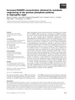
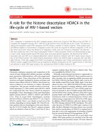
Figure 1: Cell viability analysis of rat tendon-derived stem cells (rTDSCs) in hypoxic and normoxic cultures on days 1, 4 and 7. The live and dead cells were stained
with green fluorescence and red fluorescence, respectively. Magnification: A1-D1, 40x; A2-D2 and A3-D3, 100x. n=3.
Statistics
All numerical data are expressed as mean ± SD
and analyzed by the SPSS 13.0 software. Each
experiment in this study was performed in triplicate.
When homogeneity test for variance was completed,
comparisons between normoxic culture and hypoxic
culture, between normoxic culture without U0126
treatment and normoxic culture with U0126
treatment, and between hypoxic culture without
U0126 and hypoxic culture with U0126 treatment
were analyzed by Independent-Samples T test. A
statistical
difference
was
indicated
when
p-value<0.05.
Results
Cell viability
Both in hypoxic and normoxic cultures,
osteogenic-cultured rTDSCs remained viable on days
1, 4 and 7 (Figure 1A1-3, C1-3). Generally, there were
no differences in cell viability between hypoxic
culture and normoxic culture. Inhibition of ERK1/2
signaling pathway had no effects on cell viability in
hypoxic and normoxic cultures (Figure 1B1-3, D1-3).
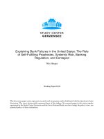
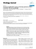
Cell proliferation
Throughout
the
7
days
of
culture,
osteogenic-cultured rTDSCs showed a consistent
proliferation potential both in the hypoxic and
normoxic cultures (Figure 2). Although there was no
significant difference in cell proliferation between the
hypoxic and normoxic cultures at day 1, proliferation
potential in hypoxic culture was significantly
increased compared with normoxic culture at days 4
and 7. Additionally, the inhibitor U0126 obviously
attenuated cell proliferation in hypoxic and normoxic
cultures on days 4 and 7.
Figure 2: Cell proliferation potential of rat tendon-derived stem cells
(rTDSCs) in hypoxic and normoxic cultures on days 1, 4 and 7. Date are
expressed as mean ± SD, n=3. #: Indicates a significant difference between
hypoxic and normoxic cultures without addition of inhibitor U0126. *: Indicates
a significant difference associated with U0126 treatment in hypoxic culture or
normoxic culture.
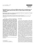
Alizarin red staining
A stronger alizarin red staining intensity was
observed in normoxic culture compared with hypoxic
culture (Figure 3). However, inhibition of ERK1/2
signaling pathway in hypoxic or normoxic culture
significantly decreased the staining intensity (Figure
3).
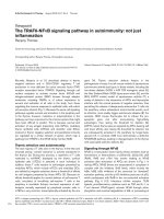
AKP activity
In normoxic culture, AKP activity of
osteogenic-cultured rTDSCs was significantly
increased compared with that in hypoxic culture on
days 14 and 21 (Figure 4). Either in hypoxic or
Int. J. Med. Sci. 2016, Vol. 13
normoxic culture, AKP activity was decreased when
the ERK1/2 signaling pathway was inhibited by
inhibitor U0126.
Gene expression
Genes of osteogenic maker were differently
expressed in hypoxic and normoxic cultures (Figure
5). In normoxic culture, expression of ALP,
osteocalcin, collagen I and RUNX2 was all
up-regulated compared with that in hypoxic culture
on days 14 and 21. However, addition of U0126 in
either hypoxic or normoxic culture inhibited gene
expression of these osteogenic markers (Figure 5).
Protein expression
In normoxic culture, protein expression of
p-ERK1/2 or RUNX2 was up-regulated compared
with hypoxic culture (Figure 6). When the expression
of p-ERK1/2 was inhibited by inhibitor U0126 in
normoxic and hypoxic cultures, expression of RUNX2
was simultaneously down-regulated (Figure 6).
Discussion
Ectopic ossification is commonly found in the
chronic tendinopathic tendon [33]. Currently, the
mechanism underlying this pathological process
633
remains unknown. Apart from the ectopic
ossification, oxygen tension may be also elevated due
to the increased vascular infiltration [24]. Considering
that TDSCs can erroneously differentiate into
non-tenocytes due to the altered micro-environments
and thus play a role in pathological conditions, we
performed this study to investigate the osteogenesis
capacity of rTDSCs in the hypoxic and normoxic
cultures. Our results showed that rTDSCs remained
viable both in hypoxic and normoxic cultures and
displayed a stronger proliferation potential in hypoxic
culture. Specially, rTDSCs in normoxic culture
possessed a promoted osteogenesis capacity
regarding alizarin red staining, AKP activity, gene
expression of osteogenesis-related markers (ALP,
osteocalcin, collagen I and RUNX2) and protein
expression of RUNX2. Additionally, we also found
that inhibition of ERK1/2 signaling pathway could
attenuate the osteogenesis potential of rTDSCs
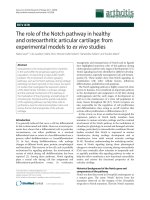
(summarized in the Figure 7). These findings
demonstrated that oxygen tension is an important
micro-environment
for
regulating
osteogenic
differentiation of TDSCs, and also indicated that
ERK1/2 signaling pathway is involved in this
regulatory process.
Figure 3: Representative photomicrographs and quantification of alizarin red staining of rat tendon-derived stem cells (rTDSCs) in hypoxic and normoxic cultures
on day 21. Magnification: 100x. n=3.
Figure 4: Alkaline phosphatase (AKP) activity of rat tendon-derived stem cells (rTDSCs) in hypoxic and normoxic cultures on days 14 and 21. Date are expressed
as mean ± SD, n=3. #: Indicates a significant difference between hypoxic and normoxic cultures without addition of inhibitor U0126. *: Indicates a significant difference
associated with U0126 treatment in hypoxic culture or normoxic culture.
Int. J. Med. Sci. 2016, Vol. 13
634
Figure 5: Real-time PCR analysis of rat tendon-derived stem cells (rTDSCs) in hypoxic and normoxic cultures on days 14 and 21. Date are expressed as mean ± SD,
n=3. #: Indicates a significant difference between hypoxic and normoxic cultures without addition of inhibitor U0126. *: Indicates a significant difference associated
with U0126 treatment in hypoxic culture or normoxic culture.
Figure 6: Western blotting analysis of rat tendon-derived stem cells (rTDSCs) in hypoxic and normoxic cultures on day 21. Date are expressed as mean ± SD, n=3.
#: Indicates a significant difference between hypoxic and normoxic cultures without addition of inhibitor U0126. *: Indicates a significant difference associated with
U0126 treatment in hypoxic culture or normoxic culture.
TDSCs are stem cells residing in tendon tissue.
Similar with other types of stem cells, TDSCs
interplay with the local micro-environment to
participate in tendon healing and tendon matrix
remodeling after injury [6]. In tendon, mechanical
loading, matrix composition, biological factors and
some other physiological factors are typical
micro-environments which can regulate biological
responses of TDSCs [34]. There are also some
evidences that aberrant micro-environments can lead
to abnormal functions of stem cells and ultimately
pathological diseases [6, 17, 34]. In chronic
tendinopathic tendon, ossification and increased
blood vessels are two common pathological features.
Hence, the raised oxygen tension resulted from the
increased blood vessels may lead to erroneous
osteogenic differentiation of TDSCs and thus some
ossification tissues in the diseased tendon. In line with
us, aberrant osteogenic differentiation of stem cells is
also previously reported in other tissues, such as
arterial calcification and skin calcification [35, 36].
Oxygen tension is low in the healthy tendon
since it has a low blood flow, while oxygen tension
may tend to rise in the tendinopathic tendon because
of the increased vascular infiltration [34, 37]. In this
study, we found that rTDSCs have an increased
alizarin red staining intensity under normoxic
condition compare with that under hypoxic condition.
Similarly, the results of ALP activity assay also
showed a similar trend to that observed in alizarin red
staining. These findings indicate that the osteogenesis
capacity of rTDSCs in normoxic culture is promoted
compared with that in hypoxic culture. In line with
us, a previous study also indicated that human TDSCs
Int. J. Med. Sci. 2016, Vol. 13
635
have a reduced osteogenic differentiation potential
but an increased proliferation capacity in hypoxic
(2%) culture [32]. Additionally, it is also reported that
osteogenic differentiation of bone mesenchymal stem
cells (BMSCs) was also attenuated in hypoxic culture
[19, 38]. However, a previous study by Zhang et al.
showed that osteogenic differentiation of human
TDSCs in hypoxic (5%) culture was increased
compared with that in normoxic (20%) culture [20].
We speculate there are several possible factors that
may be responsible for this discrepancy, such as the
different control way of oxygen tension in the
incubator, the different initial status and source of
TDSCs, and different experimental conditions [20].
Nevertheless, all these studies indicate that oxygen
tension is an important factor for regulating
osteogenesis of TDSCs.
Various osteogenesis markers, either for the
early stage or the late stage, had been identified
previously, such as ALP, collagen I, RUNX2,
osteonectin, osteocalcin and osteopontin [39]. In the
present study, we investigated expression of these
osteogenic makers from gene level or protein level.
We found that gene expression of ALP, osteocalcin,
RUNX2 and collagen I as well as protein expression of
RUNX2 are all up-regulated in the normoxic culture
compared with hypoxic osteogenic culture. This
suggests again that osteogenesis capacity of rTDSC in
normoxic culture is promoted compared with that in
hypoxic culture. Additionally, this also indirectly
implies that the ossification tissue in the chronic
tendinopathic tendon is related to osteogenesis of
TDSCs
caused
by
alteration
of
local
micro-environment, such as the elevated oxygen
tension caused by the increase in vascularization.
ERK1/2 signaling pathway is a branch of MAPK
pathways which are involved in many cell
bioactivities including cell proliferation, cell apoptosis
and cell differentiation [40]. Previously, ERK1/2
signaling pathway had been reported to participate in
inhibiting the osteogenic differentiation of BMSC
under hypoxic condition [38]. In this study, activation
of ERK1/2 signaling pathway in rTDSCs in normoxic
culture was more obvious than that in hypoxic
culture. Moreover, when ERK1/2 signaling pathway
was inhibited by inhibitor U0126, osteogenic activity
of rTDSCs regarding alizarin red staining intensity,
ALP activity and expression of the designed
osteogenesis markers was simultaneously decreased.
These results indicate that the ERK1/2 signaling
pathway is involved in the effects of altered oxygen
tension on osteogenesis capacity of rTDSCs.
Consistent with us, activation of ERK1/2 signaling
pathway is also previously reported to participate in
the osteogenesis of other types of stem cells, such as
BMSCs, Periodontal ligament stem cells and induced
pluripotent stem cells [41-43].
Previous studies demonstrated that oxygen
tension can affect cell viability and cell
proliferation of stem cells. In this study, no
differences
in
cell
viability
of
osteogenic-cultured rTDSCs were found
between hypoxic culture and normoxic
culture, indicating that rTDSCs can remain
viable in either hypoxic culture or normoxic
culture. However, proliferation capacity of
the rTDSCs in hypoxic culture was
increased compared with that in normoxic
culture. This finding confirmed previous
stem
cells-related
studies
which
demonstrated that the stemness of stem cells
is better maintained in hypoxic culture [19,
32, 44]. Apart from this, we also found that
blocking the ERK1/2signaling pathway in
hypoxic or normoxic culture inhibited cell
proliferation of rTDSCs, whereas the cell
viability was not influenced. This indicates
that ERK1/2 signaling pathway may affect
cell proliferation but not cell viability of
Figure 7: A brief graphic abstract of this study. Rat tendon-derived stem cells (rTDSCs) were
rTDSCs in different oxygen tension
cultured in normoxic (20% O2) and hypoxic (3% O2) cultures. Osteogenesis capacity of rTDSCs
conditions.
in normoxic culture was promoted compared with that in hypoxic culture, whereas inhibition
This study also has several limitations.
of ERK1/2 signaling pathway attenuated osteogenesis of rTDSCs both in normoxic and hypoxic
cultures.
First, an in vivo animal model is not used to
verify the results from the in vitro cell
Int. J. Med. Sci. 2016, Vol. 13
culture system. Second, erroneous osteogenic
differentiation of TDSCs may be resulted from a
combination of several factors including elevated
oxygen
tension,
inflammation,
mechanical
overloading and alterations in extracellular matrix
[34]. However, we just studied the effects of single
factor on osteogenesis capacity of rTDSCs in this
study. Third, because there are no reports about the
measurement of exact value of oxygen extension in
human tendon under physiological and pathological
conditions, the oxygen tension values of hypoxic and
normoxic cultures in this study were designed
according to previous studies [20, 32]. Hence, the
oxygen tension parameters used in this study may
differ from the actual oxygen tension in human
tendon under physiological and pathological
conditions.
Taken together, we can draw the conclusion that
osteogenesis capacity of rTSDCs in the normoxic
culture was increased compared with that in the
hypoxic culture, and ERK1/2 phosphorylation may
participate in this regulatory process. This study will
contribute to further understanding of the mechanism
behind the ectopic ossification in the tendinopathic
tendon and ultimately the development of effective
clinical treatment for it.
Acknowledgments
We appreciate the founding from the National
Natural Science Foundation of China (NSFC 81272029
and NSFC 81027005), Science and Technology
Achievement Transformation Fund of the Third
Military Medical University (2011XZH006).
Conflicts of Interest
The authors report no conflicts of interest.
References
1.
Vora AM, Myerson MS, Oliva F, Maffulli N. Tendinopathy of the main body of
the Achilles tendon. Foot and ankle clinics. 2005; 10: 293-308.
2. Weinreb JH, Sheth C, Apostolakos J, McCarthy MB, Barden B, Cote MP, et al.
Tendon structure, disease, and imaging. Muscles Ligaments Tendons J. 2014;
4: 66-73.
3. Way L, Scutt N, Scutt A. Cytocentrifugation: a convenient and efficient
method for seeding tendon-derived cells into monolayer cultures or 3-D tissue
engineering scaffolds. Cytotechnology. 2011; 63: 567-79.
4. Kannus P. Structure of the tendon connective tissue. Scand J Med Sci Sports.
2000; 10: 312-20.
5. Oliva F, Via AG, Maffulli N. Physiopathology of intratendinous calcific
deposition. BMC medicine. 2012; 10: 95.
6. Lui PP, Chan KM. Tendon-derived stem cells (TDSCs): from basic science to
potential roles in tendon pathology and tissue engineering applications. Stem
Cell Rev. 2011; 7: 883-97.
7. Birch HL. Tendon matrix composition and turnover in relation to functional
requirements. Int J Exp Pathol. 2007; 88: 241-8.
8. Hodgson RJ, O'Connor PJ, Grainger AJ. Tendon and ligament imaging. Br J
Radiol. 2012; 85: 1157-72.
9. Bi Y, Ehirchiou D, Kilts TM, Inkson CA, Embree MC, Sonoyama W, et al.
Identification of tendon stem/progenitor cells and the role of the extracellular
matrix in their niche. Nat Med. 2007; 13: 1219-27.
10. Salingcarnboriboon R, Yoshitake H, Tsuji K, Obinata M, Amagasa T, Nifuji A,
et al. Establishment of tendon-derived cell lines exhibiting pluripotent
mesenchymal stem cell-like property. Exp Cell Res. 2003; 287: 289-300.
636
11. Scutt N, Rolf CG, Scutt A. Glucocorticoids inhibit tenocyte proliferation and
Tendon progenitor cell recruitment. Journal of orthopaedic research : official
publication of the Orthopaedic Research Society. 2006; 24: 173-82.
12. Zhang J, Wang JH. Characterization of differential properties of rabbit tendon
stem cells and tenocytes. BMC Musculoskelet Disord. 2010; 11: 10.
13. Riley GP, Harrall RL, Constant CR, Cawston TE, Hazleman BL. Prevalence
and possible pathological significance of calcium phosphate salt accumulation
in tendon matrix degeneration. Ann Rheum Dis. 1996; 55: 109-15.
14. Maffulli N, Reaper J, Ewen SW, Waterston SW, Barrass V. Chondral
metaplasia in calcific insertional tendinopathy of the Achilles tendon. Clin J
Sport Med. 2006; 16: 329-34.
15. Fenwick S, Harrall R, Hackney R, Bord S, Horner A, Hazleman B, et al.
Endochondral ossification in Achilles and patella tendinopathy.
Rheumatology (Oxford). 2002; 41: 474-6.
16. Caplan AI, Bruder SP. Mesenchymal stem cells: building blocks for molecular
medicine in the 21st century. Trends Mol Med. 2001; 7: 259-64.
17. Rui YF, Lui PP, Chan LS, Chan KM, Fu SC, Li G. Does erroneous
differentiation of tendon-derived stem cells contribute to the pathogenesis of
calcifying tendinopathy? Chin Med J (Engl). 2011; 124: 606-10.
18. Zhang J, Wang JH. Mechanobiological response of tendon stem cells:
implications of tendon homeostasis and pathogenesis of tendinopathy. Journal
of orthopaedic research : official publication of the Orthopaedic Research
Society. 2010; 28: 639-43.
19. Fehrer C, Brunauer R, Laschober G, Unterluggauer H, Reitinger S, Kloss F, et
al. Reduced oxygen tension attenuates differentiation capacity of human
mesenchymal stem cells and prolongs their lifespan. Aging Cell. 2007; 6:
745-57.
20. Zhang J, Wang JH. Human tendon stem cells better maintain their stemness in
hypoxic culture conditions. PLoS One. 2013; 8: e61424.
21. Knobloch K, Kraemer R, Lichtenberg A, Jagodzinski M, Gossling T, Richter M,
et al. Achilles tendon and paratendon microcirculation in midportion and
insertional tendinopathy in athletes. Am J Sports Med. 2006; 34: 92-7.
22. Alfredson H, Ohberg L, Forsgren S. Is vasculo-neural ingrowth the cause of
pain in chronic Achilles tendinosis? An investigation using ultrasonography
and colour Doppler, immunohistochemistry, and diagnostic injections. Knee
Surg Sports Traumatol Arthrosc. 2003; 11: 334-8.
23. Andarawis-Puri N, Flatow EL, Soslowsky LJ. Tendon basic science:
Development, repair, regeneration, and healing. Journal of orthopaedic
research : official publication of the Orthopaedic Research Society. 2015; 33:
780-4.
24. Xu Y, Murrell GA. The basic science of tendinopathy. Clin Orthop Relat Res.
2008; 466: 1528-38.
25. Knobloch K. The role of tendon microcirculation in Achilles and patellar
tendinopathy. Journal of orthopaedic surgery and research. 2008; 3: 18.
26. Kawasaki T, Sumita Y, Egashira K, Ohba S, Kagami H, Tran SD, et al.
Transient Exposure to Hypoxic and Anoxic Oxygen Concentrations Promotes
Either Osteogenic or Ligamentogenic Characteristics of PDL Cells. Biores
Open Access. 2015; 4: 175-87.
27. Lennon DP, Edmison JM, Caplan AI. Cultivation of rat marrow-derived
mesenchymal stem cells in reduced oxygen tension: effects on in vitro and in
vivo osteochondrogenesis. J Cell Physiol. 2001; 187: 345-55.
28. Berniakovich I, Giorgio M. Low oxygen tension maintains multipotency,
whereas normoxia increases differentiation of mouse bone marrow stromal
cells. Int J Mol Sci. 2013; 14: 2119-34.
29. Xu Y, Wang Q, Li Y, Gan Y, Li P, Li S, et al. Cyclic Tensile Strain Induces
Tenogenic Differentiation of Tendon-Derived Stem Cells in Bioreactor
Culture. Biomed Res Int. 2015; 2015: 790804.
30. Ni M, Lui PP, Rui YF, Lee YW, Lee YW, Tan Q, et al. Tendon-derived stem
cells (TDSCs) promote tendon repair in a rat patellar tendon window defect
model. Journal of orthopaedic research : official publication of the
Orthopaedic Research Society. 2012; 30: 613-9.
31. Fan L, Liu R, Li J, Shi Z, Dang X, Wang K. Low oxygen tension enhances
osteogenic potential of bone marrow-derived mesenchymal stem cells with
osteonecrosis-related functional impairment. Stem Cells Int. 2015; 2015:
950312.
32. Lee WY, Lui PP, Rui YF. Hypoxia-mediated efficient expansion of human
tendon-derived stem cells in vitro. Tissue Eng Part A. 2012; 18: 484-98.
33. Oliva F, Via AG, Maffulli N. Calcific tendinopathy of the rotator cuff tendons.
Sports Med Arthrosc. 2011; 19: 237-43.
34. Lui PP. Identity of tendon stem cells--how much do we know? J Cell Mol Med.
2013; 17: 55-64.
35. Speer MY, Yang HY, Brabb T, Leaf E, Look A, Lin WL, et al. Smooth muscle
cells give rise to osteochondrogenic precursors and chondrocytes in calcifying
arteries. Circ Res. 2009; 104: 733-41.
36. Kim SY, Choi HY, Myung KB, Choi YW. The expression of molecular
mediators in the idiopathic cutaneous calcification and ossification. J Cutan
Pathol. 2008; 35: 826-31.
37. Benjamin M, Ralphs JR. Tendons and ligaments--an overview. Histol
Histopathol. 1997; 12: 1135-44.
38. Wang Y, Li J, Wang Y, Lei L, Jiang C, An S, et al. Effects of hypoxia on
osteogenic differentiation of rat bone marrow mesenchymal stem cells. Mol
Cell Biochem. 2012; 362: 25-33.
39. Vater C, Kasten P, Stiehler M. Culture media for the differentiation of
mesenchymal stromal cells. Acta Biomater. 2011; 7: 463-77.
Int. J. Med. Sci. 2016, Vol. 13
637
40. Liu L, Zhang H, Sun L, Gao Y, Jin H, Liang S, et al. ERK/MAPK activation
involves hypoxia-induced MGr1-Ag/37LRP expression and contributes to
apoptosis resistance in gastric cancer. Int J Cancer. 2010; 127: 820-9.
41. Bai B, He J, Li YS, Wang XM, Ai HJ, Cui FZ. Activation of the ERK1/2
signaling pathway during the osteogenic differentiation of mesenchymal stem
cells cultured on substrates modified with various chemical groups. Biomed
Res Int. 2013; 2013: 361906.
42. Ye G, Li C, Xiang X, Chen C, Zhang R, Yang X, et al. Bone morphogenetic
protein-9 induces PDLSCs osteogenic differentiation through the ERK and p38
signal pathways. Int J Med Sci. 2014; 11: 1065-72.
43. Zhang P, Dai Q, Ouyang N, Yang X, Wang J, Zhou S, et al. Mechanical Strain
Promotes Osteogenesis of BMSCs from Ovariectomized Rats via the ERK1/2
but not p38 or JNK-MAPK Signaling Pathways. Curr Mol Med. 2015; 15: 780-9.
44. Yoshida Y, Takahashi K, Okita K, Ichisaka T, Yamanaka S. Hypoxia enhances
the generation of induced pluripotent stem cells. Cell Stem Cell. 2009; 5:
237-41.