A prospective randomized experimental study to investigate the eradication rate of endometriosis after surgical resection versus aerosol plasma coagulation in a rat model
Bạn đang xem bản rút gọn của tài liệu. Xem và tải ngay bản đầy đủ của tài liệu tại đây (779.78 KB, 8 trang )
Int. J. Med. Sci. 2016, Vol. 13
Ivyspring
International Publisher
187
International Journal of Medical Sciences
Research Paper
2016; 13(3): 187-194. doi: 10.7150/ijms.14246
A Prospective Randomized Experimental Study to
Investigate the Eradication Rate of Endometriosis after
Surgical Resection versus Aerosol Plasma Coagulation in
a Rat Model
Ralf Rothmund1, Marcus Scharpf2, Christos Tsaousidis1, Constanze Planck1, Markus Dominik Enderle3,
Alexander Neugebauer3, Kristin Kroeker3, Daniela Nuessle3, Falko Fend2, Sara Brucker1, Bernhard
Kraemer1
1.
2.
3.
Department of Obstetrics and Gynaecology, University of Tuebingen, Calwerstr. 7, 72076 Tuebingen, Germany (Director: Prof. D. Wallwiener);
Department of Pathology, University of Tuebingen, Liebermeisterstr. 8, 72076 Tuebingen, Germany (Director: Prof. F. Fend);
Erbe Elektromedizin GmbH, Waldhoernlestr. 17, 72072 Tuebingen, Germany.
Corresponding author: PD Dr. med. Bernhard Kraemer, Department of Obstetrics and Gynaecology, University of Tuebingen, Calwerstr. 7, 72076 Tuebingen.
Tel.: +49-7071/29-86340 E-Mail:
© Ivyspring International Publisher. Reproduction is permitted for personal, noncommercial use, provided that the article is in whole, unmodified, and properly cited. See
for terms and conditions.
Received: 2015.12.20; Accepted: 2016.01.22; Published: 2016.02.18
Abstract
Purpose To investigate the eradication rate of endometriosis after surgical resection (SR) vs.
thermal ablation with aerosol plasma coagulation (AePC) in a rat model.
Methods In this prospective, randomized, controlled, single-blinded animal study endometriosis
was induced on the abdominal wall of 34 female Wistar rats. After 14 days endometriosis was
either removed by SR or ablated by AePC. 14 days later the rats were euthanized to evaluate the
eradication rate histopathologically. Intervention times were recorded.
Results Eradication rate of endometriosis after 14 days did not significantly differ between AePC
and SR (p=0.22). Intervention time per endometrial lesion was 22.1 s for AePC and 51.8 s for SR
(p<0.0001).
Conclusions This study compares the eradication rate of the new aerosol plasma coagulation device
versus standard surgical resection of endometriosis in a rat model. Despite being a thermal
method, AePC showed equality towards SR regarding eradication rate but with significantly
shorter intervention time.
Key words: aerosol plasma coagulation, endometriosis, rat model, non-contact method, thermal damage.
Introduction
Endometriosis is a benign but painful gynaecological disease which affects 10-15% of women of reproductive age [1]. Surgical removal of endometrial
lesions by laparoscopic excision is considered as the
gold standard in endometriosis therapy [2].
Common complications after surgical removal of
endometrial tissue from the pelvis result from adhesion formation due to peritoneal traumatization by
mechanical contact or thermal damage of the highly
sensitive peritoneum.
Several techniques are available for endometrio-
sis treatment, such as thermal coagulation, vaporization and excision; however their equivalence is not yet
clarified. Advantages of argon plasma coagulation
within endometriosis ablation regarding fertility have
already been presented [3].
Aerosol plasma coagulation (AePC) is a new
variation of the well-known argon plasma coagulation (APC) method which combines the argon plasma
for coagulation with a stream of fine water droplets
(water jet technology) to produce a more homogenous
tissue effect with less carbonization, less inflammation
Int. J. Med. Sci. 2016, Vol. 13
as well as the emission of surgical smoke to a minimum level in one device. This laminar flow of nebulized water reduces the issue of APC induced dessication and adhesions depending on the energy intake. AePC shows a significantly lower rate of adhesion formation compared to standard APC mainly
due to improved peritoneal conditioning [4] and
lower temperature on the tissue surface.
The aim of this experimental animal study is to
compare the eradication rate of endometriosis after
aerosol plasma coagulation with surgical resection in
a rodent model.
Material and Methods
Study design
This prospective, randomized, controlled, and
single-blinded study was approved by the Institutional Review Board (Ethics Committee of the Regional Board in Tuebingen, Germany, registration
number F 1-13). The primary objective of the study
was the eradication rate of endometriosis after aerosol
plasma coagulation compared to surgical resection.
Secondary objectives were the duration of intervention and histological findings. The number of animals
used to assess the non-inferiority in eradication rate
using aerosol plasma coagulation compared to surgical resection was prospectively calculated by the Department of Medical Biometry (University of
Tuebingen, Tuebingen, Germany). Randomization
was done by assigning each peritoneal side of a rat to
one of the two possible treatment methods by a
computer-generated randomization list.
Animals
Female Wistar rats (n = 34 animals) (Charles
River Laboratories, Sulzfeld, Germany) with an average weight of 282 ± 19 g were housed under laboratory conditions (temperature: mean 21°C ± 2°C
standard deviation, humidity: mean 55% ± 10%
standard deviation, 12:12-hour light-dark-cycle) for
ten days. Food (10 mm pellets, Provimi Kliba AG,
Kaiseraugst, Switzerland) and tap water were available ad libitum. Pre-operatively, a maximum of four
animals were kept per cage (1354G Eurostandard type
IV cages, Tecniplast Deutschland GmbH, Hohenpeissenberg, Germany) in no particular order. Cages
were lined with 5 x 5 x 1 mm wood chips (Abedd Lab
& Vet Service GmbH, Vienna, Austria). After each
surgical procedure the animals were housed in separate cages (1291H Eurostandard type III H cages,
Tecniplast Deutschland GmbH, Hohenpeissenberg,
Germany), each lined with unbleached chemical pulp
(Paul Hartmann AG, Heidenheim, Germany). After
post-operative day 2, four animals were kept per cage
(1354G Eurostandard type IV cages, Tecniplast
188
Deutschland GmbH, Hohenpeissenberg, Germany).
These cages were lined with 5 x 5 x 1 mm wood chips
(Abedd - lab & vet Service GmbH, Vienna, Austria).
Pre-operative hormone treatment
All rats were given estradiol (50 µg/kg) s.c twice
a week before endometriosis induction for hormonal
synchronization.
First surgery: induction of endometriosis
The surgical procedure was performed under
aseptic conditions in a dedicated microsurgical animal
operating theatre located at the Department of Obstetrics and Gynaecology, University of Tuebingen.
Anaesthesia was induced using inhaled isoflurane
(Abbott, Wiesbaden, Germany) with the animals
breathing spontaneously. Analgesia was provided
using a pre-operative subcutaneous injection of buprenorphine (0.05 mg/kg). The animals were placed
on a heating mat warmed to 38 °C (ThermoLux
Waermeunterlage, Witte + Sutor GmbH, Murrhardt,
Germany). After shaving with electrical clippers (Favorita II, Aesculap AG, Tuttlingen, Germany), the
surgical field was disinfected (Softasept N, B Braun,
Melsungen, Germany). Sterile covers (Cardinal
Health, Voisins le Bretonneux, France) were applied
to the surgical field. After a longitudinal midline incision, one of the two uterine horns was ligated at the
utero-tubal junction and utero-cervical junction.
Haemostasis after resection of the uterine horn was
performed by bipolar coagulation. The endometrium
was exposed by longitudinal incision and the horn
was divided into four pieces, each measuring 6 mm x
3 mm. Two pieces of the uterine horn were sewed on
each sidewall of the peritoneum respectively, with the
endometrium facing the peritoneal cavity. Implants
were documented with calibrated photos (Canon EOS
350D, Canon Inc., Tokyo, Japan).
The midline laparotomy was closed in two layers. The musculoperitoneal layer was closed with a
running suture (Vicryl 3-0, Ethicon, Norderstedt,
Germany) and the skin was closed with clips (Leukoclip SD, Smith & Nephew GmbH Wound Management, Schenefeld, Germany). After surgery the animals received analgesia with buprenorphine (0.05
mg/kg) subcutaneously every 6 hours until
post-operative day 2.
All operations were performed by the same
surgeons (C.T., C.P., B.K.).
Second surgery: treatment of endometriosis
Second surgery for endometriosis treatment was
performed 14 days after endometriosis induction according to the aforementioned methodologies for anesthesia, abdominal incision and laparotomy closure.
All implants were photodocumented before treat
Int. J. Med. Sci. 2016, Vol. 13
ment. According to randomization, both endometriotic foci on one side of the peritoneum were surgically
removed using a standard scalpel (blade no. 11,
B.Braun, Melsungen AG, Germany) Bleedings were
stopped with a bipolar coagulation clamp and the
resected tissue was evaluated histologically to ascertain the success rate of endometriosis induction. Both
endometrial grafts on the opposite sidewall of the
peritoneum were ablated with aerosol plasma coagulation. For each endometriosis site the time needed for
ablation or resection including hemostasis was measured and recorded. Lesions were photo documented
after treatment. All treatments were performed by the
same surgeons (C.T., C.P.).
Technical devices, parameters, and agents
The modular VIO generator (VIO 300D; Erbe
Elektromedizin GmbH, Tuebingen, Germany) was
used as the radiofrequency system.
For AePC, an aerosol plasma applicator as described earlier (4) was used. This hybrid instrument
combines the standard APC with a very fine spray of
sterile saline. The AePC setting was PULSED APC
effect 1 with a maximal power of 25 W. The argon
flow was set to 0.4 l/min. The tip of the APC probe
was kept at a distance of 2 to 3 mm from the peritoneal tissue in all cases. The number of radiofrequency
impulses varied between the different lesions and was
based on the experience of the surgeon to macroscopically remove the lesions. A storage oscilloscope
(LeCroy W6050A, 500 MHz, LeCroy Corp., New York,
USA) was used to measure the basic parameters
voltage (U), current (I) and application time (t), from
which the energy intake (E) by AePC can be calculated for each lesion by E = P*t = U*I*t, where (P)
stands for the electrical power.
Calibrated photos from endometriosis sites at
different stages (after transplantation, before and after
treatment) were evaluated with use of the software
AxioVision LE Rel. 4.4 (Carl Zeiss MicroImaging
GmbH, Jena, Germany).
Third surgery: evaluation of therapy success
14 days after treatment the rats were euthanized
with carbon dioxide for the evaluation of the therapeutical success. The trauma sites were photo documented. Finally the peritoneal trauma sites were excised, fixed in 4.5% phosphate buffered formalin,
embedded in paraffin and cut into 3 µm sections. The
slides were then stained with haematoxylin and eosin
and observed under a light microscope (Carl Zeiss
MicroImaging GmbH, Jena, Germany) by the same
pathologist (M.S.). Calibrated photographs were taken using a Zeiss Axio Scope microscope in combination with a AxioCam MRc camera and the ZEN 2012
189
software, Version 6.39 (Carl ZEISS, Germany). In order to ascertain the eradication rate, endometrial epithelium was assessed for its presence or absence and
categorized into five groups based on the score by
Keenan et al. [5] plus one additional category: absence
of endometrial lining (0), poorly preserved/very
rarely preserved endometrial lining (1), moderately
preserved endometrial lining and leucocyte infiltration (2), well preserved endometrial lining/ epithelial
layer (3) and fairly moderate endometrial lining (with
nuclei still present). The latter and added category is
placed between 1 and 2 of the Keenan score and was
introduced because some of the observed endometrial
epithelia were unsuitable for the given categories. It
describes moderately preserved endometrial lining
which is starting to fade away.
The degree of inflammation (acute and/or
chronic) and the degree of myonecrosis was classified
into the following groups: none, low, moderate or
high. Carbonization with or without foreign body
reactions were also examined and evaluated.
The pathologist (M.S.) was blinded to the different therapy methods.
Statistics
Data was collected and analysed by means of
descriptive statistics (mean and standard deviation),
as well as by statistical hypothesis testing. For
non-inferiority of AePC and SR in eradication rate of
endometriosis, the McNemar test for pair-by-pair
comparison of opposing lesions was used to take a
positive correlation coefficient between the two
sidewalls into consideration. A lesion pair was defined as one implant treated with AePC and one implant from the opposite sidewall which was removed
using a scalpel. Comparisons between groups were
performed by Fisher’s exact test for categorical variables (foreign bodies, carbonization). The Mann Whitney test was used for non-normally distributed variables (intervention time, scores for chronic and acute
inflammation, scores for myonecrosis, growth of endometrial implants). The t-test was used for normally
distributed variables (size of endometrial implants),
the t-test with Welch correction was used for normally
distributed variables with different variances (size of
coagulation area). All p values (p<0.05 was considered statistically significant) were two-sided and were
not adjusted for the number of parameters evaluated.
Statistical analysis was accomplished using the statistic software PRISM 5.04 (Graphpad Software, Inc, La
Jolla, USA).
For estimation of the sample size, a level of significance of 5% and a power of 90% were used to reveal statistical significance for the difference of success probabilities of 90% and 65%. The correlation
Int. J. Med. Sci. 2016, Vol. 13
coefficient between neighbouring lesions was assumed to be 0.
In each animal the side of the peritoneum for the
AePC group was randomized using a computer-generated random number procedure, whereas the
opposite side was used for the SR group.
Results
34 rats were used to perform 132 endometriosis
transplantations and a total of 64 AePC applications
and 64 surgical resections. One rat died before the first
surgery due to anaesthesia problems. One rat had to
be euthanized after the first surgery and one rat was
sacrificed after the second operation due to severe
infection as a consequence of auto-cannibalism. 31
rats tolerated the standardized procedures well. All
laparotomy sites were intact. A flow chart with the
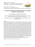
number of animals and resulting lesion pairs is summarized in figure 1.
190
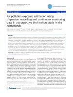
ized. Figure 2 depicts implants immediately (Fig. 2A)
and 14 days after transplantation (Fig. 2B).
The rate of successful endometriosis induction
was histologically proven after resection of the grafts.
Transplantation was successful in 90.1% (58/64). All 6
cases without endometriosis were excluded from
further evaluation.
Energy intake, number of AePC impulses and
area of coagulation
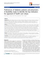
An average energy intake of 616 ± 245 J by 24.1 ±
9.7 impulses was applied for ablation of endometriosis sites with aerosol plasma coagulation. The macroscopic area of coagulation was 50.0 ± 12.7 mm2 for
AePC and 44.6 ± 23.5 mm2 for SR (p=0.16). Figure 3
displays the macroscopic tissue effect after AePC (Fig.
3A) and SR (Fig. 3B).
Duration of intervention
The surgical resection including haemostasis
was 51.8 ± 16.5 s per lesion. Interventions with AePC
were significantly faster with 22.1 ± 9.7 s (p<0.0001).
Evaluation of endometriosis therapy
Figure 1: Flow chart with the number of animals and resulting lesion pairs.
The eradication rate after endometriosis treatment was histologically determined 14 days after intervention. From 34 animals, 60 AePC and 61 SR single lesions could be achieved for histologic evaluation
resulting in a total number of 53 lesion pairs (one implant treated with AePC, the implant on the opposite
side removed using a scalpel). In 88.7% (47/53) of
lesion pairs, endometriosis was successfully removed
from both the SR and their corresponding AePC
treated sides. 1.9% (1/53) of the lesion pairs showed
remnants of endometrial implants on the resection
side only and in 9.4% (5/53) of lesion pairs remains of
endometriosis were detected on the AePC treated side
only. There were no pairs (0/53) with endometrial
residues on both sidewalls of one pair of lesions. The
McNemar test for direct comparison of pairs revealed
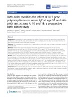
both methods to be statistically not significantly different (p=0.22). Figure 4 depicts residual endometrial
implants which remained after incomplete resection.
Table 1 summarizes the results. The histologic grading of the endometrial implants is shown in Table 2.
Table 1: Pairwise comparison of eradication by AePC or SR
(p=0.22).
Success of endometriosis induction
The size of endometriosis implants directly after
transplantation was 28.6 ± 9.3 mm2. 14 days later, implants showed a significant increase in size to 48.3 ±
19.8 mm2 (p<0.0001). Implants on the AePC side and
SR side were comparable (p=0.28). Most of the vesicular endometrial cysts were fluid-filled and vascular-
AePC
no endometriosis
no endometriosis
remnants of endometrial
implants
remnants of endometrial
implants
SR
no endometriosis
remnants of endometrial implants
no endometriosis
Frequency
47/53 (88.7%)
1/53 (1.9%)
remnants of endometrial implants
0/53 (0%)
5/53 (9.4%)
Int. J. Med. Sci. 2016, Vol. 13
Table 2: Histologic grading of endometrial lining 14 days after
treatment.
AePC
SR
Absence of Poorly preendometrial served endometrial
lining
lining
55 (91.7%) 0 (0%)
60 (98.4%) 1 (1.6%)
Fading
Moderately Well preendometrial preserved served
lining
endometrial endometrial
lining
lining
4 (6.6%)
1 (1.7%)
0 (0%)
0 (0%)
0 (0%)
0 (0%)
Further histologic results
The histological evaluation revealed an overall
moderate chronic inflammation for the SR samples
191
and an overall moderate to high chronic inflammation
for the AePC treated lesions 14 days after AePC and
SR intervention. Figure 5 depicts histological findings
of AePC treated lesions (Fig. 5A) and lesions created
by SR (Fig. 5FB). Compared to the SR group, the AePC
treated sites showed more cases graded as high for
acute inflammation and myonecrosis (p<0.0001).
Carbonization with foreign body reaction was found
in 63.3% of AePC treated lesions and in 18.0% of SR
sites as radiofrequency was only applied for coagulation purposes (p<0.0001).
Figure 2: Endometriosis implants A) immediately and B) 14 days after transplantation.
Figure 3: Macroscopic tissue effect after A) aerosol plasma coagulation and B) surgical resection.
Discussion
Figure 4: Cross section of the abdominal wall showing connective tissue with
infiltrating endometrial glands.
Therapy for endometriosis must be thoroughly
planned according to the patient`s individual symptoms and needs with pain and sterility being the main
issues. At present, the complete resection of deep infiltrating endometriosis is the therapy of choice [6]
with a positive effect on pain, quality of life and fertility [7]. Clinically, it could be demonstrated that the
success rates of spontaneous conception and assisted
reproductive methods increase after the removal of
deep infiltrating and peritoneal endometriosis [8,9].
To date, the latter can usually be achieved by minimally invasive laparoscopic techniques with various
instruments such as “cold” scissors as well as energy-based devices of which APC is a valuable option.
Int. J. Med. Sci. 2016, Vol. 13
In a clinical setting argon plasma was efficacious and
safe for the complete resection of endometriotic implants [10]; however the close combination of the least
possible adhesion formation and lateral tissue damage induced by thermal effects and the complete
therapy by total resection of affected areas remains
the major challenge for all heat induced surgical
techniques.
Figure 5: Cross sections of the abdominal wall 14 days after AePC (A) and
surgical resection (B). Both figures demonstrate an overall moderate degree of
inflammation with granulation tissue formation and myonecrosis.
Therefore, superiority of one approach has not
yet been defined and this study compares the eradication rate of sharp resection versus AePC in a rodent
model where remaining endometrial tissue after
treatment can be diagnosed histopathologically.
The design of endometriosis models is difficult
as reliable and comparable foci of interest with endometrial tissue are needed that are suitable for consecutive therapy. Our animal model is based upon
previously published literature describing the auto-transplantation of endometrium with consecutive
laparotomies [11-16]. All rats were given estradiol one
day before endometriosis induction for hormonal
synchronization. Since primary endometriosis and
recurrence are estrogen-dependent [17-19] it is unclear whether oophorectomy should be performed in
order to inhibit fluctuation of estrogen levels and to
ensure that experimental endometriotic implants
present the same activity on the peritoneal surface.
According to previous experiments [16] and to minimize the surgical trauma, we preserved the ovaries
and observed a significant growth of the transplanted
lesions (p<0.0001) that were of comparable size and
macroscopic aspect in both groups (AePC vs. SR,
192
p=0.28). Unmodified physiological ovarian estrogen
activity after the resection or coagulation of endometriosis in both groups (second look) also allows for
potential recurrence in the treated areas investigated
in the third operation (third look). At present, complete resection or destruction is the therapy of choice
if endometriosis is managed surgically [6,20]. Resection can be achieved easily in open surgery and in
areas where adjacent structures such as bowel, bladder or ureter are unlikely to be injured; however this
is not always the case in laparoscopy or if the aforementioned organs are affected directly or with the risk
of (thermal) lateral damage. We are aware of the
methodological problem that the assessment of complete resection (SR group) and destruction (AePC
group) of endometriotic lesions is difficult to compare
and ultimately dependent on the surgeon’s skills and
experience regarding the anticipated depth of the lesion. We therefore tried to reduce bias by the pairwise
comparison of the eradication sites as presented in
table 1. 1.9% of the lesion pairs showed remnants of
endometrial epithelium on the resection side only and
in 9.4% of lesion pairs, remainders of endometriosis
were detected on the AePC treated side only. This was
not statistically significant (p=0.22) indicating that
AePC and related techniques are not inferior with
respect to the eradication of endometriosis in the
presented model. We speculate that standard APC is
expected to achieve comparable eradication rates with
AePC; however the side effects such as adhesion
formation are greater as previously demonstrated [4].
With respect to operation time we could investigate that AePC was significantly faster in comparison to SR (p<0.0001). This could be hypothesized as
sharp resection of endometriosis on the peritoneal
surface requires a meticulous preparation technique
in order to completely remove the area of interest and
to reduce unwanted trauma with subsequent adhesion formation. Furthermore, the change of instruments and haemostasis of the resection area with a
bipolar coagulation clamp required additional time.
In contrast, AePC can be directly applied from a defined distance, which was 2-3 mm in this setting, to
allow for complete treatment without changing the
instrument for further haemostasis. We are aware of
the limitation in this study that both cold resection
and AePC may vary between the lesions, since the SR
and the number of radiofrequency pulses (AePC) are
based on the surgeon’s evaluation to macroscopically
remove the lesions upon second look. Clinically, laparoscopy demonstrates various benefits for endometriosis patients [21-25] and health care systems due to
shorter operation and recovery times [26]. It has to be
taken into account that AePC was applied in an open
fashion in this animal setting, however argon plasma
Int. J. Med. Sci. 2016, Vol. 13
can easily be used laparoscopically [10] with the potential to further reduce total operation time.
The animal model used in this study is not suitable to investigate adhesiogenesis as the formation of
adhesions is already triggered by a local inflammatory response inflicted on the peritoneal site after
transplantation of endometriotic foci to the rat’s peritoneum. Adhesions induced by the intervention itself
would not be distinguishable from adhesions induced
by transplantation in the endometriosis rat model.
However, we have already demonstrated in a different rat model specifically for adhesiogenesis that argon plasma itself can induce adhesion formation [27]
and consequently we could observe that the improvement of the peritoneal conditions with an aerosol in combination with APC seems to have a significant positive effect against adhesions [4].
AePC sites showed higher degrees of acute inflammation, myonecrosis and carbonization with foreign body reaction (p<0.0001). This can be explained
by the fact that in the SR group the thermal effect of
radiofrequency coagulation was only applied to
achieve haemostasis of minor bleeding of the resection area. According to Bhatta et al. [28] the histological depth of thermal lesions did not correlate with the
formation of adhesions in contrast to carbonization
and charring. The onset of the inflammatory adhesion
cascade by thermal effects and local peritoneal conditions is under investigation [29-32]. In this context, it
has to be stated that endometriosis is related to oxidative stress [33], the immunoexpression of heat
shock proteins [34] and other various inflammatory
reactions [18,35]. As the focus of this animal study is
on the complete destruction of endometriosis with SR
versus AePC representing a non-direct mode of eradication, we consider the fact that AePC is not inferior
as clinically more relevant than the histological observation of an expected acute inflammation.
Regarding the possible eradication rate of deep
infiltrating endometriosis, we assume that the water
jet technology inflicted onto the lesion with AePC
might be able to create a water cushion between the
affected tissue and the adjacent organs thus better
separating the healthy area from the spots to be
treated. This will have to be investigated in further
experiments even though it is difficult to create models that represent deep infiltrating endometriosis in
the same fashion as can be clinically found in human
patients.
Hybrid technologies, such as the combination of
waterjet with radiofrequency ablation (e.g. APC), are
easily accessible on the market and successfully applied in other medical fields; however at present, APC
does not play a major role in gynecology, with only a
few centers already using plasma energy for the
193
treatment of endometriosis. We could clearly demonstrate in our previous studies that non-contact argon
plasma coagulation, especially when combining APC
with an aerosol of liquid and gas, is significantly less
associated with adhesion side-effects compared to
other coagulation methods [4] with is very likely to
fully compensate for the additional cost of a generator
and single use probes.
Conclusion
With respect to the complete eradication of auto-transplanted endometriotic lesions, non-contact
AePC is not inferior compared to standard sharp resection (SR). The AePC treatment is significantly
faster. In some AePC cases a higher degree of inflammatory reaction could be observed histologically,
which warrants further investigations to improve
thermal side effects and peritoneal conditions of this
method that can serve as an alternative to SR in the
clinical setting of endometriosis.
Acknowledgment
The authors thank M. Eichner, M.D., Department
of Medical Biometry, University of Tuebingen,
Tuebingen, Germany, for his assistance in the statistical evaluation of the number of animals used in this
study.
Funding
This study was
ektromedizin GmbH.
supported
by
Erbe
El-
Ethical approval
This animal study was approved by the Institutional Review Board (Ethics Committee of the Regional Board in Tuebingen, Germany, registration
number F 1-13).
Authors’ contributions
Study conception and design: Ralf Rothmund,
Markus Dominik Enderle, Alexander Neugebauer,
Kristin Kroeker, Falko Fend, Sara Brucker, and Bernhard Kraemer.
Acquisition of data: Marcus Scharpf, Christos
Tsaousidis, Constanze Planck, Alexander Neugebauer, Kristin Kroeker, and Daniela Nuessle.
Analysis and interpretation of data: Ralf
Rothmund, Marcus Scharpf, Christos Tsaousidis,
Constanze Planck, Markus Dominik Enderle, Alexander Neugebauer, Kristin Kroeker, Daniela Nuessle,
and Bernhard Kraemer.
Drafting of manuscript: Marcus Scharpf, Christos Tsaousidis, Constanze Planck, Alexander
Neugebauer, Kristin Kroeker, Daniela Nuessle, and
Bernhard Kraemer.
Int. J. Med. Sci. 2016, Vol. 13
Critical revision of manuscript: Ralf Rothmund,
Marcus Scharpf, Christos Tsaousidis, Constanze
Planck, Markus Dominik Enderle, Alexander
Neugebauer, Kristin Kroeker, Daniela Nuessle, Falko
Fend, Sara Brucker, and Bernhard Kraemer.
Competing interests
The authors RR, MS, CT, CP, FF, SB and BK have
no conflicts of interest to disclose in relation to the
submitted manuscript. The authors MDE, AN, KK
and DN are employees of Erbe research department,
Germany.
References
1.
2.
3.
4.
5.
6.
7.
8.
9.
10.
11.
12.
13.
14.
15.
16.
17.
18.
19.
20.
21.
22.
23.
Macer ML, Taylor HS. Endometriosis and infertility: a review of the pathogenesis and treatment of endometriosis-associated infertility. Obstet Gynecol
Clin North Am. 2012; 39: 535–549.
Garry R. The effectiveness of laparoscopic excision of endometriosis. Curr
Opin Obstet Gynecol. 2004; 16(4): 299-303.
Roman H, Auber M, Bourdel N, et al. Postoperative recurrence and fertility
after endometrioma ablation using plasma energy: retrospective assessment of
a 3-year experience. J Minim Invasive Gynecol. 2013; 20(5): 573-582.
Kraemer B, Rothmund R, Fischer K, et al. A prospective randomized experimental study to investigate the peritoneal adhesion formation of argon plasma
coagulation (APC) versus a novel aerosol plasma in a rat model. Surg Innov.
2013; 21(4): 389-397.
Keenan JA, Williams-Boyce PK, Massey PJ, et al. Regression of endometrial
explants in a rat model of endometriosis treated with the immune modulators
loxoribine and levamisole. Fertil Steril. 1999; 72(1): 135-141.
Meuleman C, Tomassetti C, D'Hooghe TM. Clinical outcome after laparoscopic radical excision of endometriosis and laparoscopic segmental bowel
resection. Curr Opin Obstet Gynecol. 2012; 24: 245-252.
Bassi MA, Podgaec S, Dias JA Jr, et al. Quality of life after segmental resection
of the rectosigmoid by laparoscopy in patients with deep infiltrating endometriosis with bowel involvement. J Minim Invasive Gynecol. 2011; 18:
730-733.
Hart RJ, Hickey M, Maouris P, et al. Excisional surgery versus ablative surgery
for ovarian endometriomata. Cochrane Database Syst Rev. 2008;
DOI:10.1002/14651858.CD004992.pub2.
Bianchi PH, Pereira RM, Zanatta A, et al. Extensive excision of deep infiltrative
endometriosis before in vitro fertilization significantly improves pregnancy
rates. J Minim Invasive Gynecol. 2009; 16: 174-180.
Nezhat C, Kho KA, Morozov V. Use of Neutral Argon Plasma in the Laparoscopic Treatment of Endometriosis. J Soc Laparoend. 2009; 13: 479-483.
Hascalik S, Celik O, Kekilli E, et al. Novel noninvasive detection method for
endometriosis: research and development of scintigraphic survey on endometrial implants in rats. Fertil Steril. 2008; 90(1): 209–213.
Güney M, Oral B, Karahan N, et al. Regression of endometrial explants in a rat
model of endometriosis treated with melatonin. Fertil Steril. 2009; 89(4):
934–942.
Yavuz E, Oktem M, Esinler I, et al. Genistein causes regression of endometriotic implants in the rat model. Fertil Steril. 2007; 88 (Suppl 4): 1129-1134.
Akkaya P, Onalan G, Haberal N, et al. Doxycycline causes regression of
endometriotic implants: a rat model. Hum Reprod. 2009; 24(8): 1900-1908.
Vernon MW, Wilson EA. Studies on the surgical induction of endometriosis in
the rat. Fertil Steril. 1985; 44(5): 684-694.
Yildirim G, Attar R, Ficicioglu C, et al. Etanercept causes regression of endometriotic implants in a rat model. Arch Gynecol Obstet. 2011; 283(6):
1297-1302.
Vercellini P, Viganò P, Somigliana E, et al. Endometriosis: pathogenesis and
treatment. Nat Rev Endocrinol. 2014; 10(5): 261-275.
Colette S, Donnez J. Are aromatase inhibitors effective in endometriosis
treatment? Expert Opin Investig Drugs. 2011; 20(7): 917-931.
Becker CM, D'Amato RJ. Angiogenesis and antiangiogenic therapy in endometriosis. Microvasc Res. 2007; 74(2-3): 121-130.
Koninckx PR, Ussia A, Adamyan L, et al. Deep endometriosis: definition,
diagnosis, and treatment. Fertil Steril. 2012; 98(3): 564-571.
Deguara CS, Pepas L, Davis C. Does minimally invasive surgery for endometriosis improve pelvic symptoms and quality of life? Curr Opin Obstet Gynecol. 2012; 24(4): 241-244.
Touboul C, Ballester M, Dubernard G, et al. Long-term symptoms, quality of
life, and fertility after colorectal resection for endometriosis: extended analysis
of a randomized controlled trial comparing laparoscopically assisted to open
surgery. Surg Endosc. 2015; 29(7): 1879-1887.
Zanatta A, Rosin MM, Machado RL, et al. Laparoscopic Dissection and
Anatomy of Sacral Nerve Roots and Pelvic Splanchnic Nerves. J Minim Invasive Gynecol. 2014; 21(6): 982-983.
194
24. Walch K, Kernstock T, Poschalko-Hammerle G, et al. Prevalence and severity
of cyclic leg pain in women with endometriosis and in controls - effect of laparoscopic surgery. Eur J Obstet Gynecol Reprod Biol. 2014; 179: 51-57.
25. Collinet P, Leguevaque P, Neme RM, et al. Robot-assisted laparoscopy for
deep infiltrating endometriosis: international multicentric retrospective study.
Surg Endosc. 2014; 28(8): 2474-2479.
26. Lassen PD, Moeller-Larsen H, DE Nully P. Same-day discharge after laparoscopic hysterectomy. Acta Obstet Gynecol Scand. 2012; 91(11): 1339-1341.
27. Kraemer B, Rothmund R, Fischer K, et al. A prospective, randomized, experimental study to investigate the peritoneal adhesion formation of noncontact
argon plasma coagulation in a rat model. Fertil Steril. 2011; 95(4): 1328-1332.
28. Bhatta N, Isaacson K, Flotte T, et al. Injury and adhesion formation following
ovarian wedge resection with different thermal surgical modalities. Lasers
Surg Med. 1993; 13(3): 344-352.
29. Kraemer B, Scharpf M, Planck C, et al. Randomized experimental study to
investigate the peritoneal adhesion formation of conventional monopolar
contact coagulation versus noncontact argon plasma coagulation in a rat
model. Fertil Steril. 2014; 102(4): 1197-1202.
30. Binda MM, Koninckx PR. Prevention of adhesion formation in a laparoscopic
mouse model should combine local treatment with peritoneal cavity conditioning. Hum Reprod. 2009; 24(6): 1473-1479.
31. Corona R, Binda MM, Mailova K, et al. Addition of nitrous oxide to the carbon
dioxide pneumoperitoneum strongly decreases adhesion formation and the
dose-dependent adhesiogenic effect of blood in a laparoscopic mouse model.
Fertil Steril. 2013; 100(6): 1777-1783.
32. Koninckx PR, Corona R, Timmerman D, et al. Peritoneal full-conditioning
reduces postoperative adhesions and pain: a randomised controlled trial in
deep endometriosis surgery. J Ovarian Res. 2013; 6(1): 90.
33. Rosa e Silva JC, do Amara VF, Mendonça JL, et al. Serum markers of oxidative
stress and endometriosis. Clin Exp Obstet Gynecol. 2014; 41(4): 371-374.
34. Imamura T, Khan KN, Fujishita A, et al. Effect of GnRH agonist therapy on the
expression of human heat shock protein 70 in eutopic and ectopic endometria
of women with endometriosis. Eur J Obstet Gynecol Reprod Biol. 2014; 180:
16-23.
35. Greaves E, Cousins FL, Murray A, et al. A novel mouse model of endometriosis mimics human phenotype and reveals insights into the inflammatory contribution of shed endometrium. Am J Pathol. 2014; 184(7): 1930-1939.