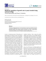
Increment of growth factors in mouse skin treated with non-thermal plasma
Bạn đang xem bản rút gọn của tài liệu. Xem và tải ngay bản đầy đủ của tài liệu tại đây (1.22 MB, 7 trang )
Int. J. Med. Sci. 2018, Vol. 15
Ivyspring
International Publisher
1203
International Journal of Medical Sciences
2018; 15(11): 1203-1209. doi: 10.7150/ijms.26342
Research Paper
Increment of growth factors in mouse skin treated with
non-thermal plasma
Byul Bo Ra Choi1,2,*, Jeong Hae Choi1,2,*, Jeong Ji2, Ki Won Song3, Hae June Lee4 and Gyoo Cheon Kim2
1.
2.
3.
4.
Feagle Co., Ltd., Yangsan 50614, Republic of Korea
Department of Oral Anatomy, School of Dentistry, Pusan National University, Yangsan 50612, Republic of Korea
Department of Biochemistry, College of Life Science and Biotechnology, Yonsei University, Seoul 03722, Republic of Korea
Department of Electrical Engineering, Pusan National University, Busan 46241, Republic of Korea
*These authors contributed equally to this work.
Corresponding author: Gyoo Cheon Kim, Department of Oral Anatomy, School of Dentistry, Pusan National University, Yangsan 626-870, Republic of Korea.
Email: ; Tel: 82-51-510-8243; fax: 82-51-510-8241.
© Ivyspring International Publisher. This is an open access article distributed under the terms of the Creative Commons Attribution (CC BY-NC) license
( See for full terms and conditions.
Received: 2018.03.28; Accepted: 2018.06.30; Published: 2018.07.30
Abstract
Non-thermal plasma (NTP) has several beneficial effects, and can be applied as a novel instrument
for skin treatment. Recently, many types of NTP have been developed for potential medical or
clinical applications, but their direct effects on skin activation remain unclear. In this study, the effect
of NTP on the alteration of mouse skin tissue was analyzed. After NTP treatment, there were no
signs of tissue damage in mouse skin, whereas significant increases in epidermal thickness and dermal
collagen density were detected. Furthermore, treatment with NTP increased the expression of
various growth factors, including TGF-α, TGF-β, VEGF, GM-CSF, and EGF, in skin tissue. Therefore,
NTP treatment on skin induces the expression of growth factors without causing damage, a
phenomenon that might be directly linked to epidermal expansion and dermal tissue remodeling.
Key words: Non-thermal plasma, Skin regeneration, Clinical application, Growth factor
Introduction
The skin is the largest organ of the body,
accounting for approximately 16% of the total body
weight of an adult [1]. Skin is constantly exposed to
external environments and serves as a protective
barrier [2], protecting the body against exogenous
hazards, including biological infection, chemical
substances, and UV [3]. The skin is composed of two
layers, the epidermis (upper layer) and the
underlying layer of dermis (lower layer). The
epidermis is mainly composed of keratinocytes
(approximately 90% of the epidermis) [4, 5]. Although
the dermis contains several functional tissues,
including nerves, hair follicles, and sweat glands [6], it
is mainly comprised of collagen, fibroblasts, and
elastin fibers that provide nutrients [7].
Maintenance of healthy skin is important not
only for anti-aging and rejuvenation, but also for
wound healing. Skin aging accompanies reduction in
collagen, decrease in various growth factors, and loss
of fibroblasts [8-10]. To protect skin from aging,
keratinocytes in the epidermis need to proliferate and
fibroblasts in the dermis need to actively produce
extracellular matrix proteins such as collagen and
elastin fibers [7, 11]. For successful healing of skin
wounds, a series of events should proceed favorably,
including coagulation, inflammation, re-epithelialization, wound contraction, extracellular matrix
rearrangement, and angiogenesis [12-14]. The skin
beauty market has been growing tremendously with
improvements in the quality of life, and healing of
skin wounds has also been of huge importance in
terms of bedsore treatment due to the acceleration of
aging. Therefore, several studies on anti-aging and
wound healing are under way.
Recently, non-thermal atmospheric plasma has
shown beneficial effects on the healing of skin
wounds; therefore, it has been considered as a
potential tool for skin treatment. According to
Int. J. Med. Sci. 2018, Vol. 15
Heinlin’s report, repeated plasma treatment for two
minutes a day on the site of venous ulcers resulted in
excellent wound healing. A previous study showed
that repeated plasma treatment performed eleven
times resulted in no bacteria in the wound area [15]. In
our previous study, treatment of skin cells with
low-temperature microwave plasma increased the
expression of collagen fibers, fibronectin, and vascular
endothelial growth factor (VEGF) genes; no thermal
damage to the cells, due to the plasma or change in
the pH of the medium, was observed [16]. These
studies clearly suggest that plasma can be a great tool
for anti-aging and healing of skin wounds. However,
there have been many studies on the phenomenon
occurring in the skin caused by plasma, but the
mechanism causing such phenomenon has been
poorly reported. In our previous studies, we have
shown that in the process of wound healing,
non-thermal plasma (NTP) inhibited E-cadherin of
keratinocytes in the epidermis, thereby β-catenin
binds to E-cadherin to migrate to the nucleus because
of the weak bond and acts as a transcription factor for
cell division [17].
In this study, we focused on the expression of
several growth factors in epidermal tissue, since along
with the contribution of E-cadherin and β-catenin, the
involvement of cell growth factors seems to be
essential for NTP-mediated biological changes in
dermal tissue. To this end, HRM2 hairless mice were
subjected to repeated treatment with NTP. After the
treatment, NTP-mediated changes in skin tissue,
along with the epidermis and dermis, was monitored.
Furthermore, NTP-mediated changes in the
expression of growth factors, including transcription
growth
factor
(TGF)-α,
TGF-β,
VEGF,
granulocyte-macrophage colony-stimulating factor
(GM-CSF), and epidermal growth factor (EGF), all of
which are well known in wound healing and dermal
tissue remodeling, were monitored. Taken together,
1204
this study suggests that NTP-mediated secretion of
several growth factors in the epidermis can be a cause
of changes in deep skin.
Methods
NTP device
The NTP device used in this study consisted of
two electrodes and an alumina tube, which is a coaxial
dielectric barrier discharge configuration. A
stainless-steel rod with 3 mm inner diameter was used
as the inner electrode and copper tape with a width of
10 mm was used as the outer electrode. An alumina
(Al2O3) tube (4 mm inner diameter and 6 mm outer
diameter) served as a dielectric, which prevented the
transition of glow to arc discharges. The argon
working gas was delivered at a flow rate of 2 standard
liters per minute using a mass flow meter. The
directions of the gas flow and the electric field are
perpendicular to each other. A sinusoidal high
voltage of 3 kV with a frequency of 15 kHz was
applied to the inner electrode and the outer copper
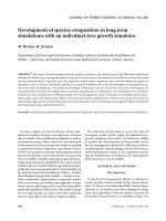
electrode was grounded (Figure 1). Non thermal
plasma generated in this device does not extend from
the nozzle like a plasma jet, but generated between
the inner electrode and the alumina tube with
discharge volume of 7.85 mm3.
Mouse experiments
Five-week-old male HRM2 melanin-possessing
hairless mice were obtained from Central Laboratory
Animal Inc. (Seoul, Korea). All experimental protocols
in this study were approved by the Animal Ethics
Committee, Pusan National University (PNU
017-1446). Mice (n = 5 per group) were subjected to
repeated treatments with gas only (GO) and NTP for a
total of six times for 5 min each. After the final
treatment at two weeks, the mice were sacrificed, and
their skin tissues were collected for histology.
Figure 1. The structure of the non-thermal plasma (NTP) device. A schematic of the NTP device and operating process in this experiment.
Int. J. Med. Sci. 2018, Vol. 15
Hematoxylin and eosin staining
Mouse skin sections were stained with
hematoxylin and eosin (H&E). Tissue sections with a
thickness of 5 µm were fixed with 10% formalin,
embedded with paraffin, cut on salinized glass slides,
deparaffinized three times with xylene, and
rehydrated
through
graded
ethanol.
After
deparaffinization, rehydration, and rinsing with
distilled water, the sections were stained with Harris
hematoxylin for 3 min, and then stained in an aqueous
solution of eosin for 30 s. The sections were
dehydrated in ethanol and cleared in xylene. The
samples were imaged using an Axio Scan Z1 slide
scanner (Goettingen, Germany).
DAPI staining
Mouse sections were stained with DAPI
(4′,6-diamidino-2-phenylindole) for nucleic acids and
mounted with ProLong™ Gold Antifade Mountant
contained in the DAPI. Fluorescent images were
acquired and analyzed using a Zeiss LSM 700 laser
scanning confocal microscope (Goettingen, Germany).
Masson's trichrome staining
The slides were treated overnight with Bouin’s
solution at 25℃, and then rinsed under tap water for
10 min to remove the yellow color. To stain the nuclei,
slices were stained with Weigert iron hematoxylin for
10 min. Then the slides were stained in Biebrich
scarlet acid fuchsin solution for 2 min, and
differentiated in phosphomolybdic-phosphotungstic
acid solution. Slides were disposed with aniline blue
for 2 min and differentiated in 1% acetic acid.
Subsequently, the slides were dehydrated and
cleared. The samples were imaged using an Axio Scan
Z1 slide scanner (Goettingen, Germany).
Immunohistochemical analysis
Mouse skin from NTP-treated groups was
prepared for immunohistochemical analysis of the
expression of TGF-α, TGF-β, VEGF, GM-CSF, and
EGF. Tissue sections (5 µm in thickness) were fixed
with 10% formalin, embedded with paraffin, cut on
slides, deparaffinized three times with xylene, and
rehydrated through graded alcohol. To diminish
non-specific staining, each section was treated with
0.3% hydrogen peroxide for 10 min and protein
blocking solution (Abcam, Cambridge, MA) for 10
min. The sections were incubated with the following
primary antibodies: rabbit polyclonal anti-TGF-α
(1:400), TGF-β (1:250), VEGF (1:100), GM-CSF (1:100),
and EGF (1:100) overnight at 4°C in Antibody Diluent
(Dako, Glostrup, Denmark), and each section was
then treated with biotinylated secondary antibody
(1:100) (Dako, Glostrup, Denmark). The sections were
1205
incubated with avidin-biotin horseradish peroxidase
complex (ABC) (Vector Laboratories, Burlingame,
CA) for 30 min. The peroxidase binding sites were
detected by staining with 3,39-diaminobenzidine
tetrahydrochloride
(DAB)
(Dako,
Glostrup,
Denmark). The samples were then counterstained
with Mayer’s hematoxylin (Dako, Glostrup,
Denmark) imaged using an Axio Scan Z1 slide
scanner (Goettingen, Germany).
Statistical analysis
One-way ANOVA and post-hoc Tukey’s test
were used to compare treatment effects for
experiments. The results are presented as mean ±
standard error (SE). p < 0.05 was considered to be
statistically significant.
Results and discussion
Histology of mouse skin after NTP treatment
The mouse tissue sections were stained with
H&E and Masson’s trichrome staining solutions to
examine the change in the general morphology of
mouse skin and dermal collagen fiber. In both
histological analyses, we found that the epidermal
layer of mouse skin treated with NTP was much
thicker than that without NTP or with gas only
(Figure 2A). The increment in epidermal thickness
was a result of the proliferation of keratinocytes in the
epidermal layer. Hoechst staining showed that the
nuclei of cells were actively dividing (Figure 2B).
These results demonstrated that NTP could induce
skin cell proliferation, as E-cadherin was inhibited by
plasma treatment so that β-catenin was released to the
cytosol and played a role as a transcriptional factor.
As shown in figure 2C, quantitative measurement
indicated that the epidermal thickness of skin treated
with NTP was twice that of non-treated and
gas-treated groups (p < 0.001). These results suggested
that NTP effectively caused the proliferation of the
epidermal layer, so that it would be useful not only
for wound healing, but also for maintaining healthy
skin by protecting skin from aging.
Collagen is a structural component of the
dermis and a very important factor in anti-aging and
skin regeneration. To further examine the histological
appearance of collagen fibers in mouse skin tissue, we
performed Masson’s trichrome staining. The blue
color indicated the expression of collagen fibers. The
density of dermal collagen fibers in the non-treated
group was lower than that in the NTP-treated group.
Furthermore, the deposition of the collagen fibers was
compact and thicker in the NTP-treated group (Figure
3). Masson’s trichrome staining showed that NTP
promoted the production of collagen in the dermal
layer. Fibroblasts in the dermal layer produce various
Int. J. Med. Sci. 2018, Vol. 15
1206
extracellular matrix components.
Thus, we investigated the expression
of various growth factors in mouse
skin after NTP treatment by means of
immunohistochemistry.
Immunohistochemical staining
of mouse skin after NTP
treatment
Immunohistochemical analysis
was performed to monitor the
changes in growth factors in the
epidermis and dermis after NTP
treatment in mouse skin. A growth
factor is a substance that stimulates
cellular growth, proliferation, and
differentiation
under controlled
conditions [20]. It is secreted by all
cell types and known to play a
significant role in anti-aging and
tissue regeneration [21]. A number of
growth factors are secreted from the
epidermis and dermis, including
TGF-α, TGF-β, VEGF, GM-CSF,
fibroblast growth factor (FGF)-2,
platelet-derived
growth
factor,
keratinocyte growth factor, and EGF
[22-24]. In the present study, after
treatment with NTP for two weeks,
the expression of TGF-α, TGF-β,
VEGF, GM-CSF, and EGF were
increased in mouse skin (Figure 4).
TGF-α is a single-chain polypeptide
that is a ligand for the EGF receptor
related to EGF [25]. The general
function of TGF-α is to activate a
signaling pathway for cell restitution,
proliferation, differentiation, and
Figure 2. The effect of NTP on mouse skin at day 14. A. After sacrificing the mouse, skin sections obtained from
development [26]. After NTP
the non-treated group (NT), gas-treated group (GO), and NTP-treated group (NTP) were stained with
hematoxylin and eosin (H&E), 100×. B. After sacrificing the mouse, skin sections obtained from the non-treated
treatment, it was strongly expressed
group (NT), gas-treated group (GO), and NTP-treated group (NTP) were stained with DAPI, 100×. C.
Quantitative analysis of epidermal thickness in the non-treated group (NT), gas-treated group (GO), and
over the epidermal layer, especially
NTP-treated group (NTP). *** p < 0.001 (ANOVA).
filling the cytosol of keratinocytes.
Considering that the thickness of the
dermal layer was increased by NTP
extracellular matrix proteins such as collagen fibers,
treatment,
keratinocytes
were stimulated by NTP and
elastin fibers, and fibronectin [18-19]. In the present
actively produced TGF-α, which supposedly caused
study, the increase in collagen fibers in the dermal
neighboring cells to proliferate.
layer was likely caused by the activation of fibroblasts
TGF-β stimulates fibroblasts and keratinocytes to
after NTP treatment. In turn, they synthesized and
induce cell migration, wound healing, and tissue
produced a large amount of collagen in the dermis.
repair [27]. In our previous study, wounded skin
Since the skin barrier function is mainly mediated by
treated with NTP healed much faster than that
the epidermal tissue, NTP treatment cannot affect
without NTP treatment [17]. In the healing of skin
fibroblasts directly. Therefore, we assumed that
wounds, re-epithelialization is very important as it is
growth factors were produced from the epidermis
essential for cell migration. Keratinocytes, Langerhans
after NTP treatment, and they then stimulated the
cells, and Merkel cells exist in the epidermis, and
fibroblasts to produce and secrete several types of
Int. J. Med. Sci. 2018, Vol. 15
1207
Figure 3. The expression of collagen on mouse skin after NTP treatment at day 14. After sacrificing the mouse, skin sections obtained from the non-treated group (NT),
gas-treated group (GO), and NTP-treated group (NTP) were stained with Masson’s trichrome stain (MTS), 100×.
among them, keratinocytes are the major component
[28]. The results of the present study showed that
TGF-β was expressed in the nuclei of several types of
cells in the epidermis. It was unclear which cells
expressed TGF-β, but it was distributed in the nuclei
of cells. Furthermore, TGF-β was also expressed in
cells of the dermis. As shown in figure 3, collagen
density was increased after NTP treatment. Collagen
fibers in the dermis are synthesized by fibroblasts,
thus the increase in TGF-β by NTP could activate
fibroblasts to produce collagen fibers.
VEGF, also known as vascular permeability
factor, is a disulfide-linked dimeric glycoprotein of
approximately 40 kDa that induces angiogenesis,
endothelial cell proliferation, and activation of
monocytes/macrophages [29-31]. In the present
study, VEGF was expressed over the epidermal layer,
but not in the basal layer, which is the lowermost
layer of keratinocytes. This result is in agreement with
the in-vitro data in our previous study, which showed
that NTP treatment on HaCaT keratinocytes and
human dermal fibroblasts increased VEGF mRNA
expression. Synthesized VEGF most likely stimulates
new blood vessel formation in NTP-treated areas in
the dermis and supplies nutrients to fibroblasts.
GM-CSF is a 23-kDa glycoprotein that plays a
significant role as an immune-modulator, activating
macrophages and granulocytes. Moreover, GM-CSF
promotes
angiogenesis,
keratinocyte
growth,
epidermis regeneration, and wound healing [32, 33].
In the present study, GM-CSF was strongly expressed
in the epidermal layer, including the basal layer,
unlike the case of VEGF. It seems that the expression
of GM-CSF after NTP treatment was strongly related
to wound healing by NTP. EGF is a small protein with
a molecular mass of 6 kDa, which increases the
renewal rate of aging cells and accelerates wound
healing in skin [34]. In addition, it stimulates the
proliferation of epidermal cells and differentiation of
skin appendages [35]. In particular, EGF has been
considered to be important in esthetics, and it has
been widely used for skin rejuvenation. In the present
study, EGF was expressed over the epidermal layer
after NTP treatment but its expression was not
observed in the basal layer. The expressed EGF by
NTP could lead to cell proliferation in the epidermis.
Above all, NTP treatment on skin not only
caused cell proliferation in the epidermis and the
increase in collagen fibers in the dermis, but also
promoted active expression of various growth factors.
These results are very promising in skin rejuvenation
and wound healing. Therefore, enhanced expression
of growth factors by treatment of NTP is thought to be
an important mechanism in wound healing. As this
study was focused on the expression of growth factors
in skin tissues by NTP, the observation was
performed after two weeks of NTP treatment. Thus, in
the next experiment, not only long-term examination
of normal tissues will be performed, but also various
wound tissues will be observed.
Conclusions
This study provides evidence for the histological
anti-aging effect and safety of the treatment of NTP in
mouse skin tissue. This treatment does not induce
thermal damage to mouse skin tissue. In addition, the
epidermal layer thickness and density of collagen in
the dermis were increased after NTP treatment.
Furthermore, we observed significant increases in the
levels of growth factors such as TGF-α, TGF-β, VEGF,
GM-CSF, and EGF after the treatment. Taken
together, since NTP not only directly activates the
proliferation of epidermal cells but also accelerates
dermal remodeling by stimulating the secretion of
several types of growth factors, this study suggests
that NTP can be an innovative tool for wound healing
and anti-aging of the skin.
Int. J. Med. Sci. 2018, Vol. 15
1208
Figure 4. The expression of growth factors on mouse skin after NTP treatment at day 14. After sacrificing the mouse, skin sections obtained from the non-treated group (NT),
gas-treated group (GO), and NTP-treated group (NTP) were subjected to immunohistochemistry (IHC) for TGF-α, TGF-β, VEGF, GM-CSF, and EGF, 100×.
Acknowledgments
This research was supported by the Bio &
Medical Technology Development Program of the
National Research Foundation (NRF) & funded by the
Korean government (2016M3A9C6918283)
Abbreviations
NTP: non-thermal plasma; VEGF: vascular
endothelial growth factor; (TGF)-α: transcription
growth factor alpha; (TGF)-β: transcription growth
factor beta; GM-CSF: granulocyte-macrophage
colony-stimulating factor; EGF: epidermal growth
factor; H&E: hematoxylin and eosin; DAPI:
4′,6-diamidino-2-phenylindole; ABC: avidin-biotin
horseradish peroxidase complex; DAB: 3,39-diaminobenzidine tetrahydrochloride.
Competing Interests
The authors have declared that no competing
interest exists.
References
1.
2.
3.
4.
RR Wickett, MO Visscher. Structure and function of the epidermal barrier.
American journal of infection control 2006.
Boer M, Duchnik E, Maleszka R, et al. Structural and biophysical
characteristics of human skin in maintaining proper epidermal barrier
function. Postepy Dermatol Alergol 2016; 33: 1-5.
Kleesz P, Darlenski R, Fluhr JW. Full-body skin mapping for six biophysical
parameters: baseline values at 16 anatomical sites in 125 human subjects. Skin
Pharmacol Physiol 2012; 25: 25-33.
Watt FM. The epidermal keratinocyte. Bioessays 1988;8: 163-167.
Int. J. Med. Sci. 2018, Vol. 15
5.
6.
7.
8.
9.
10.
11.
12.
13.
14.
15.
16.
17.
18.
19.
20.
21.
22.
23.
24.
25.
26.
27.
28.
29.
30.
31.
32.
33.
Régnier M, Patwardhan A, Scheynius A, et al. Reconstructed human
epidermis composed of keratinocytes, melanocytes and Langerhans cells. Med
Biol Eng Comput 1998;36: 821-824.
Brohem CA, Cardeal LB, Tiago M, et al. Artificial skin in perspective: concepts
and applications. Pigment Cell Melanoma Res 2011;24: 35-50.
Lee DY, Yang JM, Park KH. A dermal equivalent developed from fibroblast
culture alone: effect of EGF and insulin. Wound Repair Regen 2007;15:
936-939.
Gerber PA, Buhren BA, Schrumpf H, et al. Mechanisms of skin aging induced
by EGFR inhibitors. Support Care Cancer 2016;24: 4241-4248.
Chen J, Li Y, Zhu Q, et al. Anti-skin-aging effect of epigallocatechin gallate by
regulating epidermal growth factor receptor pathway on aging mouse model
induced by d-Galactose. Mech Ageing Dev 2017; 164: 1-7.
Aldag C, Nogueira Teixeira D, Leventhal PS. Skin rejuvenation using cosmetic
products containing growth factors, cytokines, and matrikines: a review of the
literature. Clin Cosmet Investig Dermatol 2016; 9: 411-419.
Lee DH, Oh IY, Koo KT, et al. Improvement in skin wrinkles using a
preparation containing human growth factors and hyaluronic acid serum. J
Cosmet Laser Ther 2015;17: 20-23.
Somchaichana J, Bunaprasert T, Patumraj S. Acanthus ebracteatus Vahl.
ethanol extract enhancement of the efficacy of the collagen scaffold in wound
closure: a study in a full-thickness-wound mouse model. J Biomed Biotechnol
2012; 754527.
Xue M, Jackson CJ. Extracellular Matrix Reorganization During Wound
Healing and Its Impact on Abnormal Scarring. Adv Wound Care (New
Rochelle) 2015; 4: 119-136.
Saito T, Izumi K, Shiomi A, et al. Zoledronic acid impairs re-epithelialization
through down-regulation of integrin αvβ6 and transforming growth factor
beta signalling in a three-dimensional in vitro wound healing model. Int J Oral
Maxillofac Surg 2014; 43: 373-380.
Heinlin J, Morfill G, Landthaler M, et al. Plasma medicine: possible
applications in dermatology. J Dtsch. Dermatol Ges 2010; 8: 968-976.
Choi JH, Lee HW, Lee JK, et al. Low-temperature atmospheric plasma
increases the expression of anti-aging genes of skin cells without causing
cellular damages. Arch. Dermatol Res 2013; 305: 133-140.
Choi JH, Song YS, Song K, et al. Skin renewal activity of non-thermal plasma
through the activation of β-catenin in keratinocytes. Sci Rep 2017;7: 6146.
Roh SS, Lee MH, Hwang YL, et al. Stimulation of the extracellular matrix
production in dermal fibroblasts by velvet antler extract. Ann Dermatol
2010;22: 173-179.
Jeon YK, Jang YH, Yoo DR, et al. Mesenchymal stem cells' interaction with
skin: wound-healing effect on fibroblast cells and skin tissue. Wound Repair
Regen 2010;18: 655-661.
Park JW, Hwang SR, Yoon IS. Advanced Growth Factor Delivery Systems in
Wound Management and Skin Regeneration. Molecules 2017;27: E1259.
Gibbs S, Silva Pinto AN, Murli S, et al. Epidermal growth factor and
keratinocyte growth factor differentially regulate epidermal migration,
growth, and differentiation. Wound Repair Regen 2000;8: 192-203.
Aldag C, Nogueira Teixeira D, Leventhal PS. Skin rejuvenation using cosmetic
products containing growth factors, cytokines, and matrikines: a review of the
literature. Clin Cosmet Investig Dermatol 2016; 9: 411-419
Safari M, Ghahari L, Zoroufchi MD. Effects of epidermal growth factor,
platelet derived growth factor and growth hormone on cultured rat
keratinocytes cells in vitro. Pak J Biol Sci 2014;17: 931-936.
Andreadis ST, Hamoen KE, Yarmush ML, et al. Keratinocyte growth factor
induces hyperproliferation and delays differentiation in a skin equivalent
model system. FASEB J 2001; 15: 898-906.
Borlinghaus P, Wieser S, Lamerz R. Epidermal growth factor, transforming
growth factor-alpha, and epidermal growth factor receptor content in normal
and carcinomatous gastric and colonic tissue. Clin Investig 1993;71: 903-907.
Vassar R, Fuchs E. Transgenic mice provide new insights into the role of
TGF-alpha during epidermal development and differentiation. Genes Dev
1991;5: 714-727.
Amjad SB, Carachi R, Edward M. Keratinocyte regulation of TGF-beta and
connective tissue growth factor expression: a role in suppression of scar tissue
formation. Wound Repair Regen 2007; 15: 748-755.
Zhang C, Tan CK, McFarlane C, et al. Myostatin-null mice exhibit delayed skin
wound healing through the blockade of transforming growth factor-β
signaling by decorin. Am J Physiol Cell Physiol 2012;302: C1213-1225.
Cao Y, Linden P, Farnebo J, et al. Vascular endothelial growth factor C induces
angiogenesis in vivo. Proc Natl Acad Sci USA 1998;95: 14389-14394.
Cébe-Suarez S, Zehnder-Fjällman A, Ballmer-Hofer K. The role of VEGF
receptors in angiogenesis; complex partnerships. Cell Mol Life Sci 2006; 63:
601-615.
Inoue M, Itoh H, Tanaka T, et al. Oxidized LDL regulates vascular endothelial
growth factor expression in human macrophages and endothelial cells
through activation of peroxisome proliferator-activated receptor-gamma.
Arterioscler Thromb Vasc Biol 2001;21: 560-566.
Braunstein S, Kaplan G, Gottlieb AB, et al. GM-CSF activates regenerative
epidermal growth and stimulates keratinocyte proliferation in human skin in
vivo. J Invest Dermatol 1994;103: 601-604.
Zhao J, Chen L, Shu B, et al. Granulocyte/macrophage colony-stimulating
factor influences angiogenesis by regulating the coordinated expression of
VEGF and the Ang/Tie system. PLoS. One. 2014;9: e92691.
1209
34. J.D. Zieske, H. Takahashi, A.E. Hutcheona, et al. Activation of epidermal
growth factor receptor during corneal epithelial migration. Invest Ophthalmol
Vis Sci 2000; 41: 1346-1355.
35. Tran QT, Kennedy LH, Leon Carrion S, et al. EGFR regulation of epidermal
barrier function. Physiol Genomics 2012;44: 455-469.