unsicker - cell signaling and growth factors in development
Bạn đang xem bản rút gọn của tài liệu. Xem và tải ngay bản đầy đủ của tài liệu tại đây (10.98 MB, 1,083 trang )
Cell Signaling and
Growth Factors in Development
Edited by
K. Unsicker and
K. Krieglstein
Further Titles of Interest
R.A. Meyers
Encyclopedia of Molecular Cell Biology and Molecular Medicine
2005, second edition
3-527-30542-4
D. Wedlich
Cell Migration in Development and Disease
2005
ISBN 3-527-30587-4
A. Ridley
Cell Motility
2004
ISBN 0-470-84872-3
G.S. Stein, A.B. Pardee
Cell Cycle and Growth Control
2004, second edition
ISBN 0-471-25071-6
W.W. Minuth, R. Strehl, K. Schumacher
Tissue Engineering
2005
ISBN 3-527-31186-6
E.A. Nigg
Centrosomes in Development and Disease
2004
ISBN 3-527-30980-2
M. Schliwa
Molecular Motors
2003
ISBN 3-527-30594-7
Cell Signaling and
Growth Factors in Development
From Molecules to Organogenesis
Edited by
Klaus Unsicker and
Kerstin Krieglstein
Editor
Prof. Dr. Klaus Unsicker
University of Heidelberg
Interdisciplinary Center for Neurosciences (IZN)
Department of Neuroanatomy
Im Neuenheimer Feld 307
69120 Heidelberg
Germany
Prof. Dr. Kerstin Krieglstein
University of Göttingen
Medical School
Department of Neuroanatomy
Kreuzbergring 36
37075 Göttingen
Germany
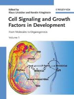
Coverillustration
The simplified signaling network (see Fig. 15.5 from
Susan Mackem) and the mouse embryo section
represent the starting point and goal of the fascinating
way from molecules to organo-genesis (mouse embryo
section reprinted from »The Atlas of Mouse
Development« edited by M.H. Kaufmann, cover page,
1992 with permission from Elsevier).
■
This book was carefully produced. Never-
theless, authors, editor and publisher do not
warrant the information contained therein to
be free of errors. Readers are advised to keep in
mind that statements, data, illustrations,
procedural details or other items may
inadvertently be inaccurate.
Library of Congress Card No.: applied for
A catalogue record for this book is available
from the British Library
Bibliographic information published by
Die Deutsche Bibliothek
Die Deutsche Bibliothek lists this publication
in the Deutsche Nationalbibliografie; detailed
bibliographic data is available in the Internet at
.
2006 WILEY-VCH Verlag GmbH & Co.
KGaA, Weinheim
All rights reserved (including those of
translation in other languages). No part of this
book may be reproduced in any form – by
photoprinting,microfilm,oranyother means–
nor transmitted or translated into machine
language without written permission from the
publishers. Registered names, trademarks,
etc. used in this book, even when not
specifically marked as such, are not to be
considered unprotected by law.
Printed in the Federal Republic of Germany.
Printed on acid-free paper.
Typesetting pagina GmbH, Tübingen
Printing betz-druck GmbH, Darmstadt
Bookbinding J. Schäffer GmbH i. G.,
Grünstadt
ISBN–13: 978-3-527-31034-0
ISBN–10: 3-527-31034-7
V
Cell Signaling and Growth Factors in Development. Edited by K. Unsicker and K. Krieglstein
Copyright 2006 WILEY-VCH Verlag GmbH & Co. KGaA, Weinheim
ISBN 3-527-31034-7
Contents
Volume 1
Preface XXVII
List of Contributors XXIX
Color Plates XXXVII
I Cell Signaling and Growth Factors in Development
1 Stem Cells
Christian Paratore and Lukas Sommer
1.1 Introduction 3
1.2 Maintenance of Stemness in Balance with Stem Cell Differen-
tiation 5
1.2.1 Wnt Signaling 7
1.2.2 Wnt Signaling Regulates ,Stemness’ in ESCs 8
1.2.3 Wnt Signaling in Hematopoietic Stem Cells 8
1.2.4 Wnt Signal Activation in the Skin 10
1.2.5 Multiple Roles of Canonical Wnt Signaling in Neural Stem
Cells 11
1.2.6 Aberrant Wnt signal activation in carcinogenesis 13
1.3 The Notch Signaling Pathway 13
1.3.1 Notch Signaling During Hematopoiesis 14
1.3.2 Notch1 Functions as a Tumor Suppressor in Mouse Skin 15
1.3.3 Notch Signaling in the Nervous System and its Role in Neural
Differentiation and Stem Cell Maintenance 15
1.3.4 Aberrant Notch Signaling 18
1.4 Signaling Pathway of the TGF
b Family Members 18
1.4.1 BMP Signaling in ESCs 20
VI Contents
1.4.2 The Influence of TGFb Family Members on MSC Differentia-
tion 20
1.4.3 Tgf
b Factors Act Instructively on NCSC Differentiation 21
1.4.4 Aberrant Growth Regulation by Mutations in the Tgf
b Signaling
Pathway 22
1.5 Shh Signaling 23
1.5.1 Hematopoiesis and T-cell Maturation 24
1.5.2 The Role of Shh in the Nervous System 24
1.5.3 Shh Signaling in Tumorigenesis 25
1.6 Conclusions 26
2 Germ Cells
Pellegrino Rossi, Susanna Dolci, Donatella Farini, and Massimo De
Felici
2.1 Introduction 39
2.2 Primordial Germ Cells 41
2.2.1 Growth Factors in PGC Commitment 41
2.2.2 Growth Factors in PGC Proliferation 42
2.2.2.1 The KL/KIT System 42
2.2.2.2 LIF/OSM/IL6 Superfamily 43
2.2.2.3 TGF
b Superfamily and Neuropeptides 44
2.3 Oocytes 45
2.3.1 Growth Factors for Fetal Oocytes 45
2.3.2 Growth Factors for Growing and Mature Oocytes 46
2.3.3 Growth Factors in the Initiation of Follicle and Oocyte
Growth 48
2.3.4 From Pre-antral to Ovulatory Follicles: Oocytes take
Control 50
2.4 Male Germ Cells 52
2.4.1 GDNF 53
2.4.2 BMPs 55
2.4.3 KL/Kit System 56
2.4.4 Control of Meiotic Progression of Spermatocytes 58
3 Implantation and Placentation
Susana M. Chuva de Sousa Lopes, Christine L. Mummery, and
So´lveig Thorsteinsdo´ttir
3.1 Introduction 73
3.1.1 Formation of the Trophectoderm, the Precursor of the Invading
Trophoblast 74
3.1.2 Initial Blastocyst-Uterine Interaction 74
3.1.3 Differentiation of Extra-embryonic Lineages During
Implantation and Gastrulation 76
Contents VII
3.1.4 Steps towards a Functional Placenta 78
3.1.5 Comparison of Mouse and Human Implantation and
Placentation 79
3.2 Molecular Mechanisms and Biological Effects 80
3.2.1 Preparation of the Blastocyst for Implantation (E3.5–E4.5) 80
3.2.2 Uterine-Embryonic Signaling and Adhesion (E4.5) 81
3.2.3 The Invasion of the Uterus (E4.5–E7.5) 85
3.2.3.1 Decidualization 85
3.2.3.2 Anchoring of Trophoblast Cells and Differentiation during
Peri-implantation 86
3.2.4 The Formation and Development of the Chorio-allantoic
Placenta (E8.5–E16.5) 88
3.3 Clinical Relevance in Humans 90
3.4 Summary 92
Acknowledgments 93
4 Cell Movements during Early Vertebrate Morphogenesis
Andrea Münsterberg and Grant Wheeler
4.1 Introduction 107
4.2 History: Classic Experiments in the Study of Gastrulation 108
4.3 Gastrulation and Neurulation in Different Vertebrate
Species 109
4.3.1 Amphibians: Xenopus laevis 109
4.3.2 The Teleost, Zebrafish 111
4.3.3 Amniotes: Chick and Mouse 112
4.4 Mechanistic Aspects, Molecules and Molecular Networks 114
4.4.1 Wnt Signaling 115
4.4.2 Convergent Extension and the Regulation of Cytoskeletal Dy-
namics 118
4.4.3 Fibroblast Growth Factors (FGF) 119
4.4.4 Regulation of Epithelial Mesenchymal Transition (EMT) 122
4.4.5 Cadherins and Protocadherins 123
4.4.6 Extracellular Matrix and Integrin Receptors 124
4.4.7 Platelet Derived Growth Factor (PDGF) 125
4.5 Conclusion and Outlook 126
5 Head Induction
Clemens Kiecker
5.1 Introduction 141
5.2 Classical Concepts for Head Induction 141
5.3 Tissues Involved in Anterior Specification 145
5.3.1 The Gastrula Organizer 145
VIII Contents
5.3.2 Primitive and Definitive Anterior Endoderm 146
5.3.3 Ectodermal Signaling Centers 149
5.3.4 Summary 149
5.4 Signaling Pathways Involved in Anterior Specification 149
5.4.1 BMP Signaling 150
5.4.2 FGF Signaling 153
5.4.3 IGF Signaling 154
5.4.4 Nodal Signaling 155
5.4.5 Retinoid Signaling 157
5.4.6 Wnt/
b-Catenin Signaling 158
5.4.7 Noncanonical Wnt Signaling 160
5.4.8 Summary 161
5.5 Mechanistic Models for Head Induction 162
5.5.1 The Two- and Three-Inhibitor Models 162
5.5.2 The Organizer-Gradient Model Dualism 162
5.6 Transcriptional Regulators in Anterior Specification 163
5.6.1 ANF/Hesx1 164
5.6.2 Blimp1 164
5.6.3 FoxA2/HNF3
b 164
5.6.4 Goosecoid 165
5.6.5 Hex 165
5.6.6 Lim1 165
5.6.7 Otx2 166
5.6.8 Six3 167
5.6.9 Transcriptional Repression in Anterior Specification 167
5.6.10 Summary 167
5.7 Conclusions and Outlook 168
Abbreviations 169
Acknowledgments 170
Notes added in proof 170
6 Anterior-Posterior Patterning of the Hindbrain: Integrating
Boundaries and Cell Segregation with Segment Formation and
Identity
Angelo Iulianella and Paul A. Trainor
6.1 Introduction 189
6.2 Hindbrain Development 190
6.2.1 Segmentation into Rhombomeres and a Blueprint for
Craniofacial Development 190
6.2.2 Segment- and Boundary-restricted Hox Gene Expression 193
6.2.3 Altering the Hox code: Hox Gene Loss- and
Gain-of-Function 194
6.2.4 Initiating Hox Gene Expression in the Hindbrain 196
Contents IX
6.2.5 Interactions between FGF and Retinoid Signaling 199
6.2.6 Krox20 and Kreisler Regulate Paralogous Groups 2 and 3 in the
Vertebrate Hindbrain 201
6.2.7 Establishing the Hox Code in the Vertebrate Hindbrain: the Role
of Auto- and Cross-regulation 204
6.2.8 A Mechanism for Establishing and Maintaining Hindbrain Seg-
mentation 206
6.2.9 Coupling Cell Lineage Restriction with Discrete Domains of
Hox Gene Expression 209
6.3 Conclusions 211
Acknowledgments 211
7 Neurogenesis in the Central Nervous System
Ve´ronique Dubreuil, Lilla Farkas, Federico Calegari, Yoichi Kosodo,
and Wieland B. Huttner
7.1 Introduction 229
7.2 Intrinsic Regulation of Neurogenesis 231
7.2.1 Neuronal Determination 231
7.2.1.1 Proneural Genes 231
7.2.1.2 Regulation of Proneural Activity 232
7.2.1.3 Proliferative Determinants 236
7.2.2 Neuronal Differentiation 236
7.3 Extrinsic Regulation of Neurogenesis 237
7.3.1 Wnt Factors 237
7.3.1.1 Wnt Pathway 238
7.3.1.2 Propagation of the Wnt Signal 238
7.3.1.3 Expression and Function during Neurogenesis 239
7.3.1.4 Effectors of the Wnt Pathway 240
7.3.1.5 Cross-talk between Signaling Pathways 241
7.3.2 Hedgehog Factors 241
7.3.2.1 Shh Pathway 241
7.3.2.2 Expression and Function during Neurogenesis 242
7.3.2.3 Eye Differentiation as a Model for Shh Activity 243
7.3.2.4 Cross-talk between Signaling Pathways 244
7.3.3 Fibroblast Growth Factors 244
7.3.3.1 Ligands, Receptors and Expression during Neurogenesis 244
7.3.3.2 Function during Neurogenesis 245
7.3.3.3 Cross-talk between Signaling Pathways 246
7.3.4 Transforming Growth Factors-
a, Neuregulins and Epidermal
Growth Factors 246
7.3.4.1 Ligands, Receptors and Expression during Neurogenesis 247
7.3.4.2 Function during Neurogenesis 247
7.3.5 Transforming Growth Factors-
b 248
X Contents
7.3.5.1 Ligands, Receptors and Expression during Neurogenesis 248
7.3.5.2 Function in Neurogenesis 249
7.3.5.3 Cross-talk between Signaling Pathways 249
7.3.5.4 BMP Family 250
7.3.6 Other Factors 251
7.3.6.1 Neurotrophins 252
7.3.6.2 Neurokines: LIF, CNTF 253
7.3.6.3 PDGF 253
7.3.6.4 GH 254
7.4 Cell Cycle Regulation and Neuronal Fate Determination 254
7.4.1 Cell Cycle of Neuroepithelial Cells 255
7.4.1.1 Interkinetic Nuclear Migration and Cell Division 255
7.4.1.2 Length of Cell Cycle of Neural Stem Cells 255
7.4.2 Cell-fate Determinants Influencing the Cell Cycle 256
7.4.2.1 Extrinsic Cell-fate Determinants Regulate the Cell Cycle 256
7.4.2.2 Intrinsic Cell-fate Determinants Regulate the Cell Cycle 257
7.4.3 Cell Cycle Regulators Influencing Cell Fate 258
7.4.3.1 Cell Cycle Regulators 258
7.4.3.2 Cell Cycle Regulators Regulate Cell-fate Determination 258
7.4.4 Model of Cell Cycle Lengthening 259
7.5 Neuron-generating Asymmetric Cell Division 260
7.5.1 Background 260
7.5.2 Asymmetric Cell Divisions of Neuroblasts in the Drosophila
CNS 260
7.5.2.1 Cell Lineage 260
7.5.2.2 Apical-Basal NB Polarity 261
7.5.2.3 Control of Spindle Orientation 262
7.5.2.4 Cell-fate Determinants 262
7.5.3 The Neuronal Cell Lineage in the Mammalian CNS 263
7.5.3.1 Neuron-generating Divisions and Cell-fate Asymmetry 263
7.5.3.2 Cell Polarity in the Mammalian Neuroepithelium 264
7.5.3.3 Neuron-generating Division and Asymmetric Cell Division in
Mammals 264
7.5.3.4 Asymmetric Distribution of Cell-fate Determinants in
Mammals 266
7.6 Conclusions 266
8 Generating Cell Diversity
Ajay Chitnis
8.1 Introduction 287
8.2 Establishing Early Compartments of the Embryo 288
8.2.1 Establishing the Three Germ Layers 288
8.2.1.1 Nodals Function as Morphogens to Define Germ Layers 288
Contents XI
8.2.1.2 Maternal Factors Regulate Nodal Signaling 290
8.2.2 Establishing the Dorsal-Ventral Axis 290
8.2.2.1 Formation of the Vertebrate Organizer 290
8.2.2.2
b-Catenin-dependent Dorsalizing Factors Position
the Organizer 291
8.2.2.3 The Vertebrate Dorsal Organizer is a Self-organizing
Tissue 291
8.2.2.4 The BMP Gradient Determines Compartments of the
Ectoderm 292
8.2.2.5 Neural Genes Prevent Differentiation and Maintain a Neural
State 293
8.2.3 Establishing the Rostral-Caudal (Anterior-Posterior) Axis 293
8.2.3.1 The Blastoderm Margin is an Early Source of Posteriorizing
Factors 293
8.2.3.2 Rostral-Caudal Patterning of the Anterior
Neuroectoderm 294
8.2.3.3 Secondary Organizers and Compartment Boundaries 295
8.2.3.4 Rostral-Caudal Patterning of the Hindbrain 296
8.2.3.5 Establishing a Rostral-Caudal FGF Gradient 296
8.2.3.6 The FGF Gradient Regulates Mesoderm and Neuroectoderm
Fate 297
8.3 Early Neurogenesis in the Neural Plate 298
8.3.1 Proneural Genes Define Neurogenic Domains 298
8.3.2 Notch Signaling Regulates Differentiation within Neurogenic
Domains 300
8.3.3 Cell Fate Decisions Regulated by Notch 300
8.3.4 A Brief Review of Notch Signaling 301
8.3.5 Factors that Influence the Outcome of Notch Signaling 302
8.3.5.1 Fringe Glycosyl Transferases 302
8.3.5.2 E3 Ubiquitin Ligases 303
8.3.5.3 Numb and Biasing Notch Signaling in the SOP 303
8.3.6 Determination of Neurogenic and Non-neurogenic Domains in
the Neuroectoderm 304
8.4 Determination of Neuronal Identities in the Spinal Cord 305
8.4.1 Cellular Organization of the Spinal Cord 305
8.4.2 Specification of Dorsal Neurons 306
8.4.3 The Notochord and Floor Plate Establish a Hedgehog
Gradient 308
8.4.4 Class I and Class II Transcription Factors Respond to a Hedge-
hog Gradient to Define Discrete Compartments of Neuron
Progenitors in the Ventral Cord 309
8.4.5 Specification of Motor Neuron Fate 310
8.4.6 Organization of the Motor Neurons and the LIM Code 311
8.5 Concluding Remarks 313
XII Contents
8.5.1 Gradients, Compartments, Boundaries and Local Interac-
tions 313
8.5.2 Three Principles of Transcription Factor Regulation 314
8.5.3 Temporal Regulation of Cell Diversity 314
9 The Molecular Basis of Directional Cell Migration
Hans Georg Mannherz
9.1 Introduction 321
9.2 How and What Moves Cells 322
9.3 The Cytoskeleton 323
9.3.1 Microtubules 325
9.3.2 Microfilaments 325
9.3.3 Intermediate Filaments 327
9.4 Motile Membrane Extensions containing Microfilaments 328
9.4.1 The Cytoskeletal Components of Transient Actin-Powered Pro-
trusive Organelles 328
9.4.2 Lamellipodial Protrusion 330
9.5 Modulation of the Structure of Transient Protusive Membrane
Extensions 333
9.6 Formation of Substratum Contacts 334
9.7 Cell Body Traction 336
9.8 Rear Detachment 337
9.9 The Role of Microtubules in Cell Migration 338
9.10 The Role of Intermediate Filaments in Cell Migration 338
9.11 Modulation of the Cytoskeletal Organization by External
Signals 338
9.12 The Cytoskeletal Organization of Neuronal Growth
Cones 341
9.13 Conclusions 343
Acknowledgement 344
10 Cell Death in Organ Development
Kerstin Krieglstein
10.1 Introduction 347
10.2 Programmed Cell Death 347
10.3 Genetics of Programmed Cell Death 348
10.4 Cell Death Receptors 349
10.5 Growth Factor Deprivation 349
10.6 Programmed Cell Death in Developing Organs 350
10.6.1 Cavitation of the Early Embryo during Implantation 350
10.6.2 Cardiovascular Development 350
10.6.3 Renal Development 351
Contents XIII
10.6.4 Nervous System Development 351
10.6.5 Inner Ear Development 352
10.6.6 Removal of Interdigital Tissue and Separation of Digits 352
10.6.7 Removal of Mal-instructed Cells of the Immune System 353
10.7 Concluding Remarks 353
Acknowledgments 353
Volume 2
II Cell Signaling and Growth Factors in Organogenesis
11 Dorso-Ventral Patterning of the Vertebrate Central Nervous
System
Elisa Martı´, Lidia Garcı´a-Campmany, and Paola Bovolenta
11.1 Introduction 361
11.2 Generating Cell Diversity in the Dorsal Neural Tube 363
11.2.1 The Neural Crest 363
11.2.1.1 Cellular and Molecular Inducers of Neural Crest 364
11.2.1.2 Molecular Identity of Newly-induced Neural Crest Cells 366
11.2.2 The Roof Plate 367
11.2.3 Signals from the Roof Plate Pattern the Dorsal Spinal
Cord 369
11.2.3.1 Transcriptional Code Defining Dorsal Spinal Cord
Interneurons 370
11.3 Generating Cell Diversity in the Ventral Neural Tube 371
11.3.1 The Floor Plate 371
11.3.2 Sonic Hedgehog Secreted from the Notochord and the Floor
Plate Patterns the Ventral Spinal Cord 373
11.4 Generation of D-V Patterning in the Anterior Neural Tube
Follows Rules Similar to those Used in the Spinal Cord 374
11.4.1 The Eye 375
11.4.1.1 D-V Patterning of the Optic Vesicle 376
11.4.1.2 D-V Patterning of the Optic Cup 379
11.4.2 D-V Patterning of the Telencephalon 381
11.5 Conclusions and Perspectives 384
XIV Contents
12 Novel Perspectives in Research on the Neural Crest and its
Derivatives
Chaya Kalcheim, Matthias Stanke, Hermann Rohrer, Kristjan
Jessen, and Rhona Mirsky
12.1 General Introduction 395
12.2 Novel Techniques to Investigate NC Development and
Fate 396
12.2.1 Cellular Tracers 396
12.2.2 Molecules Expressed by NC Cells 397
12.2.3 Investigating the Function of Molecules Expressed by NC
Cells 397
12.3 Mechanisms of Specification and Emigration of NC Cells 399
12.3.1 Specification of the NC 399
12.3.1.1 Transcription Factors in NC Formation 400
12.3.2 The Delamination of NC Progenitors from the Neural
Tube 401
12.3.2.1 A Balance between BMP and its Inhibitor Noggin Regulates NC
Delamination in the Trunk 402
12.3.2.2 BMP-dependent Genes and NC Delamination 402
12.3.2.3 The Role of the Cell Cycle in NC Delamination 403
12.4 Peripheral Neuronal Lineages: Pluripotentiality and Early
Restrictions of Migratory NC 404
12.4.1 Evidence from Cell Lineage Analysis In Vivo and In Vitro 404
12.4.2 NC Stem Cells and the PNS Lineage 406
12.5 Molecular Control of Neuron Development in Peripheral
Ganglia 408
12.5.1 Extrinsic Signals in Sensory, Sympathetic, Parasympathetic and
Enteric Neuron Development 408
12.5.1.1 Sensory Ganglia 408
12.5.1.2 Sympathetic Ganglia 408
12.5.1.3 Parasympathetic Ganglia 410
12.5.1.4 Enteric Ganglia 410
12.5.2 Transcriptional Control of Peripheral Neuron Develop-
ment 411
12.5.2.1 Sensory Neurons 411
12.5.2.2 Sympathetic and Parasympathetic Neurons 413
12.5.2.3 Enteric Neurons 414
12.6 Growth Cone Navigation and Innervation of Peripheral
Targets 414
12.6.1 Semaphorins 415
12.6.2 GDNF-family Ligands 415
12.6.3 Hepatocyte Growth Factor (HG) 416
12.6.4 Macrophage Stimulating Protein (MSP) 416
12.6.5 TGF-
b/BMP Family 416
Contents XV
12.7 Factors Controlling Neuronal Survival in the PNS 417
12.8 Glial Cell Development from the NC-Peripheral Glial Lineages:
Fate Choice and Early Developmental Events 417
12.8.1 The Main Cell Types 417
12.8.2 Molecular Markers of Early Glial Development 419
12.8.3 Transcription Factors that Control the Emergence of Glia 421
12.8.4 Inductive Signals Involved in Glial Specification 422
12.8.5 Developmental Plasticity of Early Glia 423
12.9 Signals that Control Schwann Cell-precursor Survival and
Schwann Cell Generation 423
12.9.1 Schwann Cell Precursors 423
12.9.2 Schwann Cell Generation 425
Acknowledgments 426
13 Eye Development
Filippo Del Bene and Joachim Wittbrodt
13.1 Vertebrate Eye Development: An Overview 449
13.2 Patterning of the Anterior Neural Plate 449
13.2.1 Posteriorizing Factors 450
13.2.2 Repression of Posteriorizing Factors is required for Head
Formation 451
13.2.3 Subdivision of the Anterior Neural Plate 451
13.3 Transcription Factors Function in the Establishment of Retinal
Identity 453
13.4 Patterning of the Eye Field and its Medio-lateral Separa-
tion 456
13.5 Differentiation of Neuroretina and Pigmented Retinal
Epithlium 458
13.5.1 Trigger from the Ventral Midline 458
13.5.2 Retinal Polarity 458
13.5.3 Intrinsic Signals in the Neuroretina 460
13.5.4 Six3 and Geminin 461
13.5.5 Chx10 462
13.6 Differentiation of Retinal Cell Types 463
13.6.1 Retinal Fate Determination by Basic Helix-Loop-Helix (bHLH)
and Homeobox Transcription Factors 465
13.6.2 Retinal Ganglion Cells 466
13.6.3 Amacrine Cells 467
13.6.4 Bipolar Cells 467
13.6.5 Horizontal Cells 468
13.6.6 Photoreceptor Cells 468
13.6.7 Maintenance of Retinal Stem Cells and Generation of Müller
Glia 468
XVI Contents
14 Mammalian Inner Ear Development: Of Mice and Man
Bernd Fritzsch and Kirk Beisel
14.1 Introduction 487
14.1.1 An Outline of Mammalian Ear Evolution 487
14.1.2 Human Deafness Related Mutations 488
14.2 Morphological and Cellular Events in Mammalian Ear Develop-
ment 491
14.2.1 Molecular Basis of Ear Placode Induction 493
14.2.2 Molecular Basis of Ear Morphogenesis 494
14.2.2.1 FGFs 495
14.2.2.2 The EYA/SIX/DACH Complex 497
14.2.2.3 GATA3 499
14.2.3 Molecular Biology of Otoconia, Cupula and Tectorial Membrane
Formation and Maintenance 500
14.2.4 Molecular Basis of Mammalian Ear Histogenesis 503
14.2.4.1 Sensory Neuron Development 504
14.2.4.2 Hair Cell Development 507
14.3 Postnatal Maturation 510
14.4 Aging, Hair Cell Loss and the Molecular Biology of Hair Cell
Regeneration 510
15 Limb Development
Susan Mackem
15.1 Introduction 523
15.1.1 Morphological Landmarks of the Limb 524
15.1.2 Embryological Elucidation of Inductive Interactions in Limb
Development 524
15.2 The Major Signaling Pathways in Limb Development 528
15.2.1 Retinoids 528
15.2.2 Fgfs 530
15.2.3 Wnts 535
15.2.4 Bmps 541
15.2.5 Shh 547
15.2.6 Other Players 551
15.3 Limb Initiation and Formation of the Limb ”Organizers“ 553
15.3.1 Axial Cues for Limb Initiation and AER Induction 553
15.3.2 Establishment of Early DV Polarity and AER Formation in Limb
Bud Ectoderm 556
15.3.3 AER Maturation and Maintenance 558
15.3.4 ZPA Formation 560
15.4 Function of Organizers in Regulating Pattern and Growth along
the Limb Axes 562
15.4.1 The AER-Fgfs, RA, and the PD Axis 562
Contents XVII
15.4.2 Dorsal Ectodermal Wnt7a and the DV Axis 566
15.4.3 ZPA-Shh and the AP Axis 568
15.5 Coordination of Patterning, Outgrowth and Differentiation:
Positive Co-regulation and Feedback Loops Synchronize Output
from Limb Organizers, and Antagonism between Signaling
Pathways Contributes to Polarity 571
15.6 Ongoing Late Regulation and Realization of Pattern 573
15.6.1 Condensation and Segmentation of Skeletal Elements 573
15.6.2 Apoptosis and Sculpting the Final Limb Form 576
15.7 Potential Biomedical Applications and Future Directions 578
16 Skeletal Development
William A. Horton
16.1 Introduction 619
16.2 Skeletal Morphogenesis 620
16.2.1 Pre-condensation 620
16.2.2 Membranous Skeletal Development 620
16.2.3 Endochondral Skeletal Development 621
16.3 Skeletal Growth 624
16.3.1 Growth Plate Structure and Organization 624
16.3.2 Regulation of Linear Growth 626
16.3.2.1 Ihh-PTHrP Circuit 626
16.3.2.2 FGF-FGFR3 Circuit 627
16.3.2.3 CNP-GC-B Circuit 628
16.3.2.4 VEGF-MMP9 Circuit 628
16.4 Therapeutic Considerations 629
16.5 Conclusions 631
17 Musculature and Growth Factors
Petra Neuhaus, Herbert Neuhaus, and Thomas Braun
17.1 Introduction 641
17.2 Induction of Primary Myogenesis by Cell Signaling and Growth
Factors 642
17.2.1 Wnt Molecules 643
17.2.2 Shh, Ihh 646
17.2.3 BMPs 647
17.2.4 FGFs 649
17.3 Tour Guides: How Growth Factors Guide Migration of Muscle
Precursor Cells During Embryonic Development 651
17.3.1 HGF 651
17.3.2 FGFs 652
17.4 Control of Muscle Size and Muscle Fiber Diversity by Local and
Circulating Growth Factors 653
XVIII Contents
17.4.1 Control of Muscle Size 653
17.4.1.1 Myostatin 653
17.4.1.2 IGFs 654
17.4.2 Muscle Fiber Diversity 656
17.4.2.1 Shh 656
17.4.2.2 Wnt 657
17.5 Growth Factors in Muscle Wasting and Skeletal Muscle Regen-
eration 659
17.5.1 Mediation of Muscle Wasting by Cell Signaling
Molecules 659
17.5.2 Activation of Muscle Satellite Cells by Growth Factors 660
17.5.2.1 HGF 662
17.5.2.2 FGF 662
17.6 Does Growth Factor Mediated Recruitment of Uncommitted
Stem Cells Contribute to Skeletal Muscle Cell Regenera-
tion? 664
18 Skin Development
Lydia Sorokin and Leena Bruckner-Tuderman
Abbreviations 679
18.1 Introduction 679
18.2 Morphology of the Skin and Development of Hair Follicles and
Other Adnexal Structures 681
18.2.1 Epidermis 681
18.2.2 Non-Keratinocyte Cell Types in the Epidermis 683
18.2.3 Hair Follicle 686
18.2.4 Sebaceous Glands 687
18.2.5 Nails 688
18.2.6 Eccrine and Apocrine Sweat Glands 688
18.3 Cell Adhesion and the Role of Adhesion Molecules in Develop-
ment 689
18.3.1 Cell-Cell Adhesion 689
18.3.2 Cell-Extracellular Matrix Adhesion in the Epidermis 690
18.3.2.1 Hemidesmosomes 690
18.3.2.2 Inter-Hemidesmosomal Spaces 693
18.3.3 Role of Laminins in Epidermal Basement Membrane Develop-
ment 695
18.4 Development of the Dermal Matrix 698
18.5 Epithelial-Mesenchymal Interactions and Signaling 700
18.6 Epidermal Stem Cells 703
18.7 Pathological Skin Conditions Caused by Developmental
Abnormalities 705
18.8 Future Considerations 706
Acknowledgments 707
Contents XIX
19 Tooth Development
Xiu-Ping Wang and Irma Thesleff
19.1 Introduction 719
19.2 Signaling Pathways and their Networks Regulating Tooth Devel-
opment 722
19.2.1 Fibroblast Growth Factors 722
19.2.2 TGF
b Family 725
19.2.3 Sonic Hedgehog 729
19.2.4 Wnt Family 730
19.2.5 Tumor Necrosis Factors 732
19.2.6 Other Growth Factors 733
19.2.7 Integrations between Growth Factor Signal Pathways 734
19.3 Biological Effects 735
19.3.1 Formation of Dental Placodes 736
19.3.2 Epithelial Cell Proliferation 736
19.3.3 Patterning of the Tooth Crown 738
19.3.4 Cell Differentiation 738
19.4 Biomedical Application 739
19.5 Summary 742
20 Gastrointestinal Tract
Daniel Me´nard, Jean-Franc¸ois Beaulieu, Franc¸ois Boudreau,
Nathalie Perreault, Nathalie Rivard, and Pierre H. Vachon
20.1 Introduction 755
20.2 Development of the Gastrointestinal Mucosa 756
20.2.1 Specialization of Epithelium and Functional Units 756
20.2.1.1 Esophagus 756
20.2.1.2 Stomach 756
20.2.1.3 Small Intestine 756
20.2.1.4 Colon 757
20.2.2 Mesenchymal-Epithelial Interactions 757
20.2.2.1 Growth Factors and Morphogens 758
20.2.2.2 Hox Genes 759
20.2.2.3 Epimorphin/Syntaxin 2 760
20.2.2.4 Basement Membrane Proteins 760
20.2.2.5 Mesodermal Transcription Factors 761
20.3 Gastric Cell Proliferation and Differentiation 761
20.3.1 Functional Compartmentalization of Gastric Glands 761
20.3.2 Hormones and Growth Factors 762
20.3.3 ECM and Integrins 763
20.4 Intestinal Cell Proliferation and Differentiation 765
20.4.1 Hormones and Growth Factors 765
20.4.2 ECM and Integrins 766
XX Contents
20.4.2.1 Development 768
20.4.2.2 Anteroposterior (AP) Axis 769
20.4.2.3 Crypt-Villus Axis 769
20.4.3 Cell Signaling Pathways 770
20.3.4.1 ERK-MAP Kinase Cascade 770
20.4.3.2 p38–MAP Kinase Cascade 771
20.4.3.3 Wnt Pathway 772
20.4.3.4 Phosphatidylinositol 3–Kinase Signaling Pathway 772
20.4.4 Transcription Factors 774
20.4.4.1 Transcription Factors Involved in the Determination of Intesti-
nal Epithelial Cell Lineage 775
20.4.4.2 Transcriptional Regulators of Intestinal-specific Genes 776
20.4.5 Cell Survival, Apoptosis and Anoikis 778
20.4.5.1 Crypt-Villus Axis Distinctions 779
20.4.5.2 Proximal-Distal Axis Distinctions 780
20.4.5.3 Intestinal Cell Survival and Death: Differences and Differentia-
tion 781
20.5 Biomedical Applications 782
21 Cell Signaling and Growth Factors in Lung Development
David Warburton, Saverio Bellusci, Pierre-Marie Del Moral, Stijn
DeLanghe, Vesa Kaartinen, Matt Lee, Denise Tefft, and Wei Shi
21.1 Introduction 791
21.1.1 The Stereotypic Branch Pattern of Respiratory Organs 791
21.1.2 Transduction of Candidate Growth Factor Peptide Ligand
Signals 792
21.1.3 Examples of Peptide Growth Factor Signaling Pathways 792
21.2 Growth Factors and Lung Development 792
21.2.1 Candidate Growth Factors in Lung Development 792
21.2.2 Growth Factor-mediated Epithelial-Mesenchymal Interactions
and Lung Development 793
21.2.3 FGF10 793
21.2.4 The Role of BMP4 794
21.2.5 The Role of the Vasculature and VEGF Signaling 794
21.2.6 Postnatal Lung Development 795
21.2.7 The Influence of Peptide Growth Factor Signaling on the
Correct Organization of the Matrix 795
21.3 Growth Factor Signaling Pathways in Lung Development 795
21.3.1 Critical Signaling Pathways in Lung Development 795
21.3.1.1 BMPs 795
21.3.1.2 Activin Receptor-like Kinases 796
21.3.1.3 ALKs in Pulmonary Development 796
21.3.1.4 ALKs and the Pulmonary Vasculature 797
Contents XXI
21.3.1.5 ALKs, Pulmonary Fibrosis and Inflammation 797
21.3.2 FGF Signaling Promotes Outgrowth of Lung Epithelium 798
21.3.3 FGF2b 798
21.3.4 FGF10 799
21.3.5. FGF10 Activity 799
21.3.5.1. Regulation of FGF Signaling 800
21.3.5.2. Mice and Humans Possess Several Spry Genes (Mspry1–4 and
Hspry1–4) 800
21.3.5.3. Sprouty Binds Downstream Effector Complexes 800
21.3.5.4. FGFs as Tyrosine Kinase Receptors 801
21.3.5.5. Relationship between FGF Signaling and Spry during Develop-
ment 801
21.3.5.6. Spry2 and Spry4 Share a Common Inhibitory Mecha-
nism 801
21.3.4 Sonic Hedgehog, Patched and Hip 802
21.3.4.1 Expression and Activation of TGF
b Family of Peptides 802
21.3.4.2. Developmental Specificity of the TGF
b1 Overexpression
Phenotype 803
21.3.4.3. TGF
b Signaling 804
21.3.4.4. The Bleomycin-induced Model of Lung Fibrosis 804
21.3.5. VEGF Isoform and Cognate Receptor Signaling in Lung Devel-
opment 805
21.3.5.1. Both Humans and Mice have Three Different VEGF
Isoforms 805
21.3.5.2. Vegf Knockout Mice have a Lethal Phenotype within the Early
Stages of Embryonic Development (E8.5–E9) 805
21.3.5.3. The Role of VEGF in Maintaining Alveolar Structure 806
21.3.5.4. VEGF-C and VEGF-D 806
21.3.5.5. VEGF Isoforms Induce Vasculogenesis, Angiogenesis and
Lymphoangiogenesis 806
21.3.6. Wnt Signaling 806
21.3.7. Inactivation of the
b-Catenin Gene 808
21.3.8. Dickkopf Regulates Matrix Function 808
21.4 Regulation of Signaling Networks 809
21.4.1. Growth Factor Tyrosine Kinase and TGF-
b Pathways 809
21.4.2. Mutual Regulation of Intracellular Signaling Networks 809
21.4.3. Regulation of TGF-
b signaling by EGF Signaling 810
21.4.4. Calcium Signaling and the Mitochondrial Apoptosis
Pathway 810
21.5 Developmental Modulation of Growth Factor Signaling by
Adapter Proteins 810
21.6 Morphogens and Morphogenetic Gradients 811
21.6.1. Coordination of Growth Factor Morphogenetic Signals to Deter-
mine Lung Development 811
XXII Contents
21.6.2. Action of Morphogens 811
21.6.3. Other Morphogen Gradient Systems 812
21.6.4. The APR Model 812
21.6.5. The Modified Turing Model 813
21.6.6. Expression of Fgf10 813
21.6.7. Tip-splitting Event 813
21.6.8. The Value of Hypothetical Models 814
21.6.9. Retinoic Acid Receptors 815
21.7. Conclusion 815
22 Molecular Genetics of Liver and Pancreas Development
Tomas Pieler, Fong Cheng Pan, Solomon Afelik, and Yonglong Chen
22.1 Introduction 823
22.2 From the Fertilized Egg to Primitive Endodermal Precursor
Cells 825
22.3 Commitment to Pancreas and Liver Fates in Xenopus 826
22.4 Liver and Pancreas Specification in Mouse, Chicken and
Zebrafish 826
22.5 Proliferation and Differentiation of Functionally Distinct Pan-
creatic and Hepatic Cell Populations 829
22.6 Transdifferentiation of Pancreas and Liver 830
22.7 Pancreas and Liver Regeneration 830
22.8 Generation of Pancreatic and Hepatic Cells from Pluripotent
Embryonic Precursor Cells 832
23 Molecular Networks in Cardiac Development
Thomas Brand
23.1 Introduction 841
23.2 Implications of Studying Heart Development for Adult
Cardiology 841
23.3 Model Organisms 842
23.3.1 The Mouse Embryo (Mus musculus) 842
23.3.2 The Chick Embryo (Gallus gallus) 843
23.3.3 The Frog Embryo (Xenopus laevis) 843
23.3.4 The Zebrafish Embryo (Danio rerio) 843
23.3.5 Lower Chordates 844
23.3.6 Drosophila melanogaster 844
23.3.7 Homo sapiens 844
23.4 Anatomical Description of Heart Development 845
23.5 Cardiac Induction 846
23.5.1 The Role of BMP2 in Heart Induction 847
23.5.2 Canonical Wnt Signaling Interferes with Heart Formation in
Vertebrates 849
Contents XXIII
23.5.3 FGF Cooperates with BMP2 850
23.5.4 Cripto 851
23.5.5 Shh 852
23.5.6 Notch Signaling Interferes with Myocardial Specification 852
23.6 Transcription Factor Families Involved in Early Heart
Induction 853
23.6.1 The NK Family of Homeobox Genes 853
23.6.2 The GATA Family of Zinc-finger Transcription Factors 854
23.6.3 Serum Response Factor 855
23.6.4 Synergistic Interaction of Cardiac Transcription Factors 855
23.7 Tubular Heart Formation 856
23.8 Left-Right Axis Development 857
23.8.1 Looping Morphogenesis 857
23.8.1.1 Mechanisms of L-R Axis Determination 857
23.8.1.2 Generation of Initial Asymmetry 858
23.8.1.3 Transfer of L-R Asymmetry to the Organizer Tissue 858
23.8.1.4 The Nodal Flow Model 859
23.8.1.5 Transfer of L-R Asymmetry to the Lateral Plate 861
23.8.1.6 Asymmetric Organ Morphogenesis 862
23.8.2 A-P Axis Formation in the Heart 864
23.8.3 Dorso-ventral Polarity of the Heart Tube 864
23.9 Chamber Formation 865
23.9.1 Analysis of Growth Patterns in the Heart 865
23.9.2 Reprogramming of Gene Expression at the Onset of Chamber
Development 865
23.9.3 The Secondary or Anterior Heart Field (AHF) 866
23.9.4 The Right Ventricle is a Derivative of the Anterior Heart
Field 868
23.9.5 T-box Genes Pattern the Cardiac Chambers and are Involved in
Septum Formation 869
23.9.6 Cell-Cell Interaction in Chamber Formation 869
23.9.6.1 Formation of Compact and Trabecular Layer 869
23.9.6.2 The Epicardium Controls Ventricular Compact Layer
Formation 870
23.9.6.3 Epicardial Cells Form the Coronary Vasculature after Epithelial
Mesenchymal Transition 871
23.10 Outflow Tract Patterning and the Role of the Neural Crest 872
23.10.1 Tbx1 is Mutated in the DiGeorge Syndrome 873
23.10.2 The Neural Crest Cells have an Early Role in the Heart
Tube 873
23.11 Signals Governing Valve Formation 874
23.11.1 The Tgf
b Superfamily and Valve Formation 874
23.11.2 AV Cushion Formation Requires Hyaluronic Acid 875
23.11.3 NFAT2 probably Mediates VEGF Signaling during Cushion
Formation 876
XXIV Contents
23.11.4 Wnt and Notch/Delta Signaling Pathways and Cardiac Valve
Formation 876
23.12 Epigenetic Factors 877
23.13 Outlook 877
Acknowledgments 879
24 Vasculogenesis
Georg Breier
24.1 Introduction 909
24.2 Modes of Blood Vessel Morphogenesis: Vasculogenesis,
Angiogenesis and Arteriogenesis 909
24.3 Endothelial Cell Differentiation and Hematopoiesis 910
24.4 Adult Arteriogenesis and Vasculogenesis 911
24.5 Endothelial Cell Growth and Differentiation Factors 912
24.6 VEGF and VEGF Receptors 913
24.7 Other VEGF Family Members: Involvement in Angiogenesis
and Lymphangiogenesis 914
24.7.1 Angiopoietins and Ties in Angiogenesis and
Lymphangiogenesis 915
24.7.2 Ephrins, Notch, and Arteriovenous Differentiation 916
24.8 Hypoxia-inducible Factors and Other Endothelial
Transcriptional Regulators 917
24.9 Outlook 917
25 Inductive Signaling in Kidney Morphogenesis
Hannu Sariola and Kirsi Sainio
25.1 Early Differentiation of the Kidney 925
25.2 Regulation of Ureteric Bud Branching 928
25.3 Genes Affecting Early Nephrogenesis 928
25.4. Signaling Molecules, their Receptors, and the Integrins 930
25.4.1 Glial Cell Line-derived Neurotrophic Factor 930
25.4.2 Fibroblast Growth Factors 932
25.4.3 Leukemia Inhibitory Factor 932
25.4.4 Wnt Proteins 933
25.4.5. Bone Morphogenetic Proteins 933
25.4.6. Sonic Hedgehog 934
25.4.7 Formins 934
25.4.8 Hepatocyte Growth Factor and Met Receptor Tyrosine
Kinase 934
25.4.9. Integrins 935
25.4.10. Other Molecules 935
25.5 Transcription Factors 935


