Ebook Essential clinical anatomy (5th edition): Part 2
Bạn đang xem bản rút gọn của tài liệu. Xem và tải ngay bản đầy đủ của tài liệu tại đây (2.13 MB, 20 trang )
CHAPTER
5
LOWER LIMB
BONES OF LOWER LIMB 311
Hip Bone 311
Femur 311
Patella 315
Tibia 315
Fibula 315
Tarsus, Metatarsus, and Phalanges 315
Surface Anatomy of Lower Limb
Bones 320
Clinical Box Key
FASCIA, VESSELS, AND CUTANEOUS
NERVES OF LOWER LIMB 322
Subcutaneous Tissue and Fascia 322
Venous Drainage of Lower Limb 324
Lymphatic Drainage of Lower Limb 326
Cutaneous Innervation of Lower Limb 326
THIGH AND GLUTEAL REGIONS 329
Anterior Thigh Muscles 329
Medial Thigh Muscles 330
Moore_Ch05.indd 309
NEUROVASCULAR STRUCTURES AND
RELATIONSHIPS IN ANTEROMEDIAL
THIGH 331
Femoral Triangle and Adductor Canal 331
Femoral Nerve 335
Femoral Sheath 335
Femoral Artery 336
Femoral Vein 337
Obturator Artery and Nerve 337
GLUTEAL AND POSTERIOR THIGH
REGIONS 337
Gluteal Muscles 337
Gluteal Bursae 340
Posterior Thigh Muscles 340
Nerves of Gluteal Region and Posterior
Thigh 342
Vasculature of Gluteal and Posterior
Thigh Regions 342
POPLITEAL FOSSA 346
Fascia of Popliteal Fossa 346
Vessels in Popliteal Fossa 346
Nerves in Popliteal Fossa 346
LEG 348
Anterior Compartment of Leg 348
Lateral Compartment of Leg 351
Posterior Compartment of Leg 353
FOOT 362
Deep Fascia of Foot 362
Muscles of Foot 363
Nerves of Foot 365
Arteries of Foot 365
Venous Drainage of Foot 367
Lymphatic Drainage of Foot 367
WALKING: THE GAIT CYCLE 367
JOINTS OF LOWER LIMB 369
Hip Joint 369
Knee Joint 374
Tibiofibular Joints 379
Ankle Joint 385
Joints of Foot 389
Arches of Foot 391
MEDICAL IMAGING OF LOWER
LIMB 394
Anatomical variations
Life cycle
T
Trauma
Diagnostic procedures
Surgical procedures
Pathology
309
1/11/14 3:24 AM
310
CHAPTER 5 • LOWER LIMB
The lower limbs (extremities) are specialized for locomotion,
supporting body weight, and maintaining balance. The lower
limbs are connected to the trunk by the pelvic girdle, a bony
ring composed of the sacrum and right and left hip bones
joined anteriorly at the pubic symphysis (L. symphysis
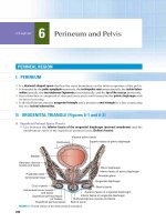
pubis). The lower limb has six major regions (Fig. 5.1):
Trunk
1. Gluteal region (L. regio glutealis) is the transitional
zone between the trunk and free lower limbs. It includes
the buttocks (L. nates, clunes) and hip region (L. regio
coxae), which overlies the hip joint and greater trochanter
of the femur.
2. Femoral region (L. regio femoris), also referred to as
the thigh, includes most of the femur, which connects
the hip and knee joints.
3. Knee region (L. regio genus) includes the distal femur,
the proximal tibia and fibula, and the patella (knee cap)
Inguinal
region
Hip joint
as well as the joints between these bony structures; the
fat-filled hollow posterior to the knee (L. poples) is called
the popliteal fossa.
4. Leg region (L. regio cruris) connects the knee and
ankle joints and includes the tibia and fibula; the calf
(L. sura) of the leg is the posterior prominence. Often,
laypersons refer incorrectly to the entire lower limb as
“the leg.”
5. Ankle or talocrural region (L. regio talocruralis)
includes the narrow distal leg and ankle (talocrural) joint.
6. Foot region (L. regio pedis), the distal part of the lower
limb, contains the tarsus, metatarsus, and phalanges (toe
bones). The superior surface is the dorsum of the foot;
the inferior, ground-contacting surface is the sole or
plantar region. The toes are the digits of the foot.
As in the hand, digit 1, the great toe (L. hallux) has only
two phalanges, and the other digits have three.
1/2 pelvic girdle
Iliac crest
Lumbar vertebra
Inguinal ligament
Hip bone
Bony
Sacrum
pelvis
Coccyx
1. Gluteal
region
(buttocks
and hip)
Pubic symphysis
Ischiopubic ramus
Greater trochanter
2. Femoral
region
(thigh)
Erector
spinae
muscles
Femur
Iliopsoas
muscle
Free lower limb
Patella
3. Knee
region
Knee joint
Rotational
axes of
pelvis,
hip joint,
and
knee joint
Center
of gravity
Tibia
4. Leg
region
5. Ankle
(talocrural)
region
Ankle
(talocrural)
joint
6. Foot
region
Fibula
Lateral and
medial malleoli
Plantar flexor
muscles
(triceps
surae)
Tarsus
Rotational
axis of
ankle joint
Metatarsus
Phalanges
(A) Anterior view
Center of
gravity
(B) Lateral view
(C) Inferior view
FIGURE 5.1. Lower limb. A. Regions and bones of lower limb. B and C. Center of gravity in a relaxed standing position.
Moore_Ch05.indd 310
1/11/14 3:24 AM
CHAPTER 5 • LOWER LIMB
BONES OF LOWER LIMB
Body weight is transferred from the vertebral column
through the sacro-iliac joints to the pelvic girdle and
from the pelvic girdle through the hip joints to the femurs
(L. femora) and then through the femurs to the knee
joints. Weight is then transferred from the knee joint to
the ankle joint by the tibia. The fibula does not articulate
with the femur and does not bear weight. At the ankle,
the weight is transferred to the talus. The talus is the
keystone of a longitudinal arch formed by the tarsal and
metatarsal bones of each foot, which distribute the weight
evenly between the heel and the forefoot when standing.
To support the erect bipedal posture better, the femurs
are oblique (directed inferomedially) within the thighs so
that when standing, the knees are adjacent and are placed
directly inferior to the trunk, returning the center of gravity to the vertical lines of the supporting legs and feet
(Figs. 5.1 and 5.2A,E). The femurs of females are slightly
more oblique than those of males, reflecting the greater
width of their pelves.
311
• Internal aspect of the body of the pubis faces almost
directly superiorly
• Acetabulum faces inferolaterally, with the acetabular
notch directed inferiorly
• Obturator foramen lies inferomedial to the acetabulum
Clinical Box
Fractures of Hip Bone
Fractures of the hip bone are “pelvic fractures.”
The term hip fracture is most commonly
applied, unfortunately, to fractures of the
femoral heads, neck, or trochanters.
Avulsion fractures of the hip bone may occur during
sports that require sudden acceleration or deceleration.
A small part of the bone with a piece of tendon or ligament
attached is “avulsed” (torn away)—for example, the anterior superior iliac spine. In older patients, pelvic fractures
often include at least two fractures of the ring of bone
formed by the pubis, pubic rami, and the acetabulum.
One cannot just break one side of a stiff ring.
Hip Bone
Each mature hip bone is formed by the fusion of three
primary bones: ilium, ischium, and pubis (Fig. 5.3A).
At puberty, these bones are still separated by a triradiate
cartilage. The cartilage disappears and the bones begin to
fuse at 15 to 17 years of age; fusion is complete between
20 and 25 years of age.
The ilium, the superior and largest part of the hip
bone, contributes to the superior part of the acetabulum
(Fig. 5.3), the cup-like cavity (socket) on the lateral aspect of
the hip bone for articulation with the head of the femur. The
ilium consists of a body, which joins the pubis and ischium
to the acetabulum, and an ala (wing), which is bordered
superiorly by the iliac crest.
The ischium forms the postero-inferior part of the
acetabulum and hip bone. The ischium consists of a body,
where it joins the ilium and superior ramus of the pubis to
form the acetabulum. The ramus of the ischium joins the
inferior ramus of the pubis to form the ischiopubic ramus
(Fig. 5.3C).
The pubis forms the anterior part of the acetabulum and
the anteromedial part of the hip bone. The right pubis has a
body that articulates with the left pubis at the pubic symphysis. It also has two rami, superior and inferior.
To place the hip bone or bony pelvis in the anatomical
position (Fig. 5.3B,C), situate it so that the
• Anterior superior iliac spine and anterosuperior aspect of
the pubis lie in the same coronal (frontal) plane
• Symphysial surface of the pubis is vertical, parallel to the
median plane
Moore_Ch05.indd 311
Femur
The femur is the longest and heaviest bone in the body. The
femur consists of a shaft (body) and superior or proximal and
inferior or distal ends (Fig. 5.2). Most of the shaft is smoothly
rounded, except for a prominent double-edged ridge on its
posterior aspect, the linea aspera, which diverges inferiorly.
The proximal end of the femur consists of a head, neck, and
greater and lesser trochanters. The head of the femur is
covered with articular cartilage, except for a medially placed
depression or pit, the fovea for the ligament of the head.
The neck of the femur is trapezoidal; the narrow end
supports the head and its broader base is continuous with
the shaft.
Where the neck joins the shaft are two large, blunt elevations—the trochanters. The conical lesser trochanter, with
its rounded tip, extends medially from the posteromedial
part of the junction of the femoral neck and shaft (Fig. 5.2A).
The greater trochanter is a large, laterally placed mass that
projects superomedially where the neck joins the shaft. The
intertrochanteric line is a roughened ridge running from
the greater to the lesser trochanter. A similar but smoother
ridge, the intertrochanteric crest, joins the trochanters
posteriorly (Fig. 5.2B).
The distal end of the femur ends in two spirally curved
femoral condyles (medial and lateral). The femoral
condyles articulate with the tibial condyles to form the
knee joint.
1/11/14 3:24 AM
312
CHAPTER 5 • LOWER LIMB
Iliac crest
Hip bone
Iliac fossa
Tubercle of
iliac crest
Iliopubic
eminence
Anterior superior
iliac spine (ASIS)
Superior ramus
of pubis
Anterior inferior
iliac spine
Greater
trochanter
Intertrochanteric
line
Lesser
trochanter
Pubic crest
Pubic tubercle
Pubic symphysis
Body of pubis
Obturator
foramen
Femur
Head of femur
Patella
Adductor
tubercle
Lateral
epicondyle
Medial
epicondyle
Lateral femoral
condyle
Medial femoral
condyle
Apex of head
Medial tibial
condyle
Intercondylar
eminence
Head
Neck
Fovea for
ligament of
head
Greater
trochanter
Head
of femur
Neck of femur
Intertrochanteric
line
Lesser
trochanter
Shaft
of femur
(B) Anterior view of proximal femur
Base
Lateral
border
Medial
border
Tuberosity
Fibula
Apex
Anterior border
Lateral surface
(C) Anterior view of patella
Medial surface
Base
Tibia
Lateral
malleolus
Calcaneus
Cuboid
Medial
malleolus
Talus
Medial
articular
surface
Lateral
articular
surface
Navicular
Cuneiforms
First metatarsal
Apex
(D) Posterior view of patella
Proximal
phalanx
Distal
phalanx
(A) Anterior view
FIGURE 5.2. Bones of lower limb. (continued)
Moore_Ch05.indd 312
1/11/14 3:24 AM
CHAPTER 5 • LOWER LIMB
Hip bone
313
Iliac crest
Posterior gluteal line
Tubercle of iliac crest
Posterior superior iliac spine
(PSIS)
Anterior gluteal line
Posterior inferior iliac spine
Inferior gluteal line
Greater sciatic notch
Ischium
Greater trochanter
Ischial spine
Head of femur
Lesser sciatic notch
Neck of femur
Ischial tuberosity
Acetabulum
Intertrochanteric crest
Lesser trochanter
Spiral line
Gluteal tuberosity
Lateral supracondylar line
Linea aspera
Medial supracondylar line
Femur
Adductor tubercle
Popliteal surface
Medial femoral condyle
Lateral femoral condyle
Intercondylar fossa
Lateral tibial condyle
Medial tibial condyle
Head
Soleal line
Vertical line
Neck
Tibia
Fibula
Medial malleolus
Lateral malleolus
Talus
Calcaneus
Navicular
Medial cuneiform
Phalanx
Cuboid
5th metatarsal
Proximal
Middle
Distal
(E) Posterior view
FIGURE 5.2. Bones of lower limb. (continued)
Moore_Ch05.indd 313
1/11/14 3:24 AM
314
CHAPTER 5 • LOWER LIMB
Iliac crest
Anterior
gluteal line
Ala
Posterior
gluteal line
Anterior superior
iliac spine (ASIS)
Body
Position of
triradiate
cartilage
Posterior
superior
iliac spine
(PSIS)
Inferior gluteal line
Anterior inferior
iliac spine
Posterior
inferior
iliac spine
Articular (lunate)
surface
Greater
sciatic notch
Acetabular fossa
Acetabulum
Acetabular notch
Ischial spine
Lesser sciatic notch
(A) Lateral aspect
Pubic crest
Body of ischium
Pubic tubercle
Parts of the hip bone
Ilium
Pubis
Ischium
£
Obturator groove
Obturator foramen
Inferior ramus of
pubis
Ischial tuberosity
*
Ramus of ischium
*Ischiopubic ramus
(C) Lateral aspect
£
Acetabulum
Iliac crest
Tuberosity of ilium
Iliac fossa
Auricular surface
of ilium
Anterior superior
iliac spine
Posterior superior
iliac spine
Anterior inferior
iliac spine
Arcuate line
(B) Medial aspect
Iliopubic
eminence
Greater
sciatic notch
Body of
ischium
Pecten pubis
Superior ramus
of pubis
Body of pubis
Posterior inferior
iliac spine
Ischial spine
Lesser sciatic notch
Obturator
foramen
Ramus of ischium*
Inferior ramus of pubis*
Ischial tuberosity
(D) Medial aspect
*Ischiopubic ramus
FIGURE 5.3. Hip bone. A and B. Parts of hip bone of a 13-year-old. C and D. Right hip bone of an adult in anatomical position. In this position, the
anterior superior iliac spine (ASIS) and the anterior aspect of the pubis lie in the same vertical plane (indicated in blue).
Moore_Ch05.indd 314
1/11/14 3:24 AM
315
CHAPTER 5 • LOWER LIMB
Axis of
femoral
head and neck
126˚
Torsion angle
of femur
12˚
Trochanteric
fossa
(A) Angle of inclination
in 3-year-old child
(B) Angle of inclination
in adult
(C) Angle of inclination
in old age
Inferior (distal)
end of femur
Transverse
axis of femoral
condyles
120˚
135˚
Long axis of
shaft of femur
Greater
trochanter
(D) Superior view demonstrating
torsion angle of femur
FIGURE 5.4. Angle of inclination and torsion angle of femur.
The proximal femur is bent, making the femur L-shaped,
so that the long axis of the head and neck project superomedially at an angle to that of the obliquely oriented shaft
(Fig. 5.4). This obtuse angle of inclination in the adult
is 115 to 140 degrees, averaging 126 degrees. The angle is
less in females because of the increased width between the
acetabula and the greater obliquity of the shaft. The angle
of inclination allows greater mobility of the femur at the hip
joint because it places the head and neck more perpendicular to the acetabulum. This is advantageous for bipedal walking; however, it imposes considerable strain on the neck of
the femur. Fractures of the neck may occur in older people
as a result of a slight stumble if the neck has been weakened
by osteoporosis.
When the femur is viewed superiorly, so that the proximal end is superimposed over the distal end (Fig. 5.4D), it
can be seen that the axis of the head and neck of the femur
and the transverse axis of the femoral condyles intersect at
the long axis of the shaft of the femur, forming the torsion
angle, or angle of declination. The mean torsion angle is
7 degrees in males and 12 degrees in females. The torsion
angle, combined with the angle of inclination, allows rotatory movements of the femoral head within the obliquely
placed acetabulum to convert into flexion and extension,
abduction and adduction, and rotational movements of
the thigh.
Patella
The patella (knee cap) is a large sesamoid bone that is
formed intratendinously after birth. This triangular bone,
located anterior to the femoral condyles, articulates with
the patellar surface of the femur (Fig. 5.2A,C). The subcutaneous anterior surface of the patella is convex; the
thick base (superior border) slopes infero-anteriorly; the
lateral and medial borders converge inferiorly to form the
pointed apex; and the articular surface (posterior surface)
Moore_Ch05.indd 315
is smooth, covered with a thick layer of articular cartilage,
and is divided into medial and lateral articular surfaces by a
vertical ridge (Fig. 5.2C,D).
Tibia
The large, weight-bearing tibia (shin bone) articulates with
the femoral condyles superiorly, the talus inferiorly, and the
fibula laterally at its proximal and distal ends (Fig. 5.2). The
distal end of the tibia is smaller than the proximal end and
has facets for articulation with the fibula and talus. The
medial malleolus is an inferiorly directed projection from
the medial side of the distal end of the tibia (Fig. 5.5A). The
large nutrient foramen of the tibia is located on the posterior aspect of the proximal third of the bone (Fig. 5.5B).
From it, the nutrient canal runs inferiorly in the tibia before
it opens into the medullary (marrow) cavity. For other bony
features, see Figure 5.5.
Fibula
The slender fibula lies posterolateral to the tibia and serves
mainly for muscle attachment (Figs. 5.2 and 5.5). At its proximal end, the fibula consists of an enlarged head superior to
a narrow neck. At its distal end, the fibula enlarges to form
the lateral malleolus, which is more prominent and more
posteriorly placed than the medial malleolus and extends
approximately 1 cm farther distally. The fibula is not directly
involved in weight bearing; however, its lateral malleolus
forms the lateral part of the socket for the trochlea of the
talus. The shafts of the tibia and fibula are connected by an
interosseous membrane throughout most of their lengths.
Tarsus, Metatarsus, and Phalanges
The bones of the foot include the tarsus, metatarsus, and
phalanges (Figs. 5.2 and 5.6).
1/11/14 3:25 AM
316
CHAPTER 5 • LOWER LIMB
Intercondylar tubercles of
intercondylar eminence
Medial tibial plateau
Lateral tibial plateau
Lateral condyle
Apex of head
Head of fibula
Neck of fibula
Anterolateral
tibial (Gerdy)
tubercle (G)
G
Medial
condyle
Anterior
intercondylar area
Tibial
tuberosity
Intercondylar
tubercles of
intercondylar
eminence
Lateral tibial
plateau
Lateral condyle
Medial tibial
plateau
Apex of head
Medial condyle
Posterior
intercondylar
area
Medial crest
Opening for
anterior tibial
vessels
Interosseous
membrane
Lateral
surface
Interosseous
border
Anterior
border
Anterior border
Medial
surface
Posterior surface
Shaft (body)
of tibia
Shaft of
tibia
Shaft of fibula
Interosseous
membrane
Soleal line
Nutrient foramen
Interosseous
border
Medial border
Medial
malleolus
(A) Anterior view (right side)
Medial
malleolus
Shaft of fibula
Posterior border
Groove for tibialis
posterior tendon
Fibular notch
of tibia occupied
by fibula
Lateral malleolus
Head of fibula
(contacting fibular
articular facet of tibia)
Fibular notch of
tibia, occupied
by fibula
Lateral
malleolus
(B) Posterior view (right side)
FIGURE 5.5. Right tibia and fibula. The shafts are connected by the interosseous membrane composed of strong obliquely oriented fibers.
TARSUS
The tarsus consists of seven bones: calcaneus, talus, cuboid,
navicular, and three cuneiforms. Only the talus articulates
with the leg bones. The calcaneus (heel bone) is the largest and strongest bone in the foot. It articulates with the
talus superiorly and the cuboid anteriorly (Fig. 5.6A). The
calcaneus transmits most of the body weight from the talus
to the ground. The sustentaculum tali (talar shelf), projecting from the superior border of the medial surface of
the calcaneus, supports the head of the talus (Fig. 5.6B).
The posterior part of the calcaneus has a large prominence,
the calcaneal tuberosity (L. tuber calcanei), which has
medial and lateral processes on its plantar aspect. More
anteriorly, there is a smaller prominence, the calcaneal
tubercle (Fig. 5.6B).
The talus (ankle bone) has a head, neck, and body
(Fig. 5.6C). The superior surface, the trochlea of the
talus, bears the weight of the body transmitted from the
tibia and articulates with the two malleoli. The talus rests
on the anterior two thirds of the calcaneus. Most of the
surface of the talus is covered with articular cartilage, and
Moore_Ch05.indd 316
thus no muscles or tendons attach to the talus. The rounded
head of talus rests partially on the sustentaculum tali of
the calcaneus and articulates anteriorly with the navicular
(Fig. 5.6B,E).
The navicular (L. little ship), a flattened, boat-shaped
bone, is located between the talar head and the cuneiforms.
The medial surface of the navicular projects inferiorly as
the tuberosity of navicular. An overly prominent tuberosity may press against the medial part of the shoe and
cause foot pain.
The cuboid is the most lateral bone in the distal row
of the tarsus. Anterior to the tuberosity of cuboid
(Fig. 5.6B), on the lateral and plantar surfaces of the bone,
is a groove for the tendon of the fibularis longus muscle
(Fig. 5.6B,C).
There are three cuneiforms: medial (first), intermediate (second), and lateral (third). Each cuneiform
(L. cuneus, wedge-shaped) articulates with the navicular
posteriorly and the base of the appropriate metatarsal
anteriorly. In addition, the lateral cuneiform articulates
with the cuboid.
1/11/14 3:25 AM
317
CHAPTER 5 • LOWER LIMB
Distal phalanx
Distal
Middle
Head
1st
metatarsal
Proximal phalanx
Phalanges
Proximal
1
2
Shaft
3
4
Base
5
M
I
L
Groove for
fibular longus
4 3
2
1
Lateral cuneiform (L)
Intermediate cuneiform (I)
Tuberosity of
5th metatarsal
Cuboid
Navicular
Tarsus
5
Talus
Tuberosity of
cuboid
Trochlea of talus
Cuboid
Medial
tubercle
Groove for tendon of
flexor hallucis longus
Lateral
tubercle
Calcaneus
Medial cuneiform (M)
Tuberosity of navicular
Head of talus
Sustentaculum tali
Calcaneal tubercle
Medial process
Calcaneal
tuberosity
(A) Dorsal view
Lateral process
*of tuberosity
(B) Plantar view
Of talus
Body
Neck Head
*
Cuboid
Navicular
Lateral cuneiform
Lateral malleolus
Cuneiforms
(lateral and intermediate)
Metatarsals (2–5)
Calcaneus
Calcaneus
Phalanges
Tuberosity of
5th metatarsal
(D) Lateral view
Cuboid
Fibular trochlea
Groove for
fibularis
longus
Base
Shaft
Tuberosity of
5th metatarsal
Head
Tubercle
(C) Lateral view
Medial malleolus
Medial cuneiform
Of talus
Neck
Body
Navicular
Head of 1st metatarsal
Medial cuneiform
1st metatarsal
Proximal
phalanx
Tuberosity of 1st metatarsal
Distal
phalanx
(E) Medial view
Cuboid
Tuberosity
of navicular
Sustentaculum tali
Calcaneus
(F) Medial view
FIGURE 5.6. Bones of foot. Blue, articular cartilage.
Moore_Ch05.indd 317
1/11/14 3:25 AM
318
CHAPTER 5 • LOWER LIMB
METATARSUS
The metatarsus consists of five long bones (metatarsals), which connect the tarsus and phalanges. They are
numbered from the medial side of the foot (Fig. 5.6B,C).
The 1st metatarsal is shorter and stouter than the others. The 2nd metatarsal is the longest. Each metatarsal
has a base (proximally), a shaft, and a head (distally).
The bases of the metatarsals articulate with the cuneiform and cuboid bones. The bases of the 1st and 5th
metatarsals have large tuberosities; the tuberosity of
the 5th metatarsal projects over the lateral margin of
the cuboid (Fig. 5.6C). The heads articulate with the
proximal phalanges.
PHALANGES
There are 14 phalanges. The 1st digit (great toe) has
two phalanges (proximal and distal); the other four digits
each have three phalanges: proximal, middle, and distal
(Fig. 5.6A,B). Each phalanx has a base (proximally), a shaft,
and a head (distally).
Clinical
C
linical B
Box
ox
Femoral Fractures
Coxa Vara and Coxa Valga
The neck of the femur is most frequently fractured,
especially in females secondary to osteoporosis.
Fractures of the proximal femur can occur at several locations—for example, transcervical and intertrochanteric
(Fig. B5.1A,B). The femoral shaft is large and strong; however, a violent direct injury, such as may be sustained in an
automobile accident, may fracture it, causing, for example, a
spiral fracture (Fig. B5.1C). Fractures of the distal femur may
be complicated by separation of the condyles, resulting in
misalignment of the knee joint.
The angle of inclination varies with age, sex, and
development of the femur (e.g., consequent to a
congenital defect in ossification of the femoral
neck). It also may change with any pathological process
that weakens the neck of the femur (e.g., rickets). When the
angle of inclination is decreased, the condition is coxa vara
(Fig. B5.2A); when it is increased, the condition is coxa valga
(Fig. B5.2B). Coxa vara causes a mild passive abduction of
the hip.
(A) Transcervical fracture
of femoral neck
Posterior views
(B) Intertrochanteric
fracture
(C) Spiral fracture
Anterior views
FIGURE B5.1. Femoral fractures.
Moore_Ch05.indd 318
(A) Coxa vara
(decreased angle
of inclination)
(B) Coxa valga
(increased angle
of inclination)
FIGURE B5.2. Coxa vara and coxa valga.
1/11/14 3:25 AM
CHAPTER 5 • LOWER LIMB
319
Tibial and Fibular Fractures
The tibial shaft is narrowest at the junction of its
inferior and middle thirds, which is the most common site of fracture. Because its anterior surface
is subcutaneous, the tibial shaft is the most frequent site of
an open fracture (compound fracture)—one in which the skin
is perforated and blood vessels are torn (Fig. B5.3A)—or a
diagonal fracture (Fig. B5.3C). Fracture of the tibia through
the nutrient canal predisposes to nonunion of the bone fragments resulting from damage to the nutrient artery. Fibular
fractures commonly occur just proximal to the lateral malleolus and often are associated with fracture–dislocations of
the ankle joint (Fig. B5.3D). When a person slips, forcing the
foot into an excessively inverted position, the ankle ligaments
tear, forcibly tilting the talus against the lateral malleolus and
shearing it off.
Bone Grafts
The fibula is a common source of bone for grafting.
Even after a segment of the fibular shaft has been
removed, walking, running, and jumping can be
normal. Free vascularized fibulas have been used to restore
skeletal integrity to limbs in which congenital bone defects
exist and to replace segments of bone after trauma or excision
of a malignant tumor. The periosteum and nutrient artery are
generally removed with the piece of bone so that the graft will
remain alive and grow when transplanted to another site. The
transplanted piece of fibula, secured in its new site, eventually
restores the blood supply of the bone to which it has been
attached.
Fractures Involving Epiphysial Plates
The primary ossification center for the
superior end of the tibia appears shortly
after birth and joins the shaft of the tibia
during adolescence (usually 16–18 years of age). Tibial fractures in children are more serious if they involve the epiphysial plates because continued normal growth of the bone may
be jeopardized. All such fractures of the immature skeleton
are routinely characterized by the Salter-Harris classification that describes the pattern of involvement. The tibial
tuberosity usually forms by inferior bone growth from the
superior epiphysial center at approximately 10 years of age,
but a separate center for the tibial tuberosity may appear at
approximately 12 years of age. Disruption of the epiphysial plate at the tibial tuberosity may cause inflammation
of the tuberosity and chronic recurring pain during adolescence (Osgood-Schlatter disease), especially in young athletes
(Fig. B5.4).
(Continued on next page)
F
F
T
T
A
A
(A) Compound (open)
fracture with
external bleeding
(B) Transverse “boot top” fracture
with shortening due to overriding of fracture fragments
Fibula (F)
Tibia (T)
F
Talus (A)
T
(C) Diagonal fracture
with shortening
Inversion
A – C Anterior views
(D) Fibular fracture with
excessive inversion of foot
Posterior view
FIGURE B5.3. Tibial and fibular fractures.
Lateral radiograph
Tibial tuberosity (ossification center, large arrow) elongated and
fragmented with overlying soft tissue swelling (small arrows)
FIGURE B5.4. Osgood-Schlatter disease.
Moore_Ch05.indd 319
1/11/14 3:25 AM
320
CHAPTER 5 • LOWER LIMB
Fractures of Foot Bones
Calcaneal fractures occur in people who fall on their
heels (e.g., from a ladder). Usually, the bone breaks
into several fragments (comminuted fracture) that disrupt the subtalar joint, where the talus articulates with the calcaneus (Fig. B5.5A). Fractures of the talar neck may occur during
severe dorsiflexion of the ankle, for example, when a person is
pressing extremely hard on the brake pedal of a car during a
head-on collision (Fig. B5.5B). Metatarsal and phalangeal fractures are a common injury in endurance athletes and may also
occur when a heavy object falls on the foot. Metatarsal fractures
are also common in dancers, especially female ballet dancers
using the demi-pointe technique. The “dancer’s fracture” usually
occurs when the dancer loses balance, putting the full body
weight on the metatarsal and fracturing the bone (Fig. B5.5C).
Talus
Dorsum of foot, fractures
of metatarsals
Lateral view, comminuted
fractures of calcaneus
(A)
Calcaneus
4th metatarsal
Fracture of talar neck
5th metatarsal
Tibia
Tuberosity of
5th metatarsal
Cuboid
Neck of talus
Talus
(C)
Calcaneus
(B)
FIGURE B5.5. Fractures of foot.
Surface Anatomy
Lower Limb Bones
Pelvic Girdle and Femur
When your hands are on your hips, they rest on the iliac crests, the
curved superior borders of the alae (wings) of the ilium (Fig. SA5.1).
The anterior third of the crest is easily palpated because it is
subcutaneous. The highest point of the crest is at the level of the
intervertebral (IV) disc between the L4 and the L5 vertebrae. The
iliac crest ends anteriorly at the pointed anterior superior iliac
spine (ASIS), which is easy to palpate, especially in thin persons,
because it is subcutaneous and often visible (Fig. SA5.1A,B). The
ASIS is used as the proximal point for measurement of leg length to
the medial malleolus of the tibia. The iliac crest ends posteriorly at
the posterior superior iliac spine (PSIS), which may be difficult
to palpate (Fig. SA5.1C). Its position is easy to locate because it lies
at the bottom of a skin dimple, approximately 4 cm lateral to the
midline, demarcating posteriorly the location of the sacro-iliac joint.
The dimple exists because the skin and fascia attach to the PSIS.
Moore_Ch05.indd 320
Highest level of iliac crest
Supracristal plane
Anterior superior iliac spine
Buttock
Site of tip of greater
trochanter of femur
Gluteal fold
Thigh
(A) Lateral view
FIGURE SA5.1.
1/11/14 3:25 AM
CHAPTER 5 • LOWER LIMB
321
Iliac crest
Iliac crest
Iliac tuberosity
Posterior superior
iliac spine
Anterior superior
iliac spine
Inguinal ligament
Head of femur
Greater trochanter
Iliac tuberosity
Pubic crest
Pubic symphysis
Greater trochanter
Pubic tubercle
Ischial tuberosity
Gluteal fold
Gluteal sulcus
Patella
Lateral epicondyle
of femur
Lateral condyle
of tibia
Anterolateral tibial
(Gerdy) tubercle
Head of fibula
Neck of fibula
Adductor tubercle
Lateral epicondyle
of femur
Medial epicondyle
of femur
Lateral condyle
of tibia
Medial condyle
of tibia
Head of fibula
Neck of fibula
Tibial tuberosity
Anterior border and medial
surface of tibia
Lateral malleolus
Tuberosity of
5th metatarsal
(B) Anterior view
Medial malleolus
Lateral malleolus
Tuberosity of navicular
Green = palpable features
of lower limb bones
Calcaneal
tuberosity
(C) Posterior view
FIGURE SA5.1. (continued)
The ischial tuberosity is easily palpated in the inferior part of
the buttock when the hip joint is flexed. It bears body weight when
sitting. The thick gluteus maximus and fat obscure the tuberosity
when the hip joint is extended. The gluteal fold, a prominent
skin fold containing fat, coincides with the inferior border of the
gluteus maximus muscle.
The greater trochanter of the femur is easily palpable on
the lateral side of the hip approximately 10 cm inferior to the
iliac crest (Fig. SA5.1B,C). Because it lies close to the skin, the
greater trochanter causes discomfort when you lie on your side on
a hard surface. In the anatomical position, a line joining the tips
of the greater trochanters normally passes through the centers of
the femoral heads and pubic tubercles. The shaft of the femur
usually is not palpable because it is covered with large muscles.
Moore_Ch05.indd 321
The medial and lateral condyles of the femur are subcutaneous and easily palpated when the knee is flexed or extended. The
patellar surface of the femur is where the patella slides during
flexion and extension of the knee joint. The lateral and medial
margins of the patella can be palpated when the knee joint is
flexed. The adductor tubercle, a small prominence of bone,
may be felt at the superior part of the medial femoral condyle.
Tibia and Fibula
The tibial tuberosity, an oval elevation on the anterior surface
of the tibia, is palpable approximately 5 cm distal (inferior) to the
apex of the patella to which it is connected by the palpable patellar ligament (Fig. SA5.1B). The subcutaneous anterior border
1/11/14 3:25 AM
322
CHAPTER 5 • LOWER LIMB
and medial surface of the tibia is also easy to palpate. The skin
covering it is freely movable. The prominence at the ankle, the
medial malleolus, is subcutaneous, and its inferior end is blunt. The
medial and lateral tibial condyles can be palpated anteriorly at
the sides of the patellar ligament, especially when the knee joint is
flexed. The head of the fibula can be palpated at the level of the
superior part of the tibial tuberosity because its knob-like head is
subcutaneous at the posterolateral aspect of the knee. The neck
of fibula can be palpated just distal to the fibular head. Only the
distal quarter of the shaft of the fibula is palpable. Feel your lateral
malleolus, noting that it is subcutaneous and that its inferior end is
sharp. Note that the tip of the lateral malleolus extends farther distally and more posteriorly than does the tip of the medial malleolus.
to slide when the 1st digit is moved passively. The tuberosity of the
5th metatarsal forms a prominent landmark on the lateral aspect
of the foot and can be palpated easily at the midpoint of the lateral
border of the foot. The shafts of the metatarsals and phalanges
can be felt on the dorsum of the foot between the extensor tendons.
Lateral malleolus
Medial malleolus
Head of talus
(indicated by thumb)
Tuberosity of
navicular
Bones of Foot
The head of talus is palpable anteromedial to the proximal part
of the lateral malleolus when the foot is inverted and anterior
to the medial malleolus when the foot is everted. Eversion of
the foot makes the head of talus more prominent as it moves
away from the navicular. The head of talus occupies the space
between the sustentaculum tali and the tuberosity of navicular.
When the foot is plantarflexed, the superior surface of the body
of the talus can be palpated on the anterior aspect of the ankle,
anterior to the inferior end of the tibia (Fig. SA5.1D).
The weight-bearing medial process of the calcaneal
tuberosity on the plantar surface of the foot is broad and large but
may not be palpable because of the thick overlying skin and subcutaneous tissue (Fig. SA5.1E). The sustentaculum tali is the only
part of the medial aspect of the calcaneus that may be palpated as
a small prominence just distal to the tip of the medial malleolus.
The tuberosity of the navicular is easily seen and palpated
on the medial aspect of the foot, infero-anterior to the tip of the
medial malleolus. Usually, palpation of bony prominences on the
plantar surface of the foot is difficult because of the thick skin,
fascia, and pads of fat. The cuboid and cuneiforms are difficult to
identify individually by palpation. The cuboid can be felt on the
lateral aspect of the foot, posterior to the base of the 5th metatarsal. The medial cuneiform can be indistinctly palpated between
the tuberosity of the navicular and the base of the 1st metatarsal.
The head of the 1st metatarsal forms a prominence on the
medial aspect of the foot. The medial and lateral sesamoid
bones, located inferior to the head of this metatarsal, can be felt
FASCIA, VESSELS, AND CUTANEOUS
NERVES OF LOWER LIMB
Subcutaneous Tissue and Fascia
The subcutaneous tissue (superficial fascia) is deep to
the skin and consists of loose connective tissue that contains a variable amount of fat, cutaneous nerves, superficial
Moore_Ch05.indd 322
(D) Dorsum of foot
Shaft of phalanx
Lateral sesamoid
Medial sesamoid
Forefoot
(metatarsals
and phalanges)
1/2
Metatarsal head
Metatarsal shaft
Tarsometatarsal line
Cuboid,
1/3
navicular
and
cuneiforms
Hindfoot
(tarsal
bones) Calcaneous
2/3
and talus
1/2
Cuneiforms
Tuberosity of 5th metatarsal
Navicular
Cuboid
Medial process
of calcaneal
tuberosity
(E) Plantar aspect of foot
FIGURE SA5.1. (continued)
veins, lymphatic vessels, and lymph nodes (Fig. 5.7). The
subcutaneous tissue of the hip and thigh is continuous with
that of the inferior part of the anterolateral abdominal wall
and buttocks. At the knee, the subcutaneous tissue loses
its fat anteriorly and laterally, and blends with the deep
fascia, but fat is present posteriorly in the popliteal fossa
and again distal to the knee in the subcutaneous tissue of
the leg.
1/11/14 3:25 AM
CHAPTER 5 • LOWER LIMB
Anterior superior
iliac spine
Inguinal ligament
Falciform
margin of
saphenous
opening
Great saphenous
vein
Subcutaneous
tissue
Fascia lata
323
Iliac crest
Iliac
tubercle
Saphenous
opening
Tensor
fasciae latae
Pubic
tubercle
Cribriform
fascia in
saphenous
opening
Level of
section in
Figure 5.8A
Iliotibial tract
Gluteus
maximus
Ischial
tuberosity
(deep to muscle
when thigh is
extended)
Iliotibial tract
Fascia lata
Bursae
Deep fascia
of leg
(crural fascia)
Patella
Level of
section in
Figure 5.8B
Tibia
Anterolateral
tibial (Gerdy)
tubercle
(B) Lateral view
Extensor
retinacula
(A) Anterior view
FIGURE 5.7. Fascia of lower limb. A. Deep fascia. B. Iliotibial tract.
The deep fascia is especially strong, investing the limb like
an elastic stocking (Fig. 5.7A). This fascia limits outward extension of contracting muscles, making muscular contraction more
efficient in compressing the veins to push blood toward the
heart. The deep fascia of the thigh is called fascia lata (L. lata,
broad). The fascia lata attaches to and is continuous with
• The inguinal ligament, pubic arch, body of pubis, and pubic
tubercle superiorly. The membranous layer of subcutaneous
tissue (Scarpa fascia) of the inferior abdominal wall also attaches to the fascia lata just inferior to the inguinal ligament.
• The iliac crest laterally and posteriorly
• The sacrum, coccyx, sacrotuberous ligament, and ischial
tuberosity posteriorly
• The superficial aspects of the bones around the knee and
the deep fascia of the leg distally
Moore_Ch05.indd 323
The fascia lata is substantial because it encloses the large
thigh muscles, especially laterally where it is thickened to
form the iliotibial tract (Fig. 5.7B). This broad band of
fibers is also the aponeurosis of the tensor fasciae latae and
gluteus maximus muscles. The iliotibial tract extends from
the iliac tubercle to the anterolateral tibial tubercle (Gerdy
tubercle) on the lateral condyle of the tibia (Fig. SA5.1).
The thigh muscles are separated into three fascial
compartments: anterior, medial, and posterior. The walls of
these compartments are formed by the fascia lata and three
fascial intermuscular septa that arise from the deep aspect of
the fascia lata and attach to the linea aspera on the posterior
aspect of the femur (Figs. 5.2A,E and 5.8A). The lateral
intermuscular septum is strong; the other two septa are
relatively weak. The iliotibial tract is continuous with the
lateral intermuscular septum.
1/11/14 3:25 AM
324
CHAPTER 5 • LOWER LIMB
Subcutaneous
tissue
Fascia lata
Anterior
Femur
Lateral
intermuscular
septum
Venous Drainage of Lower Limb
Iliotibial tract
Medial
Posterior
(A) Inferior view of transverse section of thigh
Skin
Subcutaneous
tissue
Deep (crural)
fascia
Posterior
intermuscular
septum
Fibula
Tibia
Interosseous
membrane
Anterior
Lateral
Anterior
intermuscular
septum
and the intermuscular septa divide the leg into three compartments (Fig. 5.8B): anterior (dorsiflexor), lateral (fibular),
and posterior (plantarflexor). The transverse intermuscular septum divides the plantarflexor muscles in the posterior compartment into superficial and deep parts.
or
teri
Pos eep
d
or
teri l
Pos rficia
e
sup
The lower limb has superficial and deep veins; the superficial
veins are in the subcutaneous tissue, and the deep veins are
deep to the deep fascia and accompany the major arteries.
Superficial and deep veins have valves, but they are more
numerous in deep veins.
The two major superficial veins are the great and small
saphenous veins (Fig. 5.9). The great saphenous vein
is formed by the union of the dorsal digital vein of the
great toe and the dorsal venous arch of the foot. The great
saphenous vein (Fig. 5.9A,B)
• Ascends anterior to the medial malleolus
• Passes posterior to the medial condyle of the femur (about a
hand’s breadth posterior to the medial border of the patella)
Transverse
intermuscular
septum
Patella
Deep (crural)
fascia
(B) Inferior view of transverse section of leg
Great saphenous
vein
FIGURE 5.8. Fascial compartments. A. Thigh. B. Leg. See Figure 5.7 for
level of sections.
The saphenous opening is a gap or hiatus in the fascia
lata inferior to the medial part of the inguinal ligament (Fig.
5.7A). Its medial margin is smooth, but its superior, lateral,
and inferior margins form a sharp edge, the falciform margin. The sieve-like cribriform fascia (L. cribrum, sieve) is a
localized membranous layer of subcutaneous tissue over the
saphenous opening, enclosing it. The great saphenous vein
and some lymphatics pass through the saphenous opening
and cribriform fascia to enter the femoral vein and the deep
inguinal lymph nodes, respectively.
The deep fascia of the leg or crural fascia (L. crus,
leg) is continuous with the fascia lata and attaches to the
anterior and medial borders of the tibia, where it is continuous with its periosteum (Fig. 5.7A). The crural fascia is thick
in the proximal part of the anterior aspect of the leg, where
it forms part of the proximal attachments of the underlying
muscles. Although thin in the distal part of the leg, the crural
fascia is thickened where it forms the extensor retinacula.
Anterior and posterior intermuscular septa pass from
the deep surface of the crural fascia and attach to the corresponding margins of the fibula. The interosseous membrane
Moore_Ch05.indd 324
Great saphenous
vein
Medial malleolus
Dorsal venous arch
(A) Anteromedial view
FIGURE 5.9. Superficial venous and lymphatic drainage of lower limb.
A. Normal superficial veins distended after exercise. (continued)
1/11/14 3:25 AM
CHAPTER 5 • LOWER LIMB
Superficial inguinal
lymph nodes (1)
(superior group)
Femoral vein
(5)
Deep inguinal
lymph nodes (2)
Saphenous
opening (6)
325
1
Superficial inguinal
lymph nodes (3)
(inferior group)
5
4
2
Great saphenous
vein (4)
Popliteal
fossa
3
6
Patella
Popliteal lymph
nodes
Popliteal vein
Small saphenous
vein
Great saphenous
vein (superficial
vein)
Perforating vein
Popliteal
vein
Perforating
veins
Posterior
tibial vein
Great
saphenous
vein
Fibular
vein
Dorsal digital vein
of great toe
Dorsal
venous
arch
Medial
malleolus
(B) Anteromedial view
(C) Medial view
Medial malleolus
Deep veins
Lateral malleolus
Small
saphenous vein
(D) Posterolateral view
FIGURE 5.9. Superficial venous and lymphatic drainage of lower limb. (continued) B. Great saphenous vein and superficial lymphatic drainage with inset
of saphenous opening. Arrows, superficial lymphatic drainage to the inguinal nodes. C. Perforating veins. D. Small saphenous vein and superficial lymphatic drainage (arrow) to the popliteal lymph nodes.
• Anastomoses freely with the small saphenous vein
• Traverses the saphenous opening in the fascia lata
(Fig. 5.7A)
• Empties into the femoral vein
The small saphenous vein arises on the lateral side of
the foot from the union of the dorsal digital vein of the 5th
digit with the dorsal venous arch (Fig. 5.9A,B). The small
saphenous vein (Fig. 5.9D)
• Ascends posterior to the lateral malleolus as a continuation of the lateral marginal vein
• Passes along the lateral border of the calcaneal tendon
• Inclines to the midline of the fibula and penetrates the
deep fascia
• Ascends between the heads of the gastrocnemius muscle
• Empties into the popliteal vein in the popliteal fossa
Abundant perforating veins penetrate the deep
fascia as they pass between the superficial and deep
veins (Figs. 5.9C and 5.10A). They contain valves that
allow blood to flow only from the superficial to the deep
Moore_Ch05.indd 325
veins. The perforating veins penetrate the deep fascia at
oblique angles so that when muscles contract and pressure increases inside the deep fascia, the perforating veins
are compressed, preventing blood from flowing from the
deep to the superficial veins. This pattern of venous blood
flow, from superficial to deep, is important for proper
venous return from the limb because it enables muscular
contractions to propel blood toward the heart against the
pull of gravity (musculovenous pump; see Fig. I.16A in the
Introduction chapter).
The deep veins in the lower limb accompany the major
arteries and their branches. Instead of occurring as a single vein in the limbs, the deep veins are usually paired,
frequently interconnecting accompanying veins (L. venae
comitantes) that flank the artery. They are contained within
a vascular sheath with the artery, whose pulsations also
help compress and move blood in the veins (Fig. 5.10).
The deep veins from the leg flow into the popliteal vein
posterior to the knee, which becomes the femoral vein in
the thigh. The profunda femoris vein joins the terminal
1/11/14 3:25 AM
326
CHAPTER 5 • LOWER LIMB
Superior
gluteal vein
External iliac vein
Internal iliac vein
Deep circumflex
iliac vein
Medial circumflex
femoral vein
Lateral circumflex
femoral vein
Great saphenous
vein
Obturator vein
Internal
pudendal
vein
Profunda femoris vein
(deep vein of thigh)
Inferior gluteal
vein
Femoral vein
1st perforating vein
2nd perforating vein
3rd perforating vein
Femoral vein
Femoral
vein
Medial superior
genicular vein
Genicular veins
Popliteal
vein
Medial inferior
genicular vein
Anterior tibial vein
Profunda
femoris
vein
Lateral
superior
genicular
vein
Lateral
inferior
genicular
vein
Fibular vein
Posterior
tibial vein
Dorsal venous arch
Dorsal vein:
-of great toe
-of little toe
Plantar
arch
(A) Anterior view
(B) Posterior view
Plantar
digital
veins
FIGURE 5.10. Deep venous drainage of lower limb.
portion of the femoral vein. The femoral vein passes deep
to the inguinal ligament to become the external iliac vein
in the pelvis (Fig. 5.10A).
Lymphatic Drainage of Lower Limb
The lower limb has superficial and deep lymphatic
vessels. The superficial lymphatic vessels converge on
and accompany the saphenous veins and their tributaries. The lymphatic vessels accompanying the great saphenous vein end in the superficial inguinal lymph nodes
(Fig. 5.9B). Most lymph from these nodes passes to the
external iliac lymph nodes, located along the external
iliac vein, but some lymph may also pass to the deep inguinal lymph nodes, located on the medial aspect of the
femoral vein. The lymphatic vessels accompanying the
small saphenous vein enter the popliteal lymph nodes,
which surround the popliteal vein in the fat of the popliteal fossa (Fig. 5.9D). The deep lymphatic vessels of the
leg accompany deep veins and enter the popliteal lymph
nodes. Most lymph from these nodes ascends through
deep lymphatic vessels to the deep inguinal lymph nodes.
Lymph from the deep nodes passes to the external iliac
lymph nodes.
Moore_Ch05.indd 326
Cutaneous Innervation of Lower Limb
Cutaneous nerves in the subcutaneous tissue supply the skin
of the lower limb (Fig. 5.11A,B). These nerves, except for
some in the proximal part of the limb, are branches of the
lumbar and sacral plexuses (see Chapters 3 and 4). The area
of skin supplied by cutaneous branches from a single spinal
nerve is a dermatome (Fig. 5.11C–F). Dermatomes L1–L5
extend as a series of bands from the posterior midline of the
trunk into the limbs, passing laterally and inferiorly around
the limb to its anterior and medial aspects, reflecting the
medial rotation that occurs developmentally. Dermatomes
S1 and S2 pass inferiorly down the posterior aspect of the
limb, separating near the ankle to pass to the lateral and
medial margins of the foot (Fig. 5.11F).
Although simplified into distinct zones in dermatome
maps, adjacent dermatomes overlap except at the axial
line, the line of junction of dermatomes supplied from
discontinuous spinal levels.
Two different dermatome maps are commonly used. The
pattern according to Foerster (1933) is preferred by many
because of its correlation with clinical findings (Fig. 5.11C,D)
and that of Keegan and Garrett (1948) by others for its
correlation with limb development (Fig. 5.11E,F).
1/11/14 3:25 AM
CHAPTER 5 • LOWER LIMB
Femoral branch
Genital branch
Lateral cutaneous
branch of subcostal
nerve (T12)
Superior clunial L1
nerves L2
(posterior rami) L3
Genitofemoral
nerve
Cutaneous branch
of obturator nerve
Lateral cutaneous
nerve of thigh
Inferior clunial nerves
Cutaneous branches of
obturator nerve
Anterior cutaneous
branches of
femoral nerve
Lateral cutaneous
nerve of thigh
Anterior cutaneous
branches of
femoral nerve
Posterior cutaneous
nerve of thigh
Lateral sural cutaneous
nerve (from common
fibular nerve)
Saphenous nerve
(from femoral nerve)
Infrapatellar branch
of saphenous nerve
Saphenous nerve
(from femoral nerve)
Lateral sural cutaneous
nerve (from common
fibular nerve)
Superficial fibular nerve
becoming dorsal
digital nerves
Communicating branch of
lateral sural cutaneous nerve
Medial calcaneal
branches of
tibial nerve
Deep fibular nerve
Sural nerve
Lateral plantar nerve
Medial plantar nerve
(B) Posterior view
(A) Anterior view
T10
T11
T12
S3
Medial sural cutaneous nerve
(from tibial nerve)
Branch of
saphenous nerve
Lateral dorsal cutaneous
nerve of foot (termination
of sural nerve)
Lateral cutaneous
branch of
iliohypogastric nerve
Medial clunial S1
nerves S2
(posterior rami) S3
Ilio-inguinal nerve
Lateral cutaneous
nerve of thigh
327
T10
T11
T12
S2
L1
Co
S5
S4
S3
L2
S4
S2
L1
L5
S3
L2
S2
S1
L3
L4
L5
S1
S2
S3
S4
S5
Co
L1
L2
L3
L3
S2
L2
S2
L3
Axial
line
S1
S2
L5
L3
L4
L4
L4
L5
Axial
line
L5
L4
S2
S1
S1
S2
S1
L5
(C) Anterior view
(D) Posterior view
S1
L4
L5
(E) Anterior view
(F) Posterior view
FIGURE 5.11. Cutaneous innervation of lower limb. A and B. Peripheral cutaneous nerve distribution. C–F. Dermatomes. Two different dermatome
maps are frequently used: C and D, according to Foerster (1933); E and F, according to Keegan and Garrett (1948).
Moore_Ch05.indd 327
1/11/14 3:25 AM
328
CHAPTER 5 • LOWER LIMB
Clinical Box
Abnormalities of Sensory Function
In the limbs, most cutaneous nerves are multisegmental conveying fibers from more than one segment of the spinal cord. Using a sharp object (a pin
or pinwheel), areas lacking sensation are outlined to determine whether the area of numbness matches the dermatome
pattern (Fig. 5.11C–F), indicating a segmental (spinal nerve)
lesion, or the multisegmental pattern of peripheral cutaneous nerve distribution (Fig. 5.11A,B). Because neighboring
dermatomes overlap, the area of numbness resulting from a
lesion of a single spinal nerve will be much smaller than indicated by the dermatome map.
Compartment Syndromes in Leg and
Fasciotomy
Increased pressure in a confined anatomical space adversely affects the circulation
and threatens the function and viability of
tissue within or distal to the space (compartment syndrome).
The fascial compartments of the lower limbs are generally
closed spaces, ending proximally and distally at the joints.
Trauma to muscles and/or vessels in the compartments from
burns, sustained intense use of muscles, or blunt trauma may
produce hemorrhage, edema, and inflammation of the muscles in the compartment. Because the septa and deep fascia
of the leg forming the boundaries of the leg compartments
are strong, the increased volume consequent to any of these
processes increases intracompartmental pressure.
Increased pressure in a confined space adversely affects the
circulation and threatens the function and viability of tissue
within or distally (compartment syndrome). The pressure may
reach levels high enough to compress structures significantly in
the compartment(s) concerned. The small vessels of muscles and
nerves (vasa nervorum) are particularly vulnerable to compression. Structures distal to the compressed area may become ischemic and permanently injured (e.g., muscles with compromised
blood supply and/or innervation will not function).
Loss of distal leg pulses is an obvious sign of arterial compression, as is lowering of the temperature of tissues distal to
the compression. A fasciotomy (incision of overlying fascia
or a septum) may be performed to relieve the pressure in the
compartment(s) concerned.
Varicose Veins, Thrombosis, and
Thrombophlebitis
Frequently, the great saphenous vein and its tributaries become varicose (dilated and/or tortuous so
that the cusps of their valves do not close). Varicose
veins are common in the posteromedial parts of the lower limb
and may cause discomfort (Fig. B5.6A). In a healthy vein, the
valves allow blood to flow toward the heart while preventing blood flow away from the heart (Fig B5.6B,C). Valves
also bear the weight of short columns of blood between two
valves. Valves in varicose veins, incompetent due to dilation
or rotation, no longer function properly. The resulting reverse
flow and the weight of long, unbroken columns of blood,
produces varicose veins (Fig. B5.6D).
Deep venous thrombosis (DVT) of one or more of the
deep veins of the lower limb is characterized by swelling,
warmth, and erythema (inflammation) and infection. Venous
stasis (stagnation) is an important cause of thrombus formation. Venous stasis can be caused by
• Incompetent, loose fascia that fails to resist muscle expansion,
diminishing the effectiveness of the musculovenous pump
• External pressure on the veins from bedding during prolonged institutional stays or from a tight cast, bandages,
or bands of stockings
• Muscular inactivity (e.g., during an overseas flight)
DVT with inflammation around the involved veins
(thrombophlebitis) may develop. A large thrombus that
breaks free from a lower limb vein may travel to a lung,
forming a pulmonary thromboembolism (obstruction of a
pulmonary artery). A large embolus may obstruct a main
pulmonary artery and may cause death.
(B)
(C)
(D)
Saphenous Nerve Injury
The saphenous nerve accompanies the
great saphenous vein in the leg. Should
this nerve be injured or caught by a ligature during closure of a surgical wound, the patient may complain of pain, tingling, or numbness (paresthesia) along the
medial border of the foot.
Moore_Ch05.indd 328
(A) Varicose veins
FIGURE B5.6. Varicose veins.
1/11/14 3:25 AM