Ebook BRS Gross anatomy (7th edition) Part 2
Bạn đang xem bản rút gọn của tài liệu. Xem và tải ngay bản đầy đủ của tài liệu tại đây (43.63 MB, 247 trang )
6
chapter
Perineum and Pelvis
PERINEAL REGION
I. PERINEUM
■
■
■
■
Is a diamond-shaped space that has the same boundaries as the inferior aperture of the pelvis.
Is bounded by the pubic symphysis anteriorly, the ischiopubic rami anterolaterally, the ischial tuberosities laterally, the sacrotuberous ligaments posterolaterally, and the tip of the coccyx posteriorly.
Has a floor that is composed of skin and fascia and a roof formed by the pelvic diaphragm with
its fascial covering.
Is divided into an anterior urogenital triangle and a posterior anal triangle by a line connecting
the two ischial tuberosities.
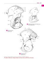
II. UROGENITAL TRIANGLE (Figures 6-1 and 6-2)
A. Superficial Perineal Space (Pouch)
■ Lies between the inferior fascia of the urogenital diaphragm (perineal membrane) and the
membranous layer of the superficial perineal fascia (Colles’s fascia).
Visceral pelvic fascia
Superior fascia of pelvic diaphragm
Peritoneum
Bladder
Obturator internus
muscle and fascia
Pelvic diaphragm
Inferior fascia of pelvic diaphragm
Pudendal canal
Ischiorectal fossa
Prostate gland
Urogenital diaphragm
Crus of penis
Ischiocavernosus muscle
and fascia
Superficial perineal fascia
(of Colles)
Superficial perineal space
Buck's fascia
Superior fascia of urogenital diaphragm
Inferior fascia of urogenital diaphragm
Bulbospongiosus muscle and fascia
Bulb of penis
FIGURE 6-1. Frontal section of the male perineum and pelvis.
244
LWBK832-C06_p244-295.indd 244
2/23/11 10:46:55 AM
Chapter 6
Perineum and Pelvis
245
Vagina
Peritoneum
Visceral pelvic fascia
Obturator internus
muscle
Superior fascia of pelvic diaphragm
Pelvic diaphragm
Pudendal canal
Ischiorectal fossa
Inferior fascia of pelvic diaphragm
Superior fascia of urogenital diaphragm
Urogenital diaphragm
Crus of clitoris
Superficial perineal fascia
Ischiocavernosus muscle
and fascia
Superficial perineal space
Vestibular bulb
Inferior fascia of urogenital diaphragm
Greater vestibular gland and orifice of its duct
Labium majus
Bulbospongiosus muscle and fascia
FIGURE 6-2. Frontal section of the female perineum and pelvis.
■
Contains the superficial transverse perineal muscle, the ischiocavernosus muscles and
crus of the penis or clitoris, the bulbospongiosus muscles and the bulb of the penis or the
vestibular bulbs, the central tendon of the perineum, the greater vestibular glands (in the
female), branches of the internal pudendal vessels, and the perineal nerve and its
branches.
1. Colles’s Fascia
■ Is the deep membranous layer of the superficial perineal fascia and forms the inferior
■
boundary of the superficial perineal pouch.
Is continuous with the dartos tunic of the scrotum, with the superficial fascia of the
penis, and with the Scarpa’s fascia of the anterior abdominal wall.
CLINICAL
CORRELATES
Extravasated urine may result from rupture of the bulbous portion of the spongy
urethra below the urogenital diaphragm; the urine may pass into the superficial
perineal space and spread inferiorly into the scrotum, anteriorly around the penis, and superiorly
into the lower part of the abdominal wall. The urine cannot spread laterally into the thigh because
the inferior fascia of the urogenital diaphragm (the perineal membrane) and the superficial fascia of
the perineum are firmly attached to the ischiopubic rami and are connected with the deep fascia
of the thigh (fascia lata). It cannot spread posteriorly into the anal region (ischiorectal fossa)
because the perineal membrane and Colles’s fascia are continuous with each other around the
superficial transverse perineal muscles. If the membranous part of the urethra is ruptured, urine
escapes into the deep perineal space and can extravasate upward around the prostate and bladder
or downward into the superficial perineal space.
2. Perineal Membrane
■ Is the inferior fascia of the urogenital diaphragm that forms the inferior boundary of
■
■
LWBK832-C06_p244-295.indd 245
the deep perineal pouch and the superior boundary of the superficial pouch.
Lies between the urogenital diaphragm and the external genitalia, is perforated by
the urethra, and is attached to the posterior margin of the urogenital diaphragm and
the ischiopubic rami.
Is thickened anteriorly to form the transverse ligament of the perineum, which spans
the subpubic angle just behind the deep dorsal vein of the penis.
2/23/11 10:46:55 AM
246
BRS Gross Anatomy
Corpus cavernosum
Corpus spongiosum
Bulbospongiosus
muscle
Ischiocavernosus
muscle
Urogenital diaphragm
Perineal body
(central tendon)
Superficial transverse
perineal muscle
External anal
sphincter muscle
Anal canal
Levator ani muscle
Anococcygeal ligament
Gluteus maximus muscle
Coccyx
FIGURE 6-3. Muscles of the male perineum.
3. Muscles of the Superficial Perineal Space (Figures 6-3 and 6-4)
a. Ischiocavernosus Muscles
■
■
■
■
Arise from the inner surface of the ischial tuberosities and the ischiopubic rami.
Insert into the corpus cavernosum (the crus of the penis or clitoris).
Are innervated by the perineal branch of the pudendal nerve.
Maintain erection of the penis by compressing the crus and the deep dorsal vein of
the penis, thereby retarding venous return.
b. Bulbospongiosus Muscles
■
■
■
■
■
Arise from the perineal body and fibrous raphe of the bulb of the penis in the male
and the perineal body in the female.
Insert into the corpus spongiosum and perineal membrane in the male and the
pubic arch and dorsum of the clitoris in the female.
Are innervated by the perineal branch of the pudendal nerve.
Compress the bulb in the male, impeding venous return from the penis and thereby
maintaining erection. Contraction (along with contraction of the ischiocavernosus)
constricts the corpus spongiosum, thereby expelling the last drops of urine or the
final semen in ejaculation.
Compress the erectile tissue of the vestibular bulbs in the female and constrict the
vaginal orifice.
c. Superficial Transverse Perineal Muscle
■
■
■
■
Arises from the ischial rami and tuberosities.
Inserts into the central tendon (perineal body).
Is innervated by the perineal branch of the pudendal nerve.
Stabilizes the central tendon.
4. Perineal Body (Central Tendon of the Perineum)
■ Is a fibromuscular mass located in the center of the perineum between the anal canal
and the vagina (or the bulb of the penis).
LWBK832-C06_p244-295.indd 246
2/23/11 10:46:56 AM
Chapter 6
247
Perineum and Pelvis
Glans clitoris
Urethral orifice
Bulbospongiosus muscle
Vaginal orifice
Ischiocavernosus muscle
Inferior fascia of
urogenital diaphragm
Perineal body
(central tendon)
Superficial transverse
perineal muscle
External anal
sphincter muscle
Anal canal
Levator ani muscle
Anococcygeal ligament
Gluteus maximus
muscle
Coccyx
FIGURE 6-4. Muscles of the female perineum.
■
Serves as a site of attachment for the superficial and deep transverse perineal, bulbospongiosus, levator ani, and external anal sphincter muscles.
5. Greater Vestibular (Bartholin’s) Glands
■
■
■
Lie in the superficial perineal space deep to the vestibular bulbs in the female.
Are homologous to the bulbourethral glands in the male.
Are compressed during coitus and secrete mucus that lubricates the vagina. Ducts
open into the vestibule between the labium minora below the hymen.
B. Deep Perineal Space (Pouch)
■
■
Lies between the superior and inferior fasciae of the urogenital diaphragm.
Contains the deep transverse perineal muscle and sphincter urethrae, the membranous
part of the urethra, the bulbourethral glands (in the male), and branches of the internal
pudendal vessels and pudendal nerve.
1. Muscles of the Deep Perineal Space
a. Deep Transverse Perineal Muscle
■
■
■
■
Arises from the inner surface of the ischial rami.
Inserts into the medial tendinous raphe and the perineal body; in the female, it
also inserts into the wall of the vagina.
Is innervated by the perineal branches of the pudendal nerve.
Stabilizes the perineal body and supports the prostate gland or the vagina.
b. Sphincter Urethrae
■
■
■
■
■
LWBK832-C06_p244-295.indd 247
Arises from the inferior pubic ramus.
Inserts into the median raphe and perineal body.
Is innervated by the perineal branch of the pudendal nerve.
Encircles and constricts the membranous urethra in the male.
Has an inferior part that is attached to the anterolateral wall of the vagina in the female,
forming a urethrovaginal sphincter that compresses both the urethra and vagina.
2/23/11 10:46:57 AM
248
BRS Gross Anatomy
2. Urogenital Diaphragm
■
■
■
■
Consists of the deep transverse perineal muscle and the sphincter urethrae and is
invested by superior and inferior fasciae.
Stretches between the two pubic rami and ischial rami but does not reach the pubic
symphysis anteriorly.
Has inferior fascia that provides attachment to the bulb of the penis.
Is pierced by the membranous urethra in the male and by the urethra and the vagina
in the female.
3. Bulbourethral (Cowper’s) Glands
■
Lie among the fibers of the sphincter urethrae in the deep perineal pouch in the male,
on the posterolateral sides of the membranous urethra. Ducts pass through the inferior fascia of the urogenital diaphragm to open into the bulbous portion of the spongy
(penile) urethra.
III. ANAL TRIANGLE
A. Ischiorectal (Ischioanal) Fossa (See Figures 6-1 and 6-2)
■
■
■
■
■
Is the potential space on either side of the anorectum and is separated from the pelvis by
the levator ani and its fasciae.
Contains ischioanal fat, which allows distention of the anal canal during defecation; the
inferior rectal nerves and vessels, which are branches of the internal pudendal vessels and
the pudendal nerve; and perineal branches of the posterior femoral cutaneous nerve
(which communicates with the inferior rectal nerve).
Contains the pudendal (Alcock’s) canal on its lateral wall. This is a fascial canal formed by
a split in the obturator internus fascia and transmits the pudendal nerve and internal
pudendal vessels.
Is occasionally the site of an abscess that can extend to other fossa by way of the communication over the anococcygeal raphe.
Has the following boundaries:
1. Anterior: the sphincter urethrae and deep transverse perineal muscles
2. Posterior: the gluteus maximus muscle and the sacrotuberous ligament
3. Superomedial: the sphincter ani externus and levator ani muscles
4. Lateral: the obturator fascia covering the obturator internus muscle
5. Floor: the skin over the anal triangle
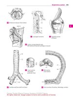
B. Muscles of the Anal Triangle (Figure 6-5)
1. Obturator Internus
■
■
■
■
Arises from the inner surface of the obturator membrane.
Has a tendon that passes around the lesser sciatic notch to insert into the medial surface of the greater trochanter of the femur.
Is innervated by the nerve to the obturator.
Laterally rotates the thigh.
2. Sphincter Ani Externus
■
■
Arises from the tip of the coccyx and the anococcygeal ligament, inserts into the central
tendon of the perineum, is innervated by the inferior rectal nerve, and closes the anus.
Is composed of three parts: subcutaneous, superficial (main part, attached to the coccyx and central tendon), and deep. Corrugator cutis ani muscle is a thin stratum of
smooth muscle fibers radiating from the superficial part of the sphincter to the deep
aspect of the perianal skin, causing puckering of that skin, which contributes to the
air-/water-tight seal of the anal canal.
3. Levator Ani Muscle
■
■
Arises from the body of the pubis, the arcus tendineus of the levator ani (a thickened
part of the obturator fascia), and the ischial spine.
Inserts into the coccyx and the anococcygeal raphe or ligament.
LWBK832-C06_p244-295.indd 248
2/23/11 10:46:57 AM
Chapter 6
Sacrum
249
Perineum and Pelvis
Sacral foramen
Iliac fossa
Piriformis muscle
Coccygeus muscle
Levator ani muscles:
Iliococccygeus
Pubococcygeus
Puborectalis
Tendinous arch
Obturator internus muscle
Obturator canal
Pubic tubercle
Anal canal
Pubic crest
Pubic symphysis
FIGURE 6-5. Muscles of the perineum and pelvis.
■
■
■
■
Is innervated by the branches of the anterior rami of sacral nerves S3 and S4 and the
perineal branch of the pudendal nerve.
Supports and raises the pelvic floor.
Consists of the puborectalis, pubococcygeus, and iliococcygeus.
Has as its most anterior fibers, which are also the most medial, the levator prostate or
pubovaginalis.
4. Coccygeus
■
■
■
■
Arises from the ischial spine and the sacrospinous ligament.
Inserts into the coccyx and the lower part of the sacrum.
Is innervated by branches of the fourth and fifth sacral nerves.
Supports and raises the pelvic floor.
C. Anal Canal (See Pelvis: VIII. B.)
IV. EXTERNAL GENITALIA AND ASSOCIATED STRUCTURES
A. Fasciae and Ligaments
1. Fundiform Ligament of the Penis
■
■
■
Arises from the linea alba and the membranous layer of the superficial fascia of the
abdomen.
Splits into left and right parts, encircles the body of the penis, and blends with the superficial penile fascia.
Enters the septum of the scrotum.
2. Suspensory Ligament of the Penis (or the Clitoris)
■
■
Arises from the pubic symphysis and the arcuate pubic ligament and inserts into the
deep fascia of the penis or into the body of the clitoris.
Lies deep to the fundiform ligaments.
3. Deep Fascia of the Penis (Buck’s Fascia)
■
■
Is a continuation of the deep perineal fascia.
Is continuous with the fascia covering the external oblique muscle and the rectus sheath.
4. Tunica Albuginea
■ Is a dense fibrous layer that envelops both the corpora cavernosa and the corpus spongiosum.
LWBK832-C06_p244-295.indd 249
2/23/11 10:46:58 AM
250
BRS Gross Anatomy
■
■
Is very dense around the corpora cavernosa, thereby greatly impeding venous return
and resulting in the extreme turgidity of these structures when the erectile tissue
becomes engorged with blood.
Is more elastic around the corpus spongiosum, which, therefore, does not become excessively turgid during erection and permits passage of the ejaculate.
5. Tunica Vaginalis
■
■
Is a serous sac of the peritoneum that covers the front and sides of the testis and epididymis.
Consists of a parietal layer that forms the innermost layer of the scrotum and a visceral
layer adherent to the testis and epididymis.
6. Processus Vaginalis
■
■
Is an embryonic diverticulum of the peritoneum that traverses the inguinal canal,
accompanying the round ligament in the female or the testis in its descent into the
scrotum and closes forming the tunica vaginalis in the male. If it does not close in
females, it forms the canal of Nuck, which is an abnormal patent pouch of peritoneum
extending into the labia majora.
Persistence of the entire processus vaginalis develops a congenital indirect inguinal
hernia, but if its middle portion persists, it develops a congenital hydrocele.
7. Gubernaculum
■
■
Is a fibrous cord that connects the fetal testis to the floor of the developing scrotum,
and its homologues in the female are the ovarian and round ligaments.
Appears to play a role in testicular descent by pulling the testis down as it migrates.
B. Male External Genitalia
1. Scrotum
■
■
■
■
■
■
Is a cutaneous pouch consisting of thin skin and the underlying dartos, which is continuous with the superficial penile fascia and superficial perineal fascia. The dartos
muscle is responsible for wrinkling the scrotal skin, and the cremaster muscle is responsible for elevating the testis.
Is covered with sparse hairs and has no fat, which is important in maintaining a temperature lower than the rest of the body for sperm production.
Contains the testis and its covering and the epididymis.
Is contracted and wrinkled when cold (or sexually stimulated) to increase its thickness
and reduce heat loss, bringing the testis into close contact with the body to conserve
heat; is relaxed when warm and hence is flaccid and distended to dissipate heat.
Receives blood from the external pudendal arteries and the posterior scrotal branches
of the internal pudendal arteries.
Is innervated by the anterior scrotal branch of the ilioinguinal nerve, the genital branch
of the genitofemoral nerve, the posterior scrotal branch of the perineal branch of the
pudendal nerve, and the perineal branch of the posterior femoral cutaneous nerve.
2. Testes (See p. 189, 263)
CLINICAL
CORRELATES
Hydrocele is an accumulation of fluid in the cavity of the tunica vaginalis (two
layers of the tunica vaginalis) of the testis or along the spermatic cord due to
an infection or injury to the testis. Hematocele is a hemorrhage into the cavity of the tunica vaginalis
due to injury to the spermatic vessels.
Varicocele is an enlargement of the pampiniform venous plexus of the spermatic cord that
appears like a “bag of worms” in the scrotum. A varicocele may cause dragging-like pain, atrophy
of the testis and/or infertility. It is more common on the left side and can be treated surgically by
removing the varicose veins.
If a man wants to have children, it is recommended that he not wear tight underwear or tight
jeans because tight clothing holds the testes close to the body wall, where higher temperatures
inhibit sperm production. Under cold conditions, the testes are pulled up toward the warm body wall,
and the scrotal skin wrinkles to increase its thickness and reduce heat loss.
LWBK832-C06_p244-295.indd 250
2/23/11 10:46:58 AM
Chapter 6
Perineum and Pelvis
251
Superficial dorsal vein
Deep dorsal vein
Dorsal artery
Dorsal nerve
Skin
Superficial fascia
Deep (Buck's) fascia
Septum penis
Deep artery of penis
Tunica albuginea
Corpus cavernosum
Corpus spongiosum
Urethra
FIGURE 6-6. Cross section of the penis.
3. Penis (Figure 6-6)
■
■
■
■
Consists of three masses of vascular erectile tissue; these are the paired corpora cavernosa and the midline corpus spongiosum, which are bounded by tunica albuginea.
Consists of a root, which includes two crura and the bulb of the penis; and the body,
which contains the single corpus spongiosum and the paired corpora cavernosa.
Has a head called the glans penis, which is formed by the terminal part of the corpus
spongiosum and is covered by a free fold of skin, the prepuce. The frenulum of the prepuce is a median ventral fold passing from the deep surface of the prepuce. The prominent margin of the glans penis is the corona, the median slit near the tip of the glans is
the external urethral orifice, and the terminal dilated part of the urethra in the glans is
the fossa navicularis.
Preputial glands are small sebaceous glands of the corona, the neck of the glans penis,
and the inner surface of the prepuce, which secrete an odoriferous substance, called
smegma.
CLINICAL
CORRELATES
Epispadias is a congenital malformation in which the spongy urethra opens as
a groove on the dorsum of the penis, frequently associated with the bladder
exstrophy (congenital eversion or turning inside out of an organ, as the bladder). Hypospadias is a
congenital malformation in which the urethra opens on the underside of the penis because of a failure of the two urethral folds to fuse completely. It is frequently associated with chordee, which is a
ventral curvature of the penis.
Circumcision is the removal of the foreskin (prepuce) that covers the glans of the penis. It is
performed as a therapeutic medical procedure for pathologic phimosis, chronic inflammations of the
penis, and penile cancer. It is also performed for cultural, religious, and medical reasons.
Phimosis is a condition in which the foreskin (prepuce) cannot be fully retracted to reveal the
glans due to a narrow opening of the prepuce. A very tight foreskin around the tip of the penis may
interfere with urination or sexual function. Paraphimosis is a painful constriction of the glans penis
caused by a tight band of constricted and retracted phimotic foreskin behind the corona. This ring of
tissue causes penile ischemia and vascular engorgement, swelling, and edema, leading to penile
gangrene.
C. Female External Genitalia
1. Labia Majora
■ Are two longitudinal folds of skin that run downward and backward from the mons pubis
and are joined anteriorly by the anterior labial commissure.
LWBK832-C06_p244-295.indd 251
2/23/11 10:46:59 AM
252
BRS Gross Anatomy
■
■
Are homologous to the scrotum of the male. Their outer surfaces are covered with pigmented skin, and after puberty, the labia majora are covered with hair.
Contain the terminations of the round ligaments of the uterus.
2. Labia Minora
■
■
Are hairless and contain no fat, unlike the labia majora.
Are divided into upper (lateral) parts, which, above the clitoris, fuse to form the prepuce
of the clitoris, and lower (medial) parts, which fuse below the clitoris to form the frenulum of the clitoris.
3. Vestibule of the Vagina (Urogenital Sinus)
■
■
Is the space or cleft between the labia minora.
Has the openings for the urethra, the vagina, and the ducts of the greater vestibular
glands in its floor.
4. Clitoris
■
■
Is homologous to the penis in the male, consists of erectile tissue, is enlarged as a result
of engorgement with blood, and is not perforated by the urethra.
Consists of two crura, two corpora cavernosa, and a glans but no corpus spongiosum.
The glans clitoris is derived from the corpora cavernosa and is covered by a sensitive
epithelium.
5. Bulbs of the Vestibule
■
■
Are the homologues of the bulb of the penis of the corpus spongiosum, a paired mass
of erectile tissue on each side of the vaginal orifice.
Are covered by the bulbospongiosus muscle, and each bulb is joined to one another
and to the undersurface of the glans clitoris by a narrow band of erectile tissue.
V. NERVE SUPPLY OF THE PERINEAL REGION (Figure 6-7)
A. Pudendal Nerve (S2–S4)
■
■
■
Passes through the greater sciatic foramen between the piriformis and coccygeus muscles.
Crosses the ischial spine and enters the perineum with the internal pudendal artery
through the lesser sciatic foramen.
Enters the pudendal canal, gives rise to the inferior rectal nerve and the perineal nerve,
and terminates as the dorsal nerve of the penis (or clitoris).
CLINICAL
CORRELATES
Pudendal nerve block is performed by injecting a local anesthetic near the
pudendal nerve. It is accomplished by inserting a needle through the posterolateral vaginal wall, just beneath the pelvic diaphragm and toward the ischial spine, thus placing the
needle around the pudendal nerve. (A finger is placed on the ischial spine and the needle is inserted
in the direction of the tip of the finger on the spine.) Pudendal block can be done subcutaneously
through the buttock by inserting the needle on the medial side of the ischial tuberosity to deposit the
anesthetic near the pudendal nerve.
1. Inferior Rectal Nerve
■
■
Arises within the pudendal canal, divides into several branches, crosses the ischiorectal fossa, and innervates the sphincter ani externus and the skin around the anus.
Communicates in the ischiorectal fossa with perineal branch of the posterior femoral cutaneous nerve, which supplies the scrotum or labium majus.
2. Perineal Nerve
■
Arises within the pudendal canal and divides into a deep branch, which supplies all
of the perineal muscles, and a superficial (posterior scrotal or labial) branch, which
supplies the scrotum or labia majora.
3. Dorsal Nerve of the Penis (or Clitoris)
■
Pierces the perineal membrane, runs between the two layers of the suspensory ligament of the penis or clitoris, and runs deep to the deep fascia on the dorsum of the
penis or clitoris to innervate the skin, prepuce, and glans.
LWBK832-C06_p244-295.indd 252
2/23/11 10:47:00 AM
Chapter 6
Dorsal nerve of penis
Perineum and Pelvis
253
Dorsal artery of penis
Scrotum
Deep artery of penis
Posterior scrotal nerves
Posterior scrotal artery
Dorsal nerve of penis
Superficial perineal branch
Deep perineal branch
Artery of bulb
Perineal artery
Perineal nerve
Inferior rectal nerve
Inferior rectal artery
Pudendal nerve
Internal pudendal
artery
FIGURE 6-7. Internal pudendal artery and pudendal nerve and branches.
VI. BLOOD SUPPLY OF THE PERINEAL REGION
(See Figure 6-7)
A. Internal Pudendal Artery
■
■
■
■
■
Arises from the internal iliac artery.
Leaves the pelvis by way of the greater sciatic foramen between the piriformis and coccygeus and immediately enters the perineum through the lesser sciatic foramen by hooking around the ischial spine.
Is accompanied by the pudendal nerve during its course.
Passes along the lateral wall of the ischiorectal fossa in the pudendal canal.
Gives rise to the following:
1. Inferior Rectal Artery
■
Arises within the pudendal canal, pierces the wall of the pudendal canal, and breaks
into several branches, which cross the ischiorectal fossa to muscles and skin around
the anal canal.
2. Perineal Arteries
■
Supply the superficial perineal muscles and give rise to transverse perineal branches
and posterior scrotal (or labial) branches.
3. Artery of the Bulb
■
LWBK832-C06_p244-295.indd 253
Arises within the deep perineal space, pierces the perineal membrane, and supplies
the bulb of the penis and the bulbourethral glands (in the male) and the vestibular
bulbs and the greater vestibular gland (in the female).
2/23/11 10:47:01 AM
254
BRS Gross Anatomy
4. Urethral Artery
■
Pierces the perineal membrane, enters the corpus spongiosum of the penis, and continues to the glans penis.
5. Deep Arteries of the Penis or Clitoris
■
■
Are terminal branches of the internal pudendal artery.
Pierce the perineal membrane, run through the center of the corpus cavernosum of
the penis or clitoris, and supply its erectile tissue.
6. Dorsal Arteries of the Penis or Clitoris
■
■
Pierce the perineal membrane and pass through the suspensory ligament of the
penis or clitoris.
Run along its dorsum on each side of the deep dorsal vein and deep to the deep fascia
(Buck’s fascia) and superficial to the tunica albuginea to supply the glans and prepuce.
B. External Pudendal Artery
■
Arises from the femoral artery, emerges through the saphenous ring, and passes medially
over the spermatic cord or the round ligament of the uterus to supply the skin above the
pubis, penis, and scrotum or labium majus.
C. Veins of the Penis
1. Deep Dorsal Vein of the Penis
■
■
Is an unpaired vein that lies in the dorsal midline deep to the deep (Buck’s) fascia and
superficial to the tunica albuginea.
Leaves the perineum through the gap between the arcuate pubic ligament and the transverse perineal ligament and drains into the prostatic and pelvic venous plexuses.
2. Superficial Dorsal Vein of the Penis
■
Runs toward the pubic symphysis between the superficial and deep fasciae and terminates
in the external (superficial) pudendal veins, which drain into the greater saphenous vein.
D. Lymph Nodes and Vessels (Figure 6-8)
1. Lymphatic Drainage of the Perineum
■
Occurs via the superficial inguinal lymph nodes, which receive lymph from the lower
abdominal wall, buttocks, penis, scrotum, labium majus, and lower parts of the vagina
Lumbar (aortic) nodes
Internal iliac nodes
Common iliac
nodes
Internal iliac
nodes
External iliac nodes
Deep inguinal nodes
Superficial
inguinal nodes
FIGURE 6-8. Lymphograph of the pelvis and lumbar region.
LWBK832-C06_p244-295.indd 254
2/23/11 10:47:01 AM
Chapter 6
Perineum and Pelvis
255
and anal canal. These nodes have efferent vessels that drain primarily into the external
iliac nodes and ultimately to the lumbar (aortic) nodes.
■ Lymph vessels from the glans penis (or clitoris) and labium minus pass to the deep
inguinal and external iliac nodes.
2. Lymphatic Drainage of the Pelvis
■
Follows the internal iliac vessels to the internal iliac nodes and subsequently to the
lumbar (aortic) nodes.
1. Internal iliac nodes receive lymph from the upper part of the rectum and vagina
and other pelvic organs, and they drain into the common iliac and then to the
lumbar (aortic) nodes. However, lymph from the uppermost part of the rectum
drains into the inferior mesenteric nodes and then to the aortic nodes.
2. Lymph from the testis and epididymis or ovary drains along the gonadal vessels
directly into the aortic nodes.
PELVIS
I. BONY PELVIS (Figures 6-9 to 6-11)
A. Pelvis
■
■
■
■
Is the basin-shaped ring of bone formed by the two hip bones, the sacrum, and the coccyx.
(The hip or coxal bone consists of the ilium, ischium, and pubis.)
Is divided by the pelvic brim or iliopectineal line into the pelvis major (false pelvis) above
and the pelvis minor (true pelvis) below.
Has an outlet that is closed by the coccygeus and levator ani muscles, which form the floor
of the pelvis.
Is normally tilted in anatomic position. Thus:
1. The anterior–superior iliac spine and the pubic tubercles are in the same vertical plane.
2. The coccyx is in the same horizontal plane as the upper margin of the pubic symphysis.
3. The axis of the pelvic cavity running through the central point of the inlet and the outlet almost parallels the curvature of the sacrum.
Iliac crest
Posterior-superior iliac spine
Anterior-superior iliac spine
Posterior sacroiliac
ligament
Anterior-inferior iliac spine
Greater sciatic foramen
Lunate (articular surface)
Acetabular fossa
Sacrospinous ligament
Acetabular notch
Lesser sciatic foramen
Superior pubic ramus
Sacrotuberous ligament
Pubic tubercle
Ischial spine
Inferior pubic ramus
Ischial tuberosity
Obturator foramen
Ramus of ischium
FIGURE 6-9. Lateral view of the hip bone.
LWBK832-C06_p244-295.indd 255
2/23/11 10:47:02 AM
256
BRS Gross Anatomy
Iliac crest
Iliac fossa
Vertebral body of L5
Lumbosacral joint
Anterior-superior iliac spine
Anterior-inferior iliac spine
Greater sciatic foramen
Sacrotuberous ligament
Sacrospinous ligament
Pubic tubercle
Coccyx
Lesser sciatic foramen
Ischial spine
Ischial tuberosity
Obturator foramen
FIGURE 6-10. Medial view of the hip bone.
B. Upper Pelvic Aperture (Pelvic Inlet or Pelvic Brim)
■ Is the superior rim of the pelvic cavity; is bounded posteriorly by the promontory of the
sacrum and the anterior border of the ala of the sacrum (sacral part), laterally by the arcuate or iliopectineal line of the ilium (iliac part), and anteriorly by the pectineal line, the
pubic crest, and the superior margin of the pubic symphysis (pubic part).
■
■
Is measured by using transverse, oblique, and anteroposterior (conjugate) diameters.
Is crossed by the ureter, gonadal vessels, middle sacral vessels, iliolumbar vessels, lumbosacral trunk, obturator nerve, spermatic cord, round ligament of the uterus, sympathetic trunk, suspensory ligament of the ovary, and so forth.
C. Lower Pelvic Aperture (Pelvic Outlet)
■ Is a diamond-shaped aperture bounded posteriorly by the sacrum and coccyx; laterally by
■
the ischial tuberosities and sacrotuberous ligaments; and anteriorly by the pubic symphysis, arcuate pubic ligament, and rami of the pubis and ischium.
Is closed by the pelvic and urogenital diaphragms.
Iliac fossa
Promontory
Body Ischial spine
Ischial spine
Body
Iliac fossa
Anterior-superior
iliac spine
Anterior-inferior
iliac spine
Iliopubic eminence
Pubic tubercle
Sacroiliac joint
Iliopectineal line
Pubic symphysis
Male pelvis
Female pelvis
FIGURE 6-11. Male and female pelvic bones.
LWBK832-C06_p244-295.indd 256
2/23/11 10:47:03 AM
Chapter 6
Perineum and Pelvis
257
D. Pelvis Major (False Pelvis)
■
Is the expanded portion of the bony pelvis above the pelvic brim.
E. Pelvis Minor (True Pelvis)
■
■
Is the cavity of the pelvis below the pelvic brim (or superior aperture) and above the pelvic
outlet (or inferior aperture).
Has an outlet that is closed by the coccygeus and levator ani muscles and the perineal
fascia, which form the floor of the pelvis.
F. Differences Between the Female and Male Pelvis
1. The bones of the female pelvis are usually smaller, lighter, and thinner than those of the
male.
2. The inlet is transversely oval in the female and heart-shaped in the male.
3. The outlet is larger in the female than in the male because of the everted ischial tuberosities in the female.
4. The cavity is wider and shallower in the female than in the male.
5. The subpubic angle or pubic arch is larger and the greater sciatic notch is wider in the
female than in the male.
6. The female sacrum is shorter and wider than the male sacrum.
7. The obturator foramen is oval or triangular in the female and round in the male.
II. JOINTS OF THE PELVIS (See Figures 6-10 and 6-11)
A. Lumbosacral Joint
■
Is the joint between vertebra L5 and the base of the sacrum, joined by an intervertebral
disk and supported by the iliolumbar ligaments.
B. Sacroiliac Joint
■ Is a synovial joint of an irregular plane type between the articular surfaces of the sacrum
■
■
and ilium.
Is covered by cartilage and is supported by the anterior, posterior, and interosseous sacroiliac ligaments.
Transmits the weight of the body to the hip bone.
C. Sacrococcygeal Joint
■ Is a cartilaginous joint between the sacrum and coccyx, reinforced by the anterior, posterior, and lateral sacrococcygeal ligaments.
D. Pubic Symphysis
■ Is a cartilaginous or fibrocartilaginous joint between the pubic bones in the median
plane.
III. PELVIC DIAPHRAGM (See Figure 6-5)
■
■
■
■
■
■
Forms the pelvic floor and supports all of the pelvic viscera.
Is formed by the levator ani and coccygeus muscles and their fascial coverings.
Lies posterior and deep to the urogenital diaphragm and medial and deep to the ischiorectal
fossa.
On contraction, raises the entire pelvic floor.
Flexes the anorectal canal during defecation and helps the voluntary control of micturition.
Helps direct the fetal head toward the birth canal at parturition.
LWBK832-C06_p244-295.indd 257
2/23/11 10:47:03 AM
258
BRS Gross Anatomy
IV. LIGAMENTS OR FOLDS AND POUCHES OF THE PELVIS
A. Broad Ligament of the Uterus (Figures 6-12 and 6-13)
■ Consists of two layers of peritoneum, extends from the lateral margin of the uterus to the
■
■
■
lateral pelvic wall, and serves to hold the uterus in position.
Contains the uterine tube, uterine vessels, round ligament of the uterus, ovarian ligament,
ureter (lower part), uterovaginal nerve plexus, and lymphatic vessels.
Does not contain the ovary but gives attachment to the ovary through the mesovarium.
Has a posterior layer that curves from the isthmus of the uterus (the rectouterine fold) to
the posterior wall of the pelvis alongside the rectum.
1. Mesovarium
■
Is a fold of peritoneum that connects the anterior surface of the ovary with the posterior layer of the broad ligament.
2. Mesosalpinx
■
Is a fold of the broad ligament that suspends the uterine tube.
3. Mesometrium
■
Is a major part of the broad ligament below the mesosalpinx and mesovarium.
B. Round Ligament of the Uterus
■
■
■
Is attached to the uterus in front of and below the attachment of the uterine tube and
represents the remains of the lower part of the gubernaculum.
Runs within the layers of the broad ligament, contains smooth muscle fibers, and holds
the fundus of the uterus forward, keeping the uterus anteverted and anteflexed.
Enters the inguinal canal at the deep inguinal ring, emerges from the superficial inguinal
ring, and becomes lost in the subcutaneous tissue of the labium majus.
C. Ovarian Ligament
■ Is a fibromuscular cord that extends from the ovary to the uterus below the uterine tube,
running within the layers of the broad ligament.
D. Suspensory Ligament of the Ovary
■ Is a band of peritoneum that extends upward from the ovary to the pelvic wall and transmits
the ovarian vessels, nerves, and lymphatics.
E. Lateral or Transverse Cervical (Cardinal or Mackenrodt’s) Ligaments of the Uterus
■ Are fibromuscular condensations of pelvic fascia from the cervix and the vagina to the pelvic walls, extend laterally below the base of the broad ligament, and support the uterus.
F. Pubocervical Ligaments
■
Are firm bands of connective tissue that extend from the posterior surface of the pubis to
the cervix of the uterus.
Mesosalpinx
Ovary
Uterine tube
Mesovarium
Mesometrium
FIGURE 6-12. Sagittal section of the broad
ligament.
LWBK832-C06_p244-295.indd 258
2/23/11 10:47:04 AM
Perineum and Pelvis
Chapter 6
Ovarian
ligament
259
Fundus of uterus
Ovary
Ovarian
artery
Uterine tube
Ampulla
Infundibulum
Fimbria
Uterine artery
Ureter
Broad ligament
Body of uterus
Isthmus of uterus
Round ligament of uterus
Vaginal artery
Internal os
Cervical canal
Cervix
External os
Vagina
FIGURE 6-13. Female reproductive organs.
G. Pubovesical (Female) or Puboprostatic (Male) Ligaments
■
Are condensations of the pelvic fascia that extend from the neck of the bladder (or the
prostate gland in the male) to the pelvic bone.
H. Sacrocervical Ligaments
■
Are firm fibromuscular bands of pelvic fascia that extend from the lower end of the sacrum to the cervix and the upper end of the vagina.
I. Inferior Pubic (Arcuate Pubic) Ligament
■
Arches across the inferior aspect of the pubic symphysis and attaches to the medial borders of the inferior pubic rami.
J. Rectouterine (Sacrouterine) Ligaments
■ Hold the cervix back and upward and sometimes elevate a shelf-like fold of peritoneum
(rectouterine fold), which passes from the isthmus of the uterus to the posterior wall of the
pelvis lateral to the rectum. It corresponds to the sacrogenital (rectoprostatic) fold in the male.
K. Rectouterine Pouch (Cul-de-sac of Douglas)
■
■
Is a sac or recess formed by a fold of the peritoneum dipping down between the rectum
and the uterus.
Lies behind the posterior fornix of the vagina and contains peritoneal fluid and some of
the small intestine.
L. Rectovesical Pouch
■
Is a peritoneal recess between the bladder and the rectum in males, and the vesicouterine
pouch is a peritoneal sac between the bladder and the uterus in females.
CLINICAL
CORRELATES
Culdocentesis is aspiration of fluid from the cul-de-sac of Douglas (rectouterine
pouch) by a needle puncture of the posterior vaginal fornix near the midline
between the uterosacral ligaments; because the rectouterine pouch is the lowest portion of the
peritoneal cavity, it can collect fluid. This procedure is done when pain occurs in the lower abdomen
and pelvic regions and when a ruptured ectopic pregnancy or ovarian cyst is suspected.
LWBK832-C06_p244-295.indd 259
2/23/11 10:47:04 AM
260
BRS Gross Anatomy
V. URETER AND URINARY BLADDER (Figures 6-14 to 6-16)
A. Ureter
■
■
■
Is a muscular tube that transmits urine by peristaltic waves.
Has three constrictions along its course: at its origin where the pelvis of the ureter joins the
ureter, where it crosses the pelvic brim, and at its junction with the bladder.
Crosses the pelvic brim in front of the bifurcation of the common iliac artery; descends
retroperitoneally on the lateral pelvic wall; and runs medial to the umbilical artery and
Urinary
bladder
Ureter
Ductus
deferens
Ampulla of
ductus
deferens
Seminal
vesicle
Prostate
Bulbourethral
(Cowper’s)
glands
Ischiopubic
ramus
Superior fascia
and muscle of
urogenital diaphragm
Bladder
A
Seminal
vesicle
Anterior
fibromuscular
stroma
Central
zone
Ejaculatory
duct
Peripheral
zone
Urethra
Transition
zone
Verumontanum
Anterior lobe
Utricle
Middle lobe
Lateral lobe
B
Urethra
Lateral lobe
Seminal
colliculus
Prostatic fascia
C
Ejaculatory
ducts
Posterior lobe
FIGURE 6-14. Male urogenital organs.
LWBK832-C06_p244-295.indd 260
2/23/11 10:47:05 AM
Chapter 6
Rectus
abdominis
Ileum
261
Perineum and Pelvis
Common iliac
artery
Descending
colon
Ureter
Common iliac
vein
Sympathetic
trunk
Femoral artery
Ureter
Psoas major
Gluteus medius
Bladder
Femoral vein
Obturator
vessels
Seminal vesicle
Rectum
Sartorius
Spermatic cord
Femoral artery
Rectus femoris
Tensor
fasciae latae
Pectineus
Prostatic
urethra
Gluteus
maximus
Pectineus
Femoral artery
Deep femoral
artery
Femoral vein
Adductor
magnus
Quadratus
femoris
Ischiorectal
fossa
Femoral vein
Adductor longus
Obturator
externus
Obturator
internus
Spermatic cord
Pectineus
Adductor longus
muscle
Ischiocavernosus
(Crus of penis)
Bulbospongiosus
(Bulb of penis)
Pudendal canal
Anal canal
FIGURE 6-15. Computed tomography scans of the male pelvis and perineum.
■
the obturator vessels and posterior to the ovary, forming the posterior boundary of the
ovarian fossa.
In females, it is accompanied in its course by the uterine artery, which runs above and
anterior to it in the base of the broad ligament of the uterus. Because of its location, the
ureter is in danger of being injured in the process of hysterectomy. It can be remembered
by the mnemonic device, “water (ureter) runs under the bridge (uterine artery).”
LWBK832-C06_p244-295.indd 261
2/23/11 10:47:05 AM
262
BRS Gross Anatomy
Bladder
Femoral artery
Femoral vein
Neck of femur
Ischial tuberosity
Obturator
internus muscle
Uterine cervix
Greater trochanter
Superior gemellus
Rectum
Gluteus maximus
Coccyx
Pubis
Labium majus
Obturator externus
Urethra
Quadratus femoris
Obturator
internus muscle
Ischial tuberosity
Vagina
Gluteus maximus
muscle
Anal canal
FIGURE 6-16. Computed tomography scans of the female pelvis and perineum.
■
■
■
Passes posterior and inferior to the ductus deferens and lies in front of the seminal vesicle
before entering the posterolateral aspect of the bladder in males.
Enters obliquely through the base of the bladder and opens by a slit-like orifice that acts
as a valve, and the circular fibers of the intramural part of the ureter act as a sphincter.
When the bladder is distended, the valve and sphincter actions prevent the reflux of urine
from the urinary bladder into the ureter.
Receives blood from the aorta and the renal, gonadal, common and internal iliac, umbilical, superior and inferior vesical, and middle rectal arteries.
CLINICAL
CORRELATES
Damage of the ureter: in the female, damage may occur during a hysterectomy
or surgical repair of a prolapsed uterus because it runs under the uterine
artery. The ureter is inadvertently clamped, ligated, or divided during a hysterectomy when the uterine artery is being ligated to control uterine bleeding.
B. Urinary Bladder
■
■
■
■
■
■
Is situated below the peritoneum and is slightly lower in the female than in the male.
Extends upward above the pelvic brim as it fills; may reach as high as the umbilicus if fully
distended.
Has the apex at the anterior end and the fundus or base as its posteroinferior triangular
portion.
Has a neck, which is the area where the fundus and the inferolateral surfaces come
together, leading into the urethra.
Has a uvula, which is a small eminence at the apex of its trigone, projecting into the orifice
of the urethra. The trigone is bounded by the two orifices of the ureters and the internal
urethral orifice, around which is a thick circular layer called the internal sphincter (sphincter vesicae).
Has bundles of smooth muscle fibers that, as a whole, are known as the detrusor muscle of
the bladder.
LWBK832-C06_p244-295.indd 262
2/23/11 10:47:06 AM
Chapter 6
■
■
Perineum and Pelvis
263
Receives blood from the superior and inferior vesical arteries (and from the vaginal artery
in females). Its venous blood is drained by the prostatic (or vesical) plexus of veins, which
empties into the internal iliac vein.
Is innervated by nerve fibers from the vesical and prostatic plexuses. The parasympathetic
nerve (pelvic splanchnic nerve originating from S2–S4) stimulates to contract the musculature (detrusor) of the bladder wall, relaxes the internal urethral sphincter, and promotes
emptying. The sympathetic nerve relaxes the detrusor of the bladder wall and constricts
the internal urethral sphincter.
CLINICAL
CORRELATES
Bladder cancer usually originates in cells lining the inside of the bladder (epithelial cells). The most common symptom is blood in the urine (hematuria).
Other symptoms include frequent urination and pain upon urination (dysuria). This cancer may be
induced by organic carcinogens that are deposited in the urine after being absorbed from the environment and also by cigarette smoking.
Tenesmus is a constant feeling of the desire to empty the bladder or bowel, accompanied by
pain, cramping, and straining due to a spasm of the urogenital diaphragm.
Interstitial cystitis is a chronic inflammatory condition of the bladder that causes frequent,
urgent, and painful urination.
C. Urethra
■
■
■
Is serves as a passage for urine from the urinary bladder to the exterior, but in male, it also
serves as a passage for semen.
Male urethra is approximately 20 cm long and consists of three parts: prostatic, membranous, and spongy. The lowest part of the membranous urethra is liable to rupture or to
penetration by a catheter.
Female urethra is approximately 4 cm long, and its external urethral orifice is situated
between the labia minora, in front of the vaginal opening but behind the glans clitoris.
D. Micturition (Urination)
■
■
■
Is initiated by stimulating stretch receptors in the detrusor muscle in the bladder wall by the
increasing volume (approximately 300 mL for adults) of urine.
Can be assisted by contraction of the abdominal muscles, which increases the intraabdominal and pelvic pressures.
Involves the following processes:
1. Sympathetic (general visceral efferent [GVE]) fibers induce relaxation of the bladder wall
and constrict the internal sphincter, inhibiting emptying. (They may also activate the
detrusor to prevent the reflux of semen into the bladder during ejaculation.)
2. General visceral afferent (GVA) impulses arise from stretch receptors in the bladder wall
and enter the spinal cord (S2–S4) via the pelvic splanchnic nerves.
3. Parasympathetic preganglionic (GVE) fibers in the pelvic splanchnic nerves synapse in
the pelvic (inferior hypogastric) plexus; postganglionic fibers to the bladder musculature induce a reflex contraction of the detrusor muscle and relaxation of the internal
urethral sphincter, enhancing the micturition.
4. General somatic efferent (GSE) fibers in the pudendal nerve cause voluntary relaxation
of the external urethral sphincter, and the bladder begins to void.
5. At the end of micturition, the external urethral sphincter contracts, and bulbospongiosus muscles in the male expel the last few drops of urine from the urethra.
VI. MALE GENITAL ORGANS (Figures 6-17 and 6-18;
See Figures 6-14 and 6-15)
A. Testis
■
■
Develops retroperitoneally and descends into the scrotum retroperitoneally.
Is covered by the tunica albuginea, which lies beneath the visceral layer of the tunica vaginalis.
LWBK832-C06_p244-295.indd 263
2/23/11 10:47:06 AM
264
BRS Gross Anatomy
Bladder
Corpus cavernosum
Ureter
Glans penis
Ejaculatory duct
Ampulla of ductus deferens
Seminal vesicle
Prostate gland
Membranous urethra
Bulbourethral gland
Corpus spongiosum
(spongy urethra)
Bulb of penis
Crus of penis
Epididymis
Ductus deferens
Testis
FIGURE 6-17. Male reproductive organs.
■
Produces spermatozoa and secretes sex hormones.
■
Is supplied by the testicular artery from the abdominal aorta and is drained by veins of the
pampiniform plexus.
Has lymph vessels that ascend with the testicular vessels and drain into the lumbar (aortic)
nodes; lymphatic vessels in the scrotum drain into the superficial inguinal nodes.
■
Sigmoid colon
Ureter
Urinary bladder
Peritoneum
Rectum
Rectovesical
pouch
Ampulla of
ductus deferens
Seminal vesicle
Ejaculatory duct
Prostate gland
Urogenital
diaphragm
External anal
sphincter muscle
Ductus deferens
Symphysis pubis
Deep dorsal vein of penis
Corpus cavernosum penis
Corpus spongiosum penis
Testicular artery and vein
Head of epididymis
Glans penis
Testis
Anal canal
Bulbourethral gland and duct
Bulb of penis
FIGURE 6-18. Sagittal section of the male pelvis.
LWBK832-C06_p244-295.indd 264
2/23/11 10:47:07 AM
Chapter 6
Perineum and Pelvis
265
CLINICAL
CORRELATES
Testicular torsion is twisting of a testis such that the spermatic cord becomes
twisted, obstructing blood supply to the testis, and causing sudden urgent pain
and swelling of the scrotum or nausea and vomiting. It is most common during adolescence and
may be caused by trauma or a spasm of the cremaster muscle. Testicular torsion requires emergency treatment and if not untwisted, testicular necrosis will occur.
Orchitis is inflammation of the testis and is marked by pain, swelling, and a feeling of heaviness
in the testis. It may be caused by the mumps, gonorrhea, syphilis, or tuberculosis. If testicular infection spreads to the epididymis, it is called epididymo-orchitis.
Testicular cancer develops commonly from the rapidly dividing early state spermatogenic cells
(seminoma or germ cell tumor). Tumor also develops from Leydig cells, which produce androgen
(Leydig cell tumor), and Sertoli cells, which support and nourish germ cells and produce androgenbinding protein and the hormone inhibin (Sertoli cell tumor). Signs and symptoms include a painless
mass or lump, testicular swelling, hardness, and a feeling of heaviness or aching in the scrotum or
lower abdomen. The cause of cancer is unknown, but the major risk factors are cryptorchidism and
Klinefelter’s syndrome (47, XXY sex chromosome, seminiferous tubule dysgenesis, gynecomastia,
and infertility). Metastasis occurs via lymph and blood vessels. It can be treated by surgical removal
of the affected testis and spermatic cord (orchiectomy), radiotherapy, and chemotherapy.
Cryptorchidism is a congenital condition in which the testis fails to descend into the scrotum
during fetal development. Undescended testes are associated with reduced fertility, increased risk
of testicular cancer, and higher susceptibility to testicular torsion and inguinal hernias. Undescended testes are brought down into the scrotum in infancy by a surgical procedure called an
orchiopexy or orchidopexy.
B. Epididymis
■
■
Consists of a head, body, and tail, and contains a convoluted duct approximately 6 m (20 ft)
long.
Functions in the maturation and storage of spermatozoa in the head and body and propulsion of the spermatozoa into the ductus deferens.
C. Ductus Deferens
■
■
■
Is a thick-walled tube that enters the pelvis at the deep inguinal ring at the lateral side of
the inferior epigastric artery.
Crosses the medial side of the umbilical artery and obturator nerve and vessels, passes
superior to the ureter near the wall of the bladder, and is dilated to become the ampulla at
its terminal part.
Contains fructose, which is nutritive to spermatozoa, and receives innervation primarily
from sympathetic nerves of the hypogastric plexus and parasympathetic nerves of the
pelvic plexus.
CLINICAL
CORRELATES
Vasectomy is surgical excision of a portion of the vas deferens (ductus deferens) through the scrotum. It stops the passage of spermatozoa but neither
reduces the amount of ejaculate greatly nor diminishes sexual desire.
D. Ejaculatory Ducts
■
■
Are formed by the union of the ductus deferens with the ducts of the seminal vesicles.
Peristaltic contractions of the muscular layer of the ductus deferens and the ejaculatory
ducts propel spermatozoa with seminal fluid into the urethra.
Open into the prostatic urethra on the seminal colliculus just lateral to the blind prostatic
utricle (see the section on urethral crest).
E. Seminal Vesicles
■
■
Are enclosed by dense endopelvic fascia and are lobulated glandular structures that are
diverticula of the ductus deferens.
Lie inferior and lateral to the ampullae of the ductus deferens against the fundus (base) of
the bladder.
LWBK832-C06_p244-295.indd 265
2/23/11 10:47:08 AM
266
■
■
■
BRS Gross Anatomy
Produce the alkaline constituent of the seminal fluid, which contains fructose and
choline.
Have lower ends that become narrow and form ducts, which join the ampullae of the
ductus deferens to form the ejaculatory ducts.
Do not store spermatozoa, as was once thought; this is done by the epididymis, the ductus
deferens, and its ampulla.
CLINICAL
CORRELATES
Seminal vesicles produce the alkaline constituent of the seminal fluid, which
contains fructose and choline. Fructose provides a forensic determination for
occurrence of rape, whereas choline crystals provide the basis for the determination of the presence of semen (Florence’s test).
F. Prostate Gland
■
■
■
■
■
■
Is located at the base of the urinary bladder and consists chiefly of glandular tissue mixed
with smooth muscle and fibrous tissue.
Has five lobes: the anterior lobe (or isthmus), which lies in front of the urethra and is
devoid of glandular substance; the middle (median) lobe, which lies between the urethra
and the ejaculatory ducts and is prone to benign hypertrophy obstructing the internal urethral orifice; the posterior lobe, which lies behind the urethra and below the ejaculatory
ducts, contains glandular tissue, and is prone to carcinomatous transformation; and the
right and left lateral lobes, which are situated on either side of the urethra and form the
main mass of the gland.
Secretes a fluid that produces the characteristic odor of semen. This fluid, the secretion
from the seminal vesicles and the bulbourethral glands, and the spermatozoa constitute
the semen or seminal fluid.
Secretes prostate-specific antigen (PSA), prostaglandins, citric acid and acid phosphatase,
and proteolytic enzymes.
Has ducts that open into the prostatic sinus, a groove on either side of the urethral crest.
Receives the ejaculatory duct, which opens into the urethra on the seminal colliculus just
lateral to the blind prostatic utricle.
CLINICAL
CORRELATES
Hypotrophy of the prostate is a benign enlargement of the prostate that affects
older men and occurs most often in the middle lobe, obstructing the internal
urethral orifice and thus leading to nocturia (excessive urination at night), dysuria (difficulty or pain
in urination), and urgency (sudden desire to urinate). Cancer occurs most often in the posterior lobe.
Transurethral resection of the prostate (TURP) is surgical removal of the prostate by means of a
cystoscope passed through the urethra. Prostatitis is inflammation of the prostate.
Prostate cancer is a slow-growing cancer that occurs particularly in the posterior lobe. It is
usually symptomless in the early stages, but it can impinge on the urethra in the late stage. Prostate
cancer spreads to the bony pelvis, pelvic lymph nodes, vertebral column, and skull via the vertebral
venous plexus, producing pain in the pelvis, the lower back, and the bones. This cancer also metastasizes to the heart and lungs through the prostatic venous plexus, internal iliac veins, and into the
inferior vena cava. It can be detected by digital rectal examination, ultrasound imaging with a device
inserted into the rectum, or PSA test. PSA concentration in the blood of normal males is less than
4.0 ng/mL.
Prostatectomy is surgical removal of a part or all of the prostate gland. Perineal prostatectomy is removal of the prostate through an incision in the perineum. Radical prostatectomy is
removal of the prostate with seminal vesicles, ductus deferens, some pelvic fasciae, and pelvic
lymph nodes through the retropubic or the perineal route. Transurethral prostatectomy is resection
of the prostate by means of a cystoscope passed through the urethra. A careful dissection of the
pelvic and prostatic nerve plexuses is required during prostatectomy to avoid loss of erection and
ejaculation.
LWBK832-C06_p244-295.indd 266
2/23/11 10:47:09 AM
Chapter 6
Perineum and Pelvis
267
G. Urethral Crest
■
■
Is located on the posterior wall of the prostatic urethra and has numerous openings for the
prostatic ducts on either side.
Has an ovoid-shaped enlargement called the seminal colliculus (verumontanum), on which
the two ejaculatory ducts and the prostatic utricle open. At the summit of the colliculus is
the prostatic utricle, which is an invagination (a blind pouch) approximately 5 mm deep;
it is analogous to the uterus and vagina in the female.
H. Prostatic Sinus
■
Is a groove between the urethral crest and the wall of the prostatic urethra and receives
the ducts of the prostate gland.
I. Erection
■
■
■
Depends on stimulation of parasympathetics from the pelvic splanchnic nerves, which
dilates the arteries supplying the erectile tissue, and thus causes engorgement of the corpora cavernosa and corpus spongiosum, compressing the veins and thus impeding
venous return and causing full erection.
Is also maintained by contraction of the bulbospongiosus and ischiocavernosus muscles,
which compresses the erectile tissues of the bulb and the crus.
Is often described using a popular mnemonic device: point (erection by parasympathetic)
and shoot (ejaculation by sympathetic).
J. Ejaculation
■
■
■
Begins with nervous stimulation. Friction to the glans penis and other sexual stimuli
result in excitation of sympathetic fibers, leading to contraction of the smooth muscle of
the epididymal ducts, the ductus deferens, the seminal vesicles, and the prostate in
turn.
Occurs as a result of contraction of the smooth muscle, thus pushing spermatozoa and the
secretions of both the seminal vesicles and prostate into the prostatic urethra, where they
join secretions from the bulbourethral and penile urethral glands. All of these secretions
are ejected together from the penile urethra because of the rhythmic contractions of the
bulbospongiosus, which compresses the urethra.
Involves contraction of the sphincter of the bladder, preventing the entry of urine into the
prostatic urethra and the reflux of the semen into the bladder.
VII. FEMALE GENITAL ORGANS (Figure 6-19;
See Figures 6-13 and 6-16)
A. Ovaries
■
■
■
■
■
■
Lie on the posterior aspect of the broad ligament on the side wall of the pelvic minor and
are bounded by the external and internal iliac vessels.
Are not covered by the peritoneum, and thus the ovum or oocyte is expelled into the peritoneal cavity and then into the uterine tube.
Are not enclosed in the broad ligament, but their anterior surface is attached to the posterior layer of the broad ligament by the mesovarium.
Have a surface that is covered by germinal (columnar) epithelium, which is modified from
the developmental peritoneal covering of the ovary.
Are supplied primarily by the ovarian arteries, which are contained in the suspensory
ligament and anastomose with branches of the uterine artery.
Are drained by the ovarian veins; the right ovarian vein joins the inferior vena cava, and
the left ovarian vein joins the left renal vein.
LWBK832-C06_p244-295.indd 267
2/23/11 10:47:09 AM
268
BRS Gross Anatomy
Suspensory ligament
Ureter
Ovary
Uterine tube
Fimbria
Isthmus of
uterus
Cervix of uterus
Rectouterine
pouch
Posterior fornix
of vagina
Rectum
Vagina
Fundus of uterus
Round ligament of uterus
Urinary bladder
Symphysis pubis
Urogenital diaphragm
Glans clitoris
Sphincter urethra
Labium majus
External anal
sphincter muscle
Anal canal
Deep transverse perineal muscle
Labium minus
FIGURE 6-19. Sagittal section of the female pelvis.
CLINICAL
CORRELATES
Ovarian cancer develops from germ cells that produce ova or eggs, stromal
cells that produce estrogen and progesterone, and epithelial cells that cover
the outer surface of the ovary. Its symptoms include a feeling of pressure in the pelvis or changes in
bowel or bladder habits. Diagnosis involves feeling a mass during a pelvic examination, visualizing it
by using an ultrasound probe placed in the vagina, or using a blood test for a protein associated with
ovarian cancer (CA-125). Some germ cell cancers release certain protein markers, such as human
chorionic gonadotropin and ␣-fetoprotein, into the blood. Cancer signs and symptoms include unusual vaginal bleeding, postmenopausal bleeding, bleeding after intercourse and pain during intercourse, pelvic pressure, abdominal and pelvic pain, back pain, indigestion, and loss of appetite.
B. Uterine Tubes
■
Extend from the uterus to the uterine end of the ovaries and connect the uterine cavity to
the peritoneal cavity.
■
■
■
Are each subdivided into four parts: the uterine part, the isthmus, the ampulla (the longest
and widest part), and the infundibulum (the funnel-shaped termination formed of fimbriae).
Convey the fertilized or unfertilized oocytes to the uterus by ciliary action and muscular contraction, which takes 3 to 4 days.
Transport spermatozoa in the opposite direction (toward the eggs); fertilization takes place
within the tube, usually in the infundibulum or ampulla. Fertilization is the process beginning with penetration of the secondary oocyte by the sperm and completed by fusion of
the male and female pronuclei.
C. Uterus
■
■
Is the organ of gestation in which the fertilized oocyte normally becomes embedded and
the developing organism grows until its birth.
Is normally anteverted (i.e., angle of 90 degrees at the junction of the vagina and cervical canal)
and anteflexed (i.e., angle of 160 to 170 degrees at the junction of the cervix and body).
LWBK832-C06_p244-295.indd 268
2/23/11 10:47:09 AM